section name header

Basic Information

AUTHOR: Narges Alipanah-Lechner, MD

Definition

Aspiration pneumonia refers to pulmonary infection of the lower airways and lung parenchyma resulting from entry of colonized oropharyngeal or upper gastrointestinal contents.1 Aspiration pneumonia is considered part of the continuum that also includes community- and hospital-acquired pneumonias.1 Chemical pneumonitis (or aspiration pneumonitis) refers to lung injury and inflammation resulting from entry of sterile substances toxic to the lower airways.

Synonyms

Pneumonia, aspiration

Aspiration pneumonitis

ICD-10CM CODE
J69.0Pneumonitis due to inhalation of food and vomit
Epidemiology & Demographics
Incidence (In U.S.)

  • 7.1 cases per 10,000 people admitted to the hospital2
  • 30.9 cases per 10,000 people admitted to the hospital >65 yr of age2
Prevalence (In U.S.)

Unknown (unreliable data)

Predominant Sex

Slight male predominance2

Predominant Age

>65 yr

Peak Incidence

Elderly patients in hospitals or nursing homes2,3

Physical Findings & Clinical Presentation

  • Symptoms develop within hours to a few days after aspiration event, though anaerobic infections can have a more subacute pre-sentation.
  • Clinical presentation ranges from minimal symptoms to fulminant respiratory failure.
  • Symptoms can include dyspnea, diffuse wheeze, cough, hypoxia, tachypnea, tachycardia, sputum production, and fever.
  • Lung exam may demonstrate wheezes, crackles, or rhonchi.
Etiology

Complex interaction of etiologies, ranging from chemical (often acid) pneumonitis after aspiration of sterile gastric contents (generally not requiring antibiotic treatment) to bacterial aspiration. Risk factors for aspiration pneumonia include vomiting, decreased consciousness, poor dentition, ineffective cough reflex or glottic closure, and gastroesophageal reflux disease.4-11Table 1 and Fig. E1 summarize risk factors for aspiration pneumonia.

TABLE 1 Risk Factors for Dysphagia and Aspiration Pneumonia

Cerebrovascular disease
Ischemic stroke
Hemorrhagic stroke
Subarachnoid hemorrhage
Degenerative neurologic disease
Alzheimer disease
Multiinfarct dementia
Parkinson disease
Amyotrophic lateral sclerosis (motor neuron disease)
Multiple sclerosis
Head and neck cancer
Oropharyngeal malignancy
Oral cavity malignancy
Esophageal malignancy
Other
Scleroderma
Diabetic gastroparesis
Reflux esophagitis
Presbyesophagus
Achalasia

From Vincent JL et al: Textbook of critical care, ed 7, Philadelphia, 2017, Elsevier.

Figure E1 Risk factors for aspiration.

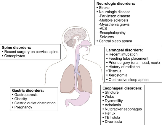
ALS, Amyotrophic lateral sclerosis; TE, tracheoesophageal.

From Broaddus VC et al: Murray & Nadel’s textbook of respiratory medicine, ed 7, Philadelphia 2022, Elsevier.

Community-Acquired Aspiration Pneumonia

  • Most patients have a mixed infection with aerobic and anaerobic bacteria. The most common bacteria are Streptococcus pneumoniae, Staphylococcus aureus, Haemophilus influenzae, and Enterobacteriaceae.12-14 Anaerobes (Peptostreptococcus, Fusobacterium nucleatum, Fusobacterium necrophorum, Prevotella, and Bacteroides species) are less frequently isolated.15
  • High-risk groups: age >65 yr; alcohol use; IV drug use; altered mental status; stroke victims; cardiac arrest; and patients with esophageal disorders, seizures, periodontal disease, or recent dental manipulations.4-11
Hospital-Acquired Aspiration Pneumonia

  • Causative organisms:
    1. Anaerobes listed above, although in many studies gram-negative aerobes (60%) and gram-positive aerobes (20%) predominate13,16-18
    2. E. coli, P. aeruginosa, S. aureus including MRSA, Klebsiella, Enterobacter, Serratia, Proteus spp., H. influenzae, S. pneumoniae, Legionella, and Acinetobacter spp. (sporadic pneumonias) in two thirds of cases
    3. Fungi, including Candida albicans, in <1%19
  • High-risk groups: Seriously ill hospitalized patients (especially patients with coma, acidosis, alcohol use disorder, uremia, diabetes mellitus, nasogastric intubation, or recent antimicrobial therapy), who are frequently colonized with aerobic gram-negative rods; patients undergoing anesthesia; those with strokes, dementia, or swallowing disorders; patients >65 yr; and those receiving antacids or H2 blockers, or proton pump inhibitors (but not sucralfate).
  • Hypoxic patients receiving concentrated O2 have diminished ciliary activity, increasing risk for aspiration pneumonia.

Diagnosis

Differential Diagnosis

  • Other necrotizing or cavitary pneumonias (especially tuberculosis, gram-negative pneumonias)
  • See “Tuberculosis, Pulmonary”
Workup

  • Chest x-ray
  • Complete blood count (CBC), blood cultures
  • Sputum Gram stain and culture
  • Consideration of tracheal aspirate or bronchoscopy sample if intubated
  • CT of chest if diagnosis is unclear or when suspecting complications
Laboratory Tests

  • CBC: Leukocytosis often present.
  • Sputum Gram stain:
    1. Often useful when carefully prepared immediately after obtaining suctioned or expectorated specimen, examined by experienced observer.
    2. Only specimens with multiple white blood cells and rare or absent epithelial cells should be examined.
    3. Unlike other bacterial pneumonias (e.g., pneumococcal), multiple organisms may be present in aspiration pneumonia.
    4. Long, slender rods suggest anaerobes, though these organisms are difficult to isolate due to frequent contamination of sputum samples with oral flora.
    5. Sputum from pneumonia caused by acid aspiration may be devoid of organisms.
    6. Cultures should be interpreted in light of morphology of visualized organisms.
Imaging Studies

  • Chest x-ray may be negative early in the disease course.20 It often reveals opacities in gravity-dependent lung regions. In an upright patient, opacities commonly affect the basal segments of the lower lobes. In a supine patient, opacities can involve the superior segment of the lower lobes, posterior right upper lobe, or the apicoposterior segment of the left upper lobe (Fig. 2). Chest x-ray can also demonstrate a pattern of diffuse lung involvement characteristic of acute respiratory distress syndrome. (See related content in “Acute Respiratory Distress Syndrome.”)
  • Aspiration pneumonia of several days’ duration (or longer) may reveal necrosis (especially community-acquired anaerobic pneumonias) and even cavitation with air-fluid levels, indicating lung abscess.
Figure 2 Anaerobic Necrotizing Pneumonia Following Aspiration of Oropharyngeal Secretions

Multiple, small (<2 cm) radiolucencies are seen throughout the posterior segment of the right upper lobe on the postero-anterior (A) and lateral (B) projections.

Courtesy Michael Gotway, MD. From Mason RJ et al: Murray & Nadel’s textbook of respiratory medicine, ed 5, Philadelphia, 2010, Saunders.

Treatment

Nonpharmacologic Therapy

  • Management to prevent repeated aspiration
  • Ventilatory support if necessary
  • Rehabilitative management: Physical, pulmonary, and dysphagia therapy combined with appropriate nutrition can reduce length of stay and mortality
  • Fig. 3 illustrates a prevention and treatment algorithm for pulmonary aspiration

Figure 3 Prevention and treatment algorithm for pulmonary aspiration.

!!flowchart!!

ARDS, Acute respiratory distress syndrome; BAL, bronchoalveolar lavage.

From Newman M et al: Perioperative medicine, ed 2, Philadelphia 2022, Elsevier.

Acute General Rx

Chemical pneumonitis: Acute aspiration of acidic gastric contents without bacteria may not require antibiotic therapy; initial treatment involves airway maintenance and management of bronchospasm and airway edema. Routine adjunctive treatment with glucocorticoids is not recommended. Empiric antibiotics may be considered in severe cases, but their ongoing use should be reassessed at 48 to 72 hr.1

  • Aspiration pneumonia: Antibiotic selection depends on the site of acquisition (long-term care facility, hospital, community), which modifies risk factors for infection with multidrug-resistant pathogens. Additionally, a history of treatment with broad-spectrum antibiotics in the past 90 days warrants empiric treatment for multidrug-resistant organisms.21,22
    1. Community-acquired aspiration pneumonia or hospital-acquired cases with low risk of multidrug-resistant pathogens: Ampicillin-sulbactam 1.5 to 3 g every 6 h IV, or amoxicillin-clavulanate PO 875 mg twice daily, or a fluoroquinolone (levofloxacin 750 mg IV or PO) is effective. Clindamycin (450 mg oral qid or 600 mg IV every 8 h) can be added to the other agent when the risk of predominantly anaerobic infection is high.
    2. Nursing home aspirations or hospital-acquired aspiration pneumonias with concerns for resistance: Broad-spectrum treatment with piperacillin-tazobactam 4.5 g q8h or 3.375 g q6h, cefepime 2 g q8h, levofloxacin 750 mg IV or PO once daily, imipenem 500 mg q6h or 1 g q8h, or meropenem 1 g q8h. The addition of vancomycin (15 mg/kg q12h IV) or linezolid (600 mg q12h PO or IV) is warranted if MRSA is suspected or known (e.g., documented nasal or respiratory colonization with MRSA).
      1. Knowledge of resident flora in the microenvironment of the aspiration within the hospital is crucial to intelligent antibiotic selection; consult infection control nurses or hospital epidemiologist.
      2. Confirmed Pseudomonas pneumonia in patients not in septic shock or at high risk of death can be treated with an antipseudomonal beta-lactam agent (piperacillin/tazobactam, cefepime, meropenem) or an antipseudomonal fluoroquinolone pending results of susceptibility testing. In severe cases, empiric two-drug combination ther-apy with an antipseudomonal beta-lactam agent and an antipseudomonal fluoroquinolone or an aminoglycoside is recommended. The choice of agents should be guided by the results of drug susceptibility testing when available.
Disposition

Repeat chest x-ray in 6 to 8 wk in most patients.

Prevention

For patients with difficulty swallowing thin liquids, adding thickening agents to provide nectar-thick, pudding-thick, and honey-thick fluids is an option. Sitting patients upright when eating (and for some time afterward), chin tucking when swallowing, eating more slowly, and use of various swallowing maneuvers are additional techniques for prevention of aspiration in elderly and debilitated patients (Box 1).

BOX 1 Reducing Risk of Aspiration and Aspiration Pneumonia in Older Adults

Hand Feeding

  • Provide a rest period (>30 min) before feeding time.
  • Have the patient sit upright at 90 degrees or highest position allowed by medical condition.
  • Avoid rushed or forced feeding; feeding by syringe is risky.
  • Alternate liquids with solids.
  • Recognize the high risks of sedatives, hypnotics, and other psychotropic medications, and try to wean or reduce dosages.
  • Speech-language therapist referral: Evaluate patient for possible benefit of chin-down position when swallowing or of adjusting liquid viscosity; thickened liquids of varying types may improve swallowing in some patients (ice cream and Jell-O are considered thin liquids).
Tube Feeding

  • Note: Both nasogastric and gastrostomy tube feeding may increase aspiration risks.
  • Consider continuous feedings rather than intermittent (bolus) feedings.
  • Keep backrest elevated at least 30 degrees during feedings, if possible.
  • Consider pump-assisted feedings rather than gravity-controlled feedings.
  • A gastric residual volume >200 ml during continuous feeding or before intermittent feedings may increase risk (but this remains controversial).23
  • Prokinetic agents such as metoclopramide or erythromycin may improve feeding tolerance, but are associated with their own serious potential side effects.
  • Placing the feeding tube tip beyond the pylorus (jejunostomy, gastrojejunostomy) may reduce aspiration in some patients.
  • Using colored dye in tube feeding is contraindicated (it was originally thought that adding coloring to liquid tube feeding formulas would help identify probable feeding aspiration if the dye was found after throat and pulmonary suctioning).24

From Fillit HM: Brocklehurst's textbook of geriatric medicine and gerontology, ed 8, Philadelphia, 2017, Elsevier.

Referral

Consultation with infectious disease and/or pulmonary specialists recommended for patients with respiratory distress, hypoxia, ventilatory support, pneumonia in more than one lobe, necrosis or cavitation on chest x-ray, or for those not clinically responding to antibiotic therapy within 2 to 3 days.

Related Content

Aspiration Pneumonia (Patient Information)

Most of these suggestions are consensus and expert opinions rather than evidence-based practices.25-28

Related Content

.

    1. Mandell L.A., Niederman M.S. : Aspiration pneumoniaN Engl J Med. ;380(7), 2019.doi:10.1056/NEJMra1714562
    2. Wu C.P. : National trends in admission for aspiration pneumonia in the United States, 2002-2012Ann Am Thorac Soc. ;14(6):874-879, 2017.doi:10.1513/AnnalsATS.201611-867OC
    3. Almirall J. : Aspiration pneumonia: a renewed perspective and practical approachRespir Med. ;185, 2021.doi:10.1016/j.rmed.2021.106485
    4. DiBardino D.M., Wunderink R.G. : Aspiration pneumonia: a review of modern trendsJ Crit Care. ;30(1):40-48, 2015.doi:10.1016/j.jcrc.2014.07.011
    5. Almirall J. : Oropharyngeal dysphagia is a risk factor for community-acquired pneumonia in the elderlyEur Respir J. ;41(4), 2013.doi:10.1183/09031936.00019012
    6. Macht M. : Swallowing dysfunction after critical illnessChest. ;146(6), 2014.doi:10.1378/chest.14-1133
    7. Taylor J. : Risk factors for aspiration in community-acquired pneumonia: analysis of a hospitalized UK cohortAm J Med. ;126(11), 2013.doi:10.1016/j.amjmed.2013.07.012
    8. van der Maarel-Wierink C. : Meta-analysis of dysphagia and aspiration pneumonia in frail eldersJ Dental Res. ;90(12), 2011.doi:10.1177/0022034511422909
    9. Herzig S. : Antipsychotics and the risk of aspiration pneumonia in individuals hospitalized for nonpsychiatric conditions: a cohort studyJ Am Geriatr Soc. ;65(12), 2017.doi:10.1111/jgs.15066
    10. Hannawi Y. : Stroke-associated pneumonia: major advances and obstaclesCerebrovasc Dis. ;35(5), 2013.doi:10.1159/000350199
    11. Perbet S et al: Early-onset pneumonia after cardiac arrest: characteristics, risk factors and influence on prognosis, Am J Respir Critical Care Med 184(9), 2011. https://doi.org/10.1164/rccm.201102-0331OC.
    12. Polverino E. : Nursing home-acquired pneumonia: a 10 year single-centre experienceThorax. ;65(4), 2010.doi:10.1136/thx.2009.124776
    13. El-Solh A. : Microbiology of severe aspiration pneumonia in institutionalized elderlyAm J Respir Crit Care Med. ;167(12), 2003.doi:10.1164/rccm.200212-1543OC
    14. Takayanagi N. : Etiology and outcome of community-acquired lung abscessRespiration. ;80(2), 2010.doi:10.1159/000312404
    15. Bartlett J. : Anaerobic bacterial infections of the lung and pleural spaceClin Infect Dis. ;16(Suppl 4), 1993.doi:10.1093/clinids/16.supple-ment_4.s248
    16. Marik P, Careau P: The role of anaerobes in patients with ventilator-associated pneumonia and aspiration pneumonia: a prospective study, Chest 115(1), 1999. https://doi.org/10.1378/chest.115.1.178.
    17. Carratalà J. : Health care-associated pneumonia requiring hospital admission: epidemiology, antibiotic therapy, and clinical outcomesArch Intern Med. ;167(13), 2007.doi:10.1001/archinte.167.13.1393
    18. Mier L. : Is penicillin G an adequate initial treatment for aspiration pneumonia? A prospective evaluation using a protected specimen brush and quantitative culturesIntensive Care Med. ;19(5), 1993.doi:10.1007/BF01690548
    19. Pappas P.G. : Clinical practice guideline for the management of candidiasis: 2016 update by the Infectious Diseases Society of AmericaClin Infect Dis. ;62(4), 2016.doi:10.1093/cid/civ933
    20. Miyashita N. : Detection failure rate of chest radiography for the identification of nursing and healthcare-associated pneumoniaJ Infect Chemother. ;21(7), 2015.doi:10.1016/j.jiac.2015.03.002
    21. Metlay J. : Diagnosis and treatment of adults with community-acquired pneumonia. An official clinical practice guideline of the American Thoracic Society and Infectious Diseases Society of AmericaAm J Respir Crit Care Med. ;200(7), 2019.doi:10.1164/rccm.201908-1581ST
    22. Kalil A. : Management of adults with hospital-acquired and ventilator-associated pneumonia: 2016 clinical practice guidelines by the Infectious Diseases Society of America and the American Thoracic SocietyClin Infect Dis. ;63(5), 2016.doi:10.1093/cid/ciw353
    23. McClave S.A. : Poor validity of residual volumes as a marker for risk of aspiration in critically ill patientsCrit Care Med. ;33:324-330, 2005.
    24. McClave S.A. : North American summit on aspiration in the critically ill patient: consensus statementJPEN J Parenter Enteral Nutr. ;26:S80-S85, 2002.
    25. Loeb M.B. : Interventions to prevent aspiration pneumonia in older adults: a systematic reviewJ Am Geriatr Soc. ;51:1018-1022, 2003.
    26. Foley N. : Dysphagia treatment post stroke: a systematic review of randomised controlled trialsAge Ageing. ;37:258-264, 2008.
    27. Palmer J.L., Metheny N.A. : Preventing aspiration in older adults with dysphagiaAm J Nurs. ;108:40-48, 2008.
    28. Metheny NA: Preventing aspiration in older adults with dysphagia. Try this: Best practices in nursing care to older adults, http://consultgerirn.org/uploads/File/trythis/try_this_20.pdf.