section name header

Basic Information

AUTHORS: Hesham Shaban, MD and Junior Uduman, MD

Definition

Rhabdomyolysis is a syndrome characterized by striated muscle lysis with resulting muscle damage and leakage of intracellular contents into the circulation. The presentation may range from an asymptomatic elevation of creatine kinase (CK) to severe muscle injury with irreversible kidney failure. In general, a 5- to 10-fold elevation of CK levels, muscle pain, and myoglobinuria in an appropriate clinical setting (see below) are sufficient criteria for the diagnosis of rhabdomyolysis. Acute kidney injury (AKI), which is not a diagnostic criterion of rhabdomyolysis, typically results from multiple factors, including volume depletion, tubular obstruction, direct heme pigment-induced proximal tubular cell injury, and renal vasoconstriction.

ICD-10CM CODES
M62.82Rhabdomyolysis (idiopathic)
T79.6Traumatic ischemia of muscle
M62.89Other specified disorders of muscle
Epidemiology & Demographics
Predominant Age

Incidence is approximately 1 per 10,000 persons in the U.S.

  • Rare in children, increased risk with age, i.e., >80 yr.
  • Reported incidence of AKI with rhabdomyolysis is 10% to 55%.
  • 7% to 10% of cases of AKI are due to rhabdomyolysis.
Mortality Rate

5% to 8%, with better prognosis when AKI is absent.

Onset

  • Evidence is limited regarding the onset of physical exertion-induced rhabdomyolysis. Exercise levels that exceed an individual’s usual exercise tolerance level commonly induce rhabdomyolysis. Extracellular volume depletion and vasoconstriction are common predisposing features. Patients with risk factors (e.g., metabolic myopathies, advanced age) develop symptoms associated with rhabdomyolysis within 2 to 6 hr after activity. Concurrent electrolyte abnormalities such as hypokalemia, hyponatremia, hypernatremia, hypomagnesemia, hypophosphatemia, and hypocalcemia increase the risk for rhabdomyolysis. Patients without risk factors generally become symptomatic 12 to 36 hr after muscle injury.
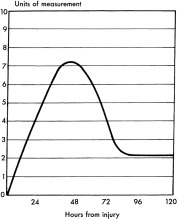
  • CK levels increase within 2 to 12 hr of the onset of muscle injury, generally peak after 24 to 72 hr, and decline 3 to 5 days after cessation of muscle injury. Peak CK levels may predict development of AKI.
  • Rhabdomyolysis from cholesterol-lowering therapy by HMG-CoA reductase inhibitors (statins) requires hospitalization in less than 0.1% of cases. The mechanism of damage is multifactorial: Bioavailability, lipophilicity, efficiency of uptake of transport proteins in hepatocytes, blood level of statins, and level of extrahepatic inhibition of mitochondrial respiration, bioavailability, and lipophilicity. Among the statins, pravastatin has a lower risk for induction of rhabdomyolysis than other statins, presumably due to lower lipid solubility. The average duration of statin therapy before onset of myopathy is 6 mo. Symptom resolution with normalization of serum CK concentrations occurs in days to weeks following drug discontinuation. The average time for onset of rhabdomyolysis after the addition of a fibrate to statin therapy is 32 days. Rare genetic variants are associated with statin-induced myopathy.
Physical Findings & Clinical Presentation

  • Classic triad of muscle pain, weakness, and dark urine from myoglobinuria
  • Muscle tenderness is present in half of cases
  • Muscle swelling occurs after intravenous fluid repletion
  • Muscular rigidity
  • Fever
  • In rhabdomyolysis secondary to long-term statin administration, fatigue (74%) is nearly as common as muscle pain (88%)
  • Oliguria or anuria with AKI
Etiology

Causes can be divided into three categories:

  • Traumatic or muscle compression
    1. High-current electrical injury
    2. Crush injury and compartment syndrome
    3. Tourniquet and limb ischemia
    4. Reperfusion after revascularization procedures for ischemia
    5. Extensive surgical (spinal) dissection, bariatric surgery
  • Nontraumatic exertional
    1. Exercise: More than 10 genetically predisposing gene sequence variants are associated with exertional rhabdomyolysis
    2. Sickle cell trait, rarely: Usually additional predisposing factors are involved (e.g., body mass index [BMI] >30 kg/m2, tobacco use, statin use, antipsychotic use, high altitude)
    3. Heat stroke
    4. Metabolic myopathies
    5. Malignant hyperthermia and neuroleptic malignant syndrome
    6. Seizure activity
  • Nontraumatic, nonexertional
    1. Drug-induced (statins alone, combinations of statins with fibrates or erythromycin, simvastatin and amiodarone, amphetamines, haloperidol, levofloxacin, macrolide antibiotics)
    2. Chronic ethanol ingestion
    3. Hypothyroidism
    4. Infectious and inflammatory myositis

Table 1 summarizes various causes of rhabdomyolysis.

TABLE 1 Causes of Rhabdomyolysis

Muscle injury/ischemiaTrauma, pressure necrosis, electric shock, burns, acute vascular disease
Myofiber exhaustionSeizures, excessive exercise, heat exhaustion
ToxinsAlcohol, cocaine, heroin, amphetamines, ecstasy, phencyclidine, snakebite
DrugsStatins, fibrates, zidovudine, neuroleptic malignant syndrome, azathioprine, theophylline, lithium, diuretics, colchicine
Electrolyte disordersHypophosphatemia, hypokalemia, excess water shifts (hyperosmolality)
InfectionsViral (influenza, HIV, coxsackievirus, Epstein-Barr virus), bacterial (Legionella, Francisella, Streptococcus pneumoniae, Salmonella, Staphylococcus aureus)
FamilialMcArdle disease, carnitine palmitoyl transferase deficiency, malignant hyperthermia
OtherHypothyroidism, polymyositis, dermatomyositis

From Floege J et al: Comprehensive clinical nephrology, ed 4, Philadelphia, 2010, Saunders.

Diagnosis

Differential Diagnosis

“Creatine Kinase Elevation” in Section IV describes a clinical algorithm for the evaluation of CK elevation.

Laboratory Tests

  • Creatine kinase: Usually CK is 5 to 10 times the upper limit of the normal range and typically peaks 24 to 72 hr after the initial insult (Fig. 1). Levels >15,000 IU/L are more likely associated with AKI. However, in patients with concomitant risk factors such as hypokalemia or volume depletion, CK levels as low as 5000 U/L may be associated with AKI.
  • Myoglobin: Filtration into urine produces a “port wine” color at concentrations of 100 to 300 mg/dl. Myoglobinuria can be suspected by a positive urine dipstick test for blood within urine or by light microscopy detection of red blood cells. Due to its rapid hepatic metabolism, myoglobin lacks sensitivity for detection and diagnosis of rhabdomyolysis. Therefore serum and/or urine myoglobin measurement is not recommended to establish the diagnosis of rhabdomyolysis. Blood urea nitrogen and plasma creatinine concentrations are used to monitor severity of AKI.
  • Potassium, calcium, phosphorus, and uric acid are released from damaged muscle, and levels should be monitored. Hyperkalemia is less common in nontraumatic rhabdomyolysis.
  • Calcium: Hypocalcemia from deposition of calcium phosphate complexes in damaged muscle tissue. Hypercalcemia may follow resolution of rhabdomyolysis from liberation of calcium from damaged muscle into the circulation and increased gut calcium absorption from enhanced vitamin D production.
  • Anion gap metabolic acidosis may occur from release of organic acids and phosphates from damaged muscle. Metabolic acidosis is less common in nontraumatic rhabdomyolysis.
  • Urinalysis: Myoglobin is detected as blood on dipstick, but red blood cells are absent on microscopy. Microscopic identification of pigmented tubular casts establishes acute tubular necrosis.
  • Rhabdomyolysis-induced acute tubular necrosis may occur with a low fractional excretion of sodium (FENa <1%).
  • Box 1 summarizes laboratory abnormalities observed with rhabdomyolysis.

BOX 1 Laboratory Abnormalities Observed With Rhabdomyolysis

Potassium

Elevated

Risk for acute kidney injury

Bicarbonate

Decreased (20 mEq/L)

Metabolic acidosis

Uric Acid

Elevated (>7 mg/dl)

Marker of acute renal failure

Sodium

Usually normal

Can decrease with mannitol therapy

Use serum osmolality values as a guide

Phosphate

Elevated

Risk for precipitation of calcium phosphate

May need phosphate binders if phosphate >7 mg/dl

Creatine Kinase

Elevated

Associated with creatine kinase level of 15 to 75,000

Blood Urea Nitrogen

Elevated (>20 mg/dl)

Creatinine

Elevated

Calcium

Initially low

Rebound phase may demonstrate hypercalcemia

Liver Function Tests

Occasionally elevated

Serum aspartate transaminase, lactate dehydrogenase, aldolase, muscle enzyme levels elevated

Troponin

Normal

Suspect myocardial damage as a cause (or effect) if elevated

7% false-positive rate for troponin I

Anion Gap

Sometimes elevated

May predict acute kidney injury

Prothrombin Time, Partial Thromboplastin Time, D-Dimer

Disseminated intravascular coagulation in up to 30% of severe cases

Associated with greater mortality

From Adams JG et al: Emergency medicine, clinical essentials, ed 2, Philadelphia, 2013, Elsevier.

Figure 1 Typical creatine kinase elimination curve.

Treatment

Acute General Rx

  • Identify precipitating factor(s) and discontinue potentially contributory drugs or toxins.
  • Early, aggressive, high-volume intravenous fluid replacement with normal saline. Extracellular fluid volume-loading and diuresis reduce the risk for renal damage by elimination of urate and phosphate that can precipitate in the kidneys. Fig. 2 describes a treatment algorithm for rhabdomyolysis.
  • Fasciotomy is indicated in compartment syndrome for preservation of muscle and nerve function.
  • Initiate volume repletion with normal saline at a rate of 200 to 1000 ml/hr, depending on clinical circumstances and severity of muscle damage. Titrate the infusion rate to maintain a urine output of at least 200 ml/hr. Consider treatment with mannitol (up to 200 g per day with cumulative dose up to 800 g) to enhance urine flow rate. Typically, a 20% mannitol infusion at a dose of 0.5 g/kg is given over a 15-min interval followed by an infusion at 0.1 gram/kg per hr. Discontinue mannitol and volume resuscitation if a diuresis threshold of >20 ml/hr is not established. Maintain volume repletion until myoglobinuria stops (negative urine dipstick blood test) or plasma CK levels decrease to <5000 U/L.
  • Correct hypocalcemia if symptomatic or hyperkalemia that produces electrocardiographic changes.
  • Treatment of all electrolyte imbalances.
  • Urine alkalinization: Maintain urine pH at 6 to 7 and plasma pH at 7.50. This recommendation is controversial. Early volume resuscitation and expansion are the most important treatments.
  • Initiation of renal replacement therapy is determined by severity of kidney injury and/or electrolyte imbalances. Continuous renal replacement therapy and specialized hemodialysis membranes to enhance myoglobin clearance have not been systematically studied or proven superior to intermittent hemodialysis.

Figure 2 Early goal-directed therapy for rhabdomyolysis.

!!flowchart!!

CK, Creatine kinase; CVP, central venous pressure; D5NL bicarb, 5% dextrose in normal sodium bicarbonate solution; EGDT, early goal-directed therapy; IV, intravenous; IVF, intravenous fluid; NS, normal saline; RRT, renal replacement therapy; UOP, urinary output.

From Adams JG et al: Emergency medicine, clinical essentials, ed 2, Philadelphia, 2013, Elsevier.

Disposition

Early diagnosis and management are required to prevent AKI.

Referral

Renal consultation and surgical consultation if compartment syndrome develops

Pearls & Considerations

Comments

  • Statin-induced rhabdomyolysis occurs 12 times more frequently when statins are combined with fibrates than when used alone.
  • Short-term, high-dose glucocorticoid steroid administration (500 to 1000 mg methylprednisolone) has been used for treatment of alcohol-induced rhabdomyolysis that is refractory to volume repletion. This treatment may be efficacious in cases of severe rhabdomyolysis by reducing secondary leukocyte inflammatory muscle injury.
Related Content

Rhabdomyolysis (Patient Information)

Statin-Induced Muscle Syndrome (Related Key Topic)