section name header

Basic Information ⬇

Definition

Progressive multifocal leukoencephalopathy (PML) is an uncommon and often fatal subacute demyelinating disease of the white matter, caused by reactivation of the JC virus (JCV) infection, which has tropism for oligodendrocytes, in an immunocompromised individual. PML originally was described half a century ago by Astrom et al in patients with chronic lymphoid leukemia and Hodgkin lymphoma. JCV was isolated in 1971 and is named for the initials of the first patient from whose brain it was isolated.

Synonym

PML

ICD-10CM CODE
A81.2Progressive multifocal leukoencephalopathy
Epidemiology & Demographics
Incidence & Prevalence

  • Seroepidemiologic studies have shown that asymptomatic primary infection with JCV occurs at an early age, and by adult life 30% to 90% of the population is seropositive. It becomes dormant in tissues such as kidney, bone marrow, tonsils, and lymphoreticular tissue.
  • The PML era can be broadly divided into four epochs: Pre-HIV, HIV (pre-ART), HIV (post-ART), and modern era.
  • Prior to the HIV epidemic in the early 1980s, PML was rare and seen almost exclusively in patients with lymphoproliferative and myeloproliferative disorders.
  • HIV: A fiftyfold increase in PML cases was reported with HIV epidemic. During the HIV precombined antiretroviral therapy (cART) era, 3% to 5% of patients with HIV developed PML. PML is an AIDS-defining illness. cART has led to some decline in PML cases.
  • Modern era: HIV still accounts for a large percentage of PML incidence but needs to be considered in anyone whose cellular immunity is compromised with an underlying disorder or secondary to iatrogenic immunosuppression or immunomodulation. Bone marrow transplant, lupus, rheumatoid arthritis, multiple sclerosis, as well as solid or hematologic malignancies are all at risk.
  • Natalizumab is the medication associated with the highest incidence of PML and is dependent on JCV antibody status, history of prior immunosuppression, and length of treatment.
Predominant Sex & Age

Males and females are equally affected.

Increasing age is associated with higher risk of PML.

Risk Factors

Typically seen in patients with profound immunosuppression and impaired cell-mediated immunity. These include patients with AIDS, lymphoproliferative disorders, chronic infections such as tuberculosis, and, more recently, patients on immunosuppressive or immunomodulating medications such as corticosteroids, natalizumab, rituximab, ocrelizumab, alemtuzumab, efalizumab, brentuximab vedotin, cyclophosphamide, mycophenolate mofetil, anti-TNF-alpha agents, and oral agents used in the treatment of multiple sclerosis (dimethyl fumarate and fingolimod). In rare instances, it may be seen in the absence of apparent immunologic abnormality.

Genetics

Unknown.

Physical Findings & Clinical Presentation

  • Subacute neurologic deficits are common; can also present acutely as a stroke mimic.
  • The clinical course spans weeks to months leading to severe disability and death.
  • PML lesions usually involve cerebral hemispheres; cerebellar and brain stem involvement may be seen in AIDS. Multifocal asymmetric white matter involvement is common; rarely there can be gray matter involvement. Usually the optic nerve and spinal cord are not involved.
  • Non-AIDS-associated PML: Early lesions involve occipital subcortical white matter and cause visual-field deficits (homonymous hemianopia) or cortical blindness. Motor weakness and altered mentation may be seen, while headache, seizures, and extrapyramidal syndromes are rare.
  • AIDS-related PML: Motor weakness is more common. Abnormalities of speech, cognition, gait, sensation, and visual impairment are also seen.
  • Natalizumab-associated PML: Frontal lobe is commonly involved, leading to cognitive impairment, neurobehavioral changes, motor disorders, language disorders, and visual defects.
  • Immune reconstitution inflammatory syndrome (IRIS): A paradoxical clinical deterioration that occurs during immunologic recovery in previously immunocompromised patients, such as antiretroviral therapy-treated HIV-seropositive patients or cessation of immunomodulating treatment, such as natalizumab. An increase in the number or size of lesions on neuroimaging with contrast enhancement of brain lesions and brain edema may be seen.
Etiology & Pathogenesis

  • JCV infection may be acquired through respiratory tissue or oropharyngeal route; seroconversion usually occurs in childhood.
  • A cytotoxic-specific response against JCV acts as a containing mechanism for PML. Immunosuppression leads to development of PML either as primary infection of the CNS following immunosuppression or reactivation of a dormant infection. The virus produces lytic infection of the oligodendrocytes and causes oligodendrocyte death and demyelination. Neuronal infection does not usually occur except in granule cell neurons of the cerebellum. Infection of the astrocytes is abortive, leading to characteristic bizarre, enlarged cells.

Diagnosis ⬆ ⬇

Differential Diagnosis

  • Multiple sclerosis
  • Acute disseminated encephalomyelitis
  • Vasculitis
  • Fungal meningitis
  • HIV encephalitis
  • Mitochondrial encephalopathies (e.g., mitochondrial encephalopathy, lactic acidosis, and stroke-like episodes [MELAS])
  • Posterior reversible encephalopathy syndrome
Workup

  • Diagnosis is based on compatible clinical presentation, MRI brain features, and detection of JCV in cerebrospinal fluid (CSF).
  • The two diagnostic approaches employed include:
    1. Demonstration of typical histopathologic findings (multifocal demyelination, enlarged bizarre astrocytes with lobulated hyperchromatic nuclei, and enlarged oligodendroglial nuclei) and JCV in tissue specimen (electron microscopy, immunohistochemistry, or polymerase chain reaction [PCR] techniques); or
    2. Demonstration of JCV in the CSF of patients who fulfill clinical and radiographic criteria
Laboratory Tests

  • CSF examination: Usually normal; may show mild lymphocytic pleocytosis (20 to 25 leukocytes/ml) and elevated protein (usually <65 mg/dl).
  • CSF PCR for JCV has a sensitivity of 95% and specificity of 99% using ultrasensitive PCR techniques but may be lower depending on the techniques used.
Imaging Studies

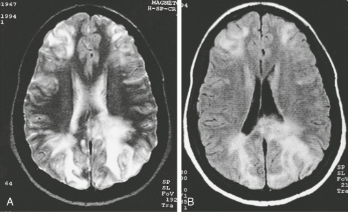
  • MRI brain (Fig. E1) is the neuroimaging of choice and shows demyelinating lesions that are hyperintense on T2, and fluid-attenuated inversion recovery (FLAIR) sequences are hypointense on T1. U-fibers can be involved, but edema is usually minimal.
  • Only 10% to 15% of lesions enhance with gadolinium in patients with AIDS unless associated with IRIS. However, in drug-related PML, contrast enhancement is present in up to 40% and may be related to IRIS from medication withdrawal.
  • Computed tomography of the head may show subcortical hypodensities.
  • MR spectroscopy will demonstrate features of demyelination-decreased N-acetyl acetate, increased choline, and increased lactate.

Figure E1 Progressive multifocal leukoencephalopathy.

Axial T2-weighted (A) and fluid-attenuated inversion recovery (FLAIR) (B) images of patient with human immunodeficiency virus (HIV). Multiple areas of white matter disease are identified; FLAIR image clarifies the involvement of periventricular white matter. These findings in an HIV-positive patient strongly suggest progressive multifocal leukoencephalopathy.

From Vincent JL et al: Textbook of critical care, ed 7, Philadelphia, 2017, Elsevier.

Treatment ⬆ ⬇

Acute General Rx

There is no specific treatment for PML. Treatment is aimed at reversal of the underlying causes of immunosuppression.

  • HIV-positive patients: Antiretroviral therapy should be optimized.
  • HIV-negative patients: Immunosuppressive medication should be discontinued; plasma exchange may be considered for natalizumab-treated patients.
  • IRIS: Treatment with steroids should be considered.
Chronic Rx

No specific pharmacologic therapy is proven for chronic treatment. Multiple medications have been tried but have been unsuccessful in trials.

There have been recent case series reporting successful treatment of PML using checkpoint inhibitors (pembrolizumab, nivolumab) or filgrastim. These therapies are under further investigation.

Disposition

Of all HIV-related cerebral disorders, PML probably has the worst prognosis. Pre-AIDS era survival was 6 mo. With ART, incidence and prognosis of PML has improved. A lower CSF JCV load and better CD4 counts have better prognoses. Natalizumab-treated patients have worse prognosis, with longer time to diagnosis, presence of widespread disease, and brain stem involvement.

Referral

  • Neurology if patients have neurologic deficits of uncertain etiology, especially in the setting of AIDS or immunosuppression
  • Infectious disease especially if new diagnosis of HIV

Pearls & Considerations ⬆ ⬇

Comments

PML is a fatal opportunistic cerebral infection that should be considered in all immunosuppressed individuals presenting with neurologic deficits and demyelinating cerebral lesions. Early referral to a center with neurologic expertise, especially neuroimmunology or neuroinfectious diseases, should be considered.

Suggested Readings ⬆

  1. Berger J.R. : The clinical features of PMLCleve Clin J Med. ;2(78):S8-S12, 2011.
  2. Cortese I. : Pembrolizumab treatment for progressive multifocal leukoencephalopathyNew Engl J Med. ;380(17):1597-1605, 2019.
  3. Grebenciucova E., Berger J.R. : Progressive multifocal leukoencephalopathyNeurol Clin. ;36(4):739-750, 2018.