section name header

Basic Information

AUTHOR: B. Shoshana Zha, MD, PhD

Definition

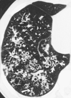
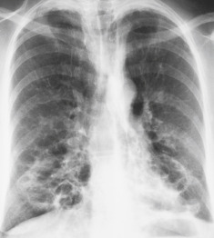
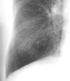
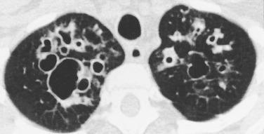
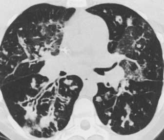
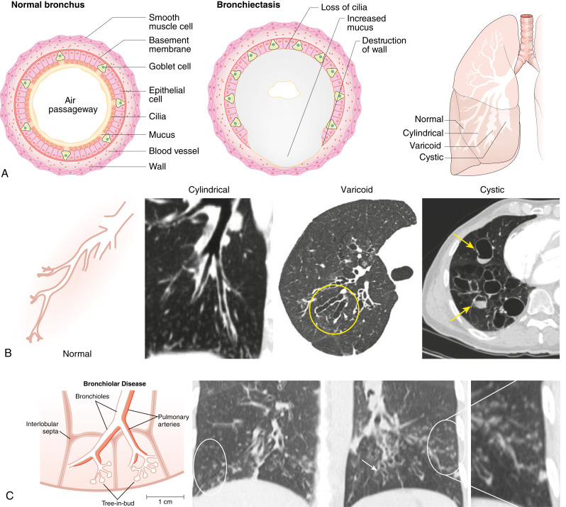
Bronchiectasis is a chronic lung disease resulting from dilatation of bronchi due to a variety of causes and propagated through an interplay of host factors, environment, and respiratory pathogens. Clinically, it is marked by a syndrome of cough, sputum production, and recurrent respiratory infections. It is confirmed radiographically by a lack of bronchial tapering and a bronchus-to-arterial ratio greater than 1.5. It can be divided into cylindric, varicose, and cystic subtypes, which often overlap and coexist, but may aid in diagnosis and overall prognosis.

ICD-10CM CODES
J47.0Bronchiectasis with acute lower respiratory infection
J47.1Bronchiectasis with (acute) exacerbation
J47.9Bronchiectasis, uncomplicated
Q33.4Congenital bronchiectasis
Epidemiology & Demographics

  • The incidence and prevalence of bronchiectasis is steadily rising in the U.S. and Europe.1-4 Prevalence increases markedly with age, with peak incidence in those older than age 65. In the U.S., the average annual prevalence was approximately 700 per 100,000 persons among Medicare beneficiaries between 2012 and 2014,5 with likely higher prevalence given noted escalations of incidence.
  • Bronchiectasis is more common in women than men.
  • There are multiple identified etiologies that can vary by geographic region, yet idiopathic remains the most frequent diagnosis worldwide.6
  • Table 1 summarizes conditions associated with bronchiectasis.

TABLE 1 Conditions Associated with Bronchiectasis

Postinfectious Conditions
Childhood lower respiratory tract infections
Granulomatous infections
Necrotizing pneumonias in adults
Other respiratory infections
Primary Immune Disorders
Humoral defects
Cellular and/or mixed disorders
Neutrophil dysfunction
Other
Cystic Fibrosis (CF)
Classic CF
Variants of CF
Young syndrome
Alpha1-Antitrypsin Abnormalities
Deficiencies
Anomalies
Heritable Structural Abnormalities
Primary ciliary dyskinesia
Williams-Campbell syndrome
Mounier-Kuhn syndrome
Marfan syndrome
Sequestration, agenesis, hypoplasia
Idiopathic Inflammatory Disorders
Sarcoidosis
Rheumatoid arthritis
Ankylosing spondylitis
Systemic lupus erythematosus
Sjögren syndrome
Inflammatory bowel disease
Relapsing polychondritis
Inhalation and Obstruction
Gastroesophageal reflux/aspiration
Pneumonia
Toxic inhalation/thermal injury
Postobstruction accident
Foreign body
Tumors, benign and malignant
Extrinsic airway compression
Allergic bronchopulmonary aspergillosis/mycosis
Miscellaneous
Human immunodeficiency virus infection
Yellow nail syndrome
Radiation injury

From Broaddus VC et al: Murray & Nadel’s textbook of respiratory medicine, ed 7, Philadelphia, 2022, Elsevier.

Physical Findings & Clinical Presentation

  • Chronic cough, typically with expectoration of purulent sputum
  • Frequent bronchitis or pneumonias requiring antibiotic treatment
  • Generalized malaise and/or weight loss
  • Fever and/or night sweats
  • Hemoptysis
  • Crackles, wheezing, or squeaks on lung exam
  • Halitosis
Etiology

  • Postinfectious (e.g., previous pneumonia, lung abscess, tuberculosis, nontuberculous mycobacterial infections, fungal infections, viral infections)
  • Cystic fibrosis
  • Ciliary dysfunction (primary ciliary dyskinesia, Kartagener syndrome)
  • Chronic obstructive pulmonary diseases (e.g., chronic obstructive pulmonary disease [COPD], asthma)
  • Impaired host defense (e.g., hypogammaglobulinemia, acquired immunodeficiency syndrome, chemotherapy)
  • Localized airway obstruction (aspiration, congenital structural defects, foreign bodies, neoplasms)
  • Inflammation (chronic gastroesophageal reflux disease [GERD], inflammatory pneumonitis, granulomatous lung disease, allergic aspergillosis)
  • Congenital disorders such as tracheobronchomegaly (Mounier-Kuhn syndrome), cartilage deficiency (Williams-Campbell syndrome, Marfan syndrome), alpha-1 anti-trypsin deficiency
  • Connective tissue and autoimmune diseases (Sjogren disease, ulcerative colitis, rheumatoid arthritis)
  • Cellular pathophysiologic mechanism of bronchiectasis is illustrated in Fig. E1

Figure E1 Cellular pathophysiologic mechanism of bronchiectasis.

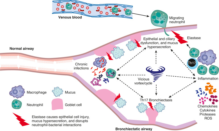
Cross-sectional view of the bronchiectatic airway demonstrating the components that play a role in the pathogenesis, including neutrophils, neutrophil elastase, goblet cells that produce mucus, and the chemokines and proinflammatory cytokines produced by macrophages and T helper type 17 cells (Th17). The epithelial and ciliary dysfunction, mucus hypersecretion, inflammation with unchecked protease (elastase) activity, chronic infection, and resultant bronchiectasis are intertwined and help perpetuate each other, creating the paradigm of the “vicious vortex/cycle” central to bronchiectasis pathogenesis. ROS, Reactive oxygen species.

From Broaddus VC et al: Murray & Nadel’s textbook of respiratory medicine, ed 7, Philadelphia, 2022, Elsevier.

Diagnosis

Differential Diagnosis

  • Chronic bronchitis or chronic rhinosinusitis
  • Chronic obstructive pulmonary disease
  • Asthma
  • Tuberculosis
  • Interstitial fibrosis
  • Chronic lung abscess
  • Foreign body aspiration
  • Cystic fibrosis
  • Lung carcinoma
  • Gastroesophageal reflux disease
  • Diffuse panbronchiolitis
  • Bronchiolitis obliterans
Laboratory Tests

  • Pulmonary function tests with bronchodilators
  • Sputum for Gram stain, culture and susceptibilities, and acid-fast bacteria
  • Serum immunoglobulins (total immunoglobulin A [IgA], IgM, IgG and subclasses)
  • Testing for allergic bronchopulmonary aspergillosis (serum IgE, precipitating IgG antibodies [precipitins] to Aspergillus, specific IgE antibodies to Aspergillus, Aspergillus skin test)
  • CBC with differential (leukocytosis with left shift, anemia)
  • Alpha-1 antitrypsin level and phenotype
  • Sweat test in patients with suspected cystic fibrosis
  • Primary ciliary dyskinesia evaluation when suspected
  • Antinuclear antibody, rheumatoid factor, and SSA/SSB when driven by history or exam
  • Serum anti-Pseudomonas aeruginosa (PA) IgG antibody testing is highly accurate to detect chronic PA colonization in bronchiectasis patients7 and may be a marker of treatment response
Imaging Studies

  • Chest x-ray examination (Fig. 2 and Fig. E3) may be normal or have crowded lung markings, “tram-track” sign of dilated bronchi, or small cystic spaces at the base of the lungs.
  • High-resolution computed tomography (CT) scan of the chest (Fig. 4 and Figs. E5 to E7 to ) is the gold standard of diagnosis.
    1. The CT study should be noncontrast with the use of a 1- to 1.5-mm window every 1 cm with acquisition time of 1 sec.
    2. Findings include enlarged bronchoarterial ratio of >1.5 (normal 0.7), lack of tapering of an airway toward the periphery, ballooned cysts at the end of bronchi, and airway mucus impaction.
    3. Radiographic examples of different forms of bronchiectasis are illustrated in Fig. E8.
  • Bronchoscopy may be helpful to evaluate hemoptysis, rule out obstructive lesions, removal of impacted mucus plugs, and obtain microbiologic data on respiratory pathogens when expectorated or induced sputum is not attainable.
  • Table 2 summarizes diagnostic studies for the classification and management of patients with bronchiectasis.

TABLE 2 Diagnostic Studies for the Classification and Management of Patients with Bronchiectasis

TestComments
Routine, Universal Studies
Computed tomography lung scan (CTLS)If bronchiectasis is suspected, CTLS is the definitive test. Thin-section, high-resolution images may help detect subtle airway dilation before bronchial walls are grossly thickened. Contrast is generally not helpful and may, in fact, compromise the overall resolution of the study. CTLS may also identify esophageal abnormalities.
Pulmonary function tests (PFTs)Comprehensive PFTs, including spirometry, bronchodilator responsiveness, lung volumes, and diffusion capacity, are important studies that aid in management, prognosis, may provide useful hints regarding predisposing conditions.
Complete blood countAnemia may reflect effects of chronic infection or blood loss (consider inflammatory bowel disorders).
Leukocytosis may mark severity of infection.
Eosinophilia may suggest ABPA/M.
ESR, C-reactive proteinNonspecific markers of inflammation; very high levels may suggest underlying connective tissue disease or vasculitis. Can aid during treatment of exacerbations.
Routine sputum cultureAntibiotic therapy in bronchiectasis should generally be directed against specific pathogens and guided by in vitro susceptibility. The presence of mucoid strains of Pseudomonas aeruginosa and Staphylococcus aureus may raise suspicions for CF. Stenotrophomonas maltophilia, Alcaligenes xylosoxidans, and Burkholderia cepacia are gram-negative bacilli that may prove problematic pathogens in patients with long-standing bronchiectasis. Isolation of B. cepacia and Helicobacter pylori requires special laboratory techniques.
Mycobacterial sputum cultureEnvironmental mycobacteria such as Mycobacterium avium complex and M. abscessus are increasingly common. Presence can worsen symptoms and progress disease.
Fungal sputum cultureIn patients with an asthmatic component, the presence of Aspergillus species (or other molds including Pseudallescheria or Penicillium) may be suggestive of etiology.
CT scan of sinusesMany patients with bronchiectasis also suffer chronic rhinosinusitis. The presence of extensive sinus involvement suggests possible CF, immunoglobulin deficiencies, or ciliary disorders. Also, optimal management often entails aggressive sinus care.
Specific, Directed Studies
Sweat chloride, CF genotyping, and nasal potential differencesFor bronchiectasis patients with bilateral disease, recurrent sinusitis, and no other identified risk factor, mild variants of CF appear to be relatively common. Sweat chloride is regarded as the primary screening test for CF, but a considerable portion of adults with CF have borderline or normal results. Nasal potential difference may be useful for identifying CF in equivocal cases.
Alpha-1 antitrypsin (AAT) levels and phenotypeAAT anomalies appear to be a substantial risk factor for bronchiectasis, especially with white females. Abnormal proteinase inhibitor (Pi) phenotypes, even heterozygous patterns such as MS, appear to confer risk even with normal levels of AAT. Repletion of AAT may enhance resistance to lower respiratory tract infections.
Immunoglobulin (Ig) levelsDeficiencies of IgG or IgA may promote bronchiectasis; IgG subclass deficiencies may also be a factor. Elevated levels of IgE may suggest ABPA/M or Job syndrome. Hyper-IgM may be associated, as well, with chronic infections.
Ciliary morphology or functionFor individuals with suggestive stories, a nasal ciliated epithelium biopsy with transmission electron microscopy may identify primary ciliary dyskinesia. Other studies including ex vivo ciliary activity, the saccharine test, or spermatozoa analysis may aid in this diagnosis.
Nasal nitric oxide (NNO) levelsPatients with documented PCD have significantly lower levels of NNO than normal or patients with CF. Although not universally available, such testing may prove highly useful in identifying PCD. Paradoxically, exhaled NO levels have been elevated in bronchiectasis of diverse etiologies except CF.
Barium swallow (BaS)The BaS may detect disturbed deglutition, esophageal diverticula, obstructing lesions (tumors or strictures), hypomotility, achalasia, hiatal hernias, or lower esophageal sphincter (LES) incompetence with reflux. The absence of reflux on a BaS, however, does not exclude this problem (see “pH probe”).
pH probeFor patients suspected of gastroesophageal reflux, an 18- to 24-h study with a transnasal pH probe may identify, quantitate, and characterize reflux. Medications that inhibit acid production must be stopped before such tests.
Esophageal manometryFor patients being considered for surgical repair of the LES, manometry should be performed to determine that the esophagus generates sufficient pressure to propel food and liquids through the tightened sphincter.
Tailored hypopharyngography (TH)TH is useful in detecting abnormalities of the initial phase of swallowing, deglutition. Persons particularly prone to problems include those with prior strokes, Parkinson disease, bulbar disorders including postpolio syndrome, and those with prior laryngeal or pharyngeal surgery. Note that some patients have gross aspiration without clinical manifestations (choking, coughing); this may occur in individuals with none of the above risk factors.
Less Common, Exotic Studies
Collagen vascular disease (CVD) serologiesVarious CVDs may contribute to the risk for bronchiectasis, including RA, ankylosing spondylitis, and systemic lupus erythematosus. Thus, for patients with compatible histories or physical findings, assays for rheumatoid factor, HLA-B27, and ANA may provide insight into predisposing conditions. CVD serologies may also suggest the diagnosis of Sjögren syndrome, particularly SSA/Ro and/or SSB/La.
Schirmer testFor patients with histories suggestive of “sicca syndrome” (dry eyes, dry mouth, oral ulcers), a positive Schirmer test may indicate the presence of either primary or secondary (associated with a CVD) Sjögren syndrome.

ABPA/M, Allergic bronchopulmonary aspergillosis/other mycoses; ANA, antinuclear antibody; CF, cystic fibrosis; CT, computed tomography; ESR, erythrocyte sedimentation rate; HLA, human leukocyte antigen; MS, multiple sclerosis; NO, nitric oxide; PCD, primary ciliary dyskinesia; RA, rheumatoid arthritis.

Adapted from Mason RJ: Murray & Nadel’s textbook of respiratory medicine, ed 5, Philadelphia, 2010, Saunders.

Figure 4 Bronchiectasis

Computed Tomography Demonstrating Dilated Subsegmental Bronchi. The Bronchi are Larger than the Accompanying Vessels with Some Demonstrating the “signet Ring” Sign (Arrows). Plugging of Peripheral Bronchi is Also Evident (Curved Arrow).

From Grant LA: Grainger & Allison’s diagnostic radiology essentials, ed 2, Philadelphia, 2019, Elsevier.

Figure 2 Bronchiectasis

Multiple Ring Shadows, Many Containing Air-Fluid Levels, are Present Throughout the Lower Zones of This Patient with Cystic Bronchiectasis.

From Grant LA: Grainger & Allison’s diagnostic radiology essentials, ed 2, Philadelphia, 2019, Elsevier.

Figure E3 Bronchiectasis.

Targeted image of a right lower lung base demonstrating tramlines and ring opacities.

From Grant LA: Grainger & Allison’s diagnostic radiology essentials, ed 2, Philadelphia, 2019, Elsevier.

Figure E5 Varicose bronchiectasis.

Targeted left side on a chest computed tomography demonstrating beaded configuration of varicose bronchiectasis.

From Grant LA: Grainger & Allison’s diagnostic radiology essentials, ed 2, Philadelphia, 2019, Elsevier.

Figure E6 Cystic bronchiectasis.

Computed tomography demonstrates multiple ring shadows due to irregularly dilated bronchi.

From Grant LA: Grainger & Allison’s diagnostic radiology essentials, ed 2, Philadelphia, 2019, Elsevier.

Figure E7 Cylindrical bronchiectasis.

The bronchi fail to taper and have irregular thickened walls.

From Grant LA: Grainger & Allison’s diagnostic radiology essentials, ed 2, Philadelphia, 2019, Elsevier.

Figure E8 Radiographic examples of different forms of bronchiectasis.

A, The three top figures show a cross-section diagram of a normal airway, a bronchiectatic airway, and the three different forms of bronchiectasis on longitudinal view. B, The middle panel shows a diagram of a normal bronchial tree demonstrating usual tapering as the airway branches distally (far left) and axial computed tomography images showing cylindrical bronchiectasis (middle left), varicoid bronchiectasis (middle right, yellow circle), and cystic bronchiectasis (arrows, far right). C, The bottom panel shows a diagram of bronchiolitis (left) and a coronal computed tomography image (right) of inflammatory bronchiolitis manifested as tree-in-bud opacification (white ovals) (enlarged at far right) and bronchiectasis (arrow) in a patient with nontuberculous mycobacterial lung disease.

From Broaddus VC et al: Murray & Nadel’s textbook of respiratory medicine, ed 7, Philadelphia, 2022, Elsevier.

Treatment

Nonpharmacologic Therapy

  • Mucus clearance is essential and central to therapy: Low risk and shown to improve chronic symptoms of cough, breathlessness, and may reduce frequency of exacerbations. Techniques include positive expiratory pressure (e.g., Acapella, Aerobika), chest wall oscillation (i.e., vest therapy), and chest percussion (e.g., caregiver cupping hand with aggressive patting).
  • Exercise, particularly aerobic and/or active breathing (e.g., yoga)
  • Supplemental oxygen for hypoxemia
  • Nocturnal positive pressure for hypercarbia in advanced disease
Acute General Rx

  • Antibiotic therapy should be based on the results of sputum gram stain, culture, and susceptibility testing, and continued for 14 days of therapy. In those with inadequate or inconclusive sputum test results, therapy can be based on knowledge of past culture growth, or empiric with doxycycline, fluoroquinolone, or amoxicillin/clavulanate for broader coverage.
  • Intravenous antibiotics should be used in patients who require hospitalization, such as signs of respiratory distress, systemic symptoms, or inability to improve with oral outpatient therapy.
    1. Hospital admission should focus on mucus clearance and a nutrition assessment.
    2. Evaluate for hypoxemia and need for supplemental supplies prior to discharge.
  • Bronchodilators are useful in patients with demonstrable airflow obstruction.
    1. Avoid inhaled corticosteroids if able as there is a strong association of nontuberculous mycobacterium colonization with their use.
Complementary & Alternative Medicine

  • Avoidance of tobacco
  • Maintenance of proper nutrition and hydration
  • Daily exercise
Chronic Rx

  • Infections should be promptly identified and treated.
  • Adults with bronchiectasis with a new isolation of P. aeruginosa should be offered eradication antibiotic treatment. This should not be offered to adults with bronchiectasis following new isolation of pathogens other than P. aeruginosa.8
    1. Measurement of serum anti-Pseudomonas aeruginosa (PA) IgG antibody may help predict success.7
  • Bronchodilators should be used before physiotherapy, including inhaled mucoactive drugs and inhaled antibiotics, in those with evidence of obstructive ventilatory defect or bronchospasm. This increases tolerability and optimizes delivery of medications in diseased areas of the lungs.
  • Inhaled corticosteroids should not be offered to adults with isolated bronchiectasis. However, the diagnosis of bronchiectasis should not affect the use of inhaled corticosteroid in patients with comorbid asthma.
  • Inhaled hyperosmolar agents (e.g., hypertonic saline) should be considered in conjunction with airway clearance in patients who have difficulty expectorating sputum.
  • Inhaled mucolytics can be considered in those with difficulty expectorating sputum, poor quality of life, or where standard airway clearance has failed to control symptoms. However, rhDNase should not be offered to patients with bronchiectasis not related to cystic fibrosis as there is an associated decline in forced expiratory volume in the first second (FEV1) and forced vital capacity in this population.9
  • Macrolide thrice weekly should be offered for adults with bronchiectasis who have three or more exacerbations per year.10
  • Immunoglobulin replacement should be considered in patients with selective immunoglobulin deficiency.
Disposition

  • Prognosis is variable with severity of the disease and underlying etiology of bronchiectasis. The bronchiectasis severity index is one helpful scoring system.11,12
  • Pulmonary function declines at a faster rate in those with Pseudomonas colonization or frequent exacerbations.
  • Concomitant rheumatoid arthritis or COPD is associated with worse prognosis.
Referral

  • Surgical partial lung resection may be an option in patients with localized disease, severe disease unresponsive to medical therapy, or in patients with massive hemoptysis. Surgical resection of localized bronchiectasis is safe and improves quality of life.
  • Embolization should be considered for recurrent hemoptysis.
  • Referral for lung transplant should be considered similarly to patients with cystic fibrosis.

Pearls & Considerations

Comments

  • The activity and quantity of neutrophil serine proteases are increased in the sputum of patients with bronchiectasis at baseline and increase further during exacerbations. A phase 2 trial revealed a reduction of neutrophil serine protease activity with brensocatib, an oral reversible inhibitor of dipeptidyl peptidase 1 (DPP-1), an enzyme responsible for the activation of neutrophil serine proteases.13 There is ongoing clinical investigation of this agent.
  • Other potential therapeutics being considered in bronchiectasis include novel inhibitors of dipeptidyl peptidase 1, antagonism of CXC chemokine receptor 2, and immunomodulatory drugs.14
  • Patients with bronchiectasis are susceptible to nontuberculous mycobacterium pulmonary infection. Consider periodically screening using acid fast bacillus sputum cultures, and directly testing when there is a change in symptoms or clinical course.
Related Content

Bronchiectasis (Patient Information)

Related Content

    1. Henkle E. : Population-based incidence of pulmonary nontuberculous mycobacterial disease in Oregon 2007 to 2012Ann Am Thorac Soc. ;12(5):642-647, 2015.doi:10.1513/AnnalsATS.201412-559OC
    2. Quint J.K. : Changes in the incidence, prevalence and mortality of bronchiectasis in the UK from 2004 to 2013: a population-based cohort studyEur Respir J. ;47(1):186-193, 2016.doi:10.1183/13993003.01033-2015
    3. Aksamit T.R. : Adult patients with bronchiectasis: a first look at the US Bronchiectasis Research RegistryChest. ;151:982-992, 2017.doi:10.1016/j.chest.2016.10.055
    4. Seitz A.E. : Trends in bronchiectasis among Medicare beneficiaries in the United States, 2000 to 2007Chest. ;142(2):432-439, 2012.doi:10.1378/chest.11-2209
    5. Henkle E. : Characteristics and health-care utilization history of patients with bronchiectasis in US Medicare enrollees with prescription drug plans2006 to 2014, Chest. ;154(6):1311-1320, 2018.doi:10.1016/j.chest.2018.07.014
    6. Chandrasekaran R. : Geographic variation in the aetiology, epidemiology and microbiology of bronchiectasisBMC Pulm Med. ;18(1), 2018.doi:10.1186/s12890-018-0638-0
    7. Suarez-Cuartin G. : Anti-Pseudomonas aeruginosa IgG antibodies and chronic airway infection in bronchiectasisRespir Med. ;128:1-6, 2017.doi:10.1016/j.rmed.2017.05.001
    8. Polverino E. : European Respiratory Society guidelines for the management of adult bronchiectasisEur Respir J. ;50, 2017.doi:10.1183/13993003.00629-2017
    9. Tarrant B.J. : Mucoactive agents for chronic, non-cystic fibrosis lung disease: a systematic review and meta-analysisRespirology. ;22(6):1084-1092, 2017.doi:10.1111/resp.13047
    10. Chalmers J.D. : Long-term macrolide antibiotics for the treatment of bronchiectasis in adults: an individual participant data meta-analysisLancet Respir Med. ;7(10):845-854, 2019.doi:10.1016/S2213-2600(19)30191-2
    11. Quittner A.L. : Quality of Life Questionnaire-Bronchiectasis: final psychometric analyses and determination of minimal important difference scoresThorax. ;70(1):12-20, 2015.doi:10.1136/thoraxjnl-2014-205918
    12. Chalmers J.D. : The bronchiectasis severity index an international derivation and validation studyAm J Respir Crit Care Med. ;189(5):576-585, 2014.doi:10.1164/rccm.201309-1575OC
    13. Chalmers J.D. : Phase 2 trial of the DPP-1 inhibitor brensocatib in bronchiectasisN Engl J Med. ;383(22):2127-2137, 2020.doi:10.1056/nejmoa2021713
    14. O'Donnell A.E. : Bronchiectasis - a clinical reviewN Engl J Med. ;387(6):533-545, 2022.