Table E107 describes features of the peripheral blood smear. Table E108 summarizes peripheral blood film evaluation in a patient with red cell membrane disorder. See Fig. E51 for useful peripheral blood and RBC features in the evaluation of anemia.
TABLE E107 Features of the Peripheral Blood Smear
| Red Blood Cell Morphology | Definition | Interpretation |
|---|---|---|
| Polychromasia | Large, bluish RBCs lacking normal central pallor on peripheral blood smear; bluish stain is the result of residual ribonucleic acid | Rapid production and release of RBCs from BM; elevated reticulocyte count; most commonly seen in any hemolytic anemia and states of increased RBC turnover |
| Basophilic stippling | Many small bluish dots in portion of erythrocytes; comes from staining of clustered polyribosomes in young circulating RBCs | Seen in a variety of erythropoietic disorders, including acquired (e.g., myelodysplasia) and congenital hemolytic anemias and occasionally in lead poisoning |
| Pappenheimer bodies | Several grayish, irregularly shaped inclusions in a portion of erythrocytes visible on peripheral smear; composed of aggregates of ribosomes, ferritin, and mitochondria | Erythropoietic malfunction in congenital anemias such as hemoglobinopathies, particularly with splenic hypofunction or acquired anemias such as megaloblastic anemia |
| Heinz bodies | Several grayish, round inclusions visible after supravital staining with methyl crystal violet of the peripheral blood smear, often in the context of bite cells; represent aggregates of denatured hemoglobin | Indicative of oxidative injury to the erythrocyte, such as occurs in G6PD deficiency and other RBC enzymopathies or unstable hemoglobins |
| Howell-Jolly bodies | Usually one or at most a few purplish inclusions in the erythrocyte visible on the routine peripheral blood smear; represent residual fragments of nuclei containing chromatin | Associated with states of splenic hypofunction, splenic atrophy, splenic thrombosis, or after splenectomy |
| Schistocytes | RBCs that are fragmented into a variety of shapes and sizes, including helmet-shaped cells; indicative of shearing of the erythrocyte within the circulation | Associated with microangiopathic hemolytic anemias, including DIC, TTP, HUS, or aHUS, as well as other mechanical causes of hemolysis, such as prosthetic heart valves or severe cardiac valvular stenosis |
| Spherocytes | RBCs that have lost their central pallor and appear spherical; indicative of loss of cytoskeletal integrity from internal or external causes | Associated with hereditary spherocytosis, autoimmune hemolytic anemia; may also be observed in addition to schistocytes in the presence of microangiopathic hemolytic anemia |
| Teardrop cells | Pear-shaped erythrocytes visible on peripheral blood smear; indicative of mechanical stress on the RBC during release from the BM or passage through the spleen | Seen in a variety of conditions along with other poikilocytes, including severe iron deficiency anemia, congenital anemias such as thalassemias, hemoglobinopathies, and acquired disorders such as megaloblastic anemia. As isolated poikilocyte, teardrop RBCs may be initial changes of myelophthisis (BM replacement or infiltration), e.g., myelodysplastic syndrome or myelofibrosis |
| Burr cells (echinocytes) | RBCs that have smooth undulations present on the surface circumferentially; pathogenesis unknown | Indicative of uremia when present on a properly made peripheral blood smear |
| Spur cells (acanthocytes) | RBCs that have spiny points present on the surface circumferentially; reflective of abnormal lipid composition of RBC membrane | Most commonly indicative of hemolytic anemia of advanced liver disease when present in significant numbers; also seen in abetalipoproteinemia and in RBCs lacking the Kell blood group antigen |
aHUS, Atypical hemolytic uremic syndrome; BM, bone marrow; DIC, disseminated intravascular coagulation; G6PD, glucose-6-phosphate dehydrogenase; HUS, hemolytic uremic syndrome; RBC, red blood cell; TTP, thrombotic thrombocytopenic purpura.
From Hoffman R: Hematology: basic principles and practice, ed 7, Philadelphia, 2018, Elsevier.
TABLE E108 Peripheral Blood Film Evaluation in a Patient With Red Cell Membrane Disorder
| Shape | Pathobiology | Diagnosis |
|---|---|---|
| Microspherocytes | Loss of membrane lipids leading to a reduction of surface area resulting from deficiencies of spectrin, ankyrin, or band 3 and protein 4.2; removal of membrane material from antibody-coated red cells by macrophages; removal of membrane-associated Heinz bodies, with the adjacent membrane lipids, by the spleen | HS immunohemolytic anemias; Heinz body hemolytic anemias |
| Elliptocytes | Permanent red cell deformation resulting from a weakening of skeletal protein interactions (such as the spectrin dimer-dimer contact). This facilitates disruption of existing protein contacts during shear stress-induced elliptical deformation. Subsequently, new protein contacts are formed that stabilize elliptical shape; unknown | Mild common HE; iron deficiency, megaloblastic anemias, myelofibrosis, myelophthisic anemias, myelodysplastic syndrome, thalassemias |
| Poikilocytes/Fragments | Weakening of skeletal protein contacts resulting from skeletal protein mutations; unknown | Hemolytic HE/HPP; iron deficiency, megaloblastic anemias, myelofibrosis, myelophthisic anemias, myelodysplastic syndrome, thalassemias |
| Schistocytes, fragmented red cells | Red cells “torn” by mechanical trauma (fibrin strands, turbulent flow) | “Microangiopathic” hemolytic anemia associated with disseminated intravascular coagulation, thrombotic thrombocytopenic purpura, vasculitis, heart valve prostheses |
| Acanthocytes | Uptake of cholesterol and its preferential accumulation in the outer leaflet of the lipid bilayer; selective accumulation of sphingomyelin in the outer lipid leaflet; unknown | Spur cell hemolytic anemia in severe liver disease; abetalipoproteinemia; chorea-acanthocytosis syndrome, malnutrition, hypothyroidism; McLeod phenotype |
| Echinocytes | Expansion of the surface area of the outer hemileaflet of lipid bilayer relative to the inner hemileaflet; unknown | Hemolytic anemia associated with hypomagnesemia and hypophosphatemia in malnourished patients, pyruvate kinase deficiency; in vitro artifact of low blood storage (ATP depletion), contact with glass or elevated pH Hemolysis in long-distance runners, renal failure |
| Stomatocytes | Expansion of the surface area of the inner hemileaflet of the bilayer relative to the outer leaflet; unknown | Exposure of red cells to cationic anesthetics in vitro; in vivo the drug concentrations may not be sufficient to produce similar effect; alcoholism, inherited disorders of membrane permeability (hereditary stomatocytosis) |
| Target cells | Absolute excess of membrane lipids (both cholesterol and phospholipids: “Symmetric” lipid gain), followed by an increase of cell surface area; relative excess of surface area because of a decrease in cell volume | Obstructive jaundice, liver disease with intrahepatic cholestasis; thalassemias and some hemoglobinopathies (C, D, E) |
ATP,Adenosine triphosphate; HE, hereditary elliptocytosis; HPP, hereditary pyropoikilocytosis; HS, hereditary spherocytosis.
From Hoffman R et al: Hematology: basic principles and practice, ed 7, Philadelphia, 2018, Saunders.
Figure E51 Useful peripheral blood and red blood cell features in the evaluation of anemia.
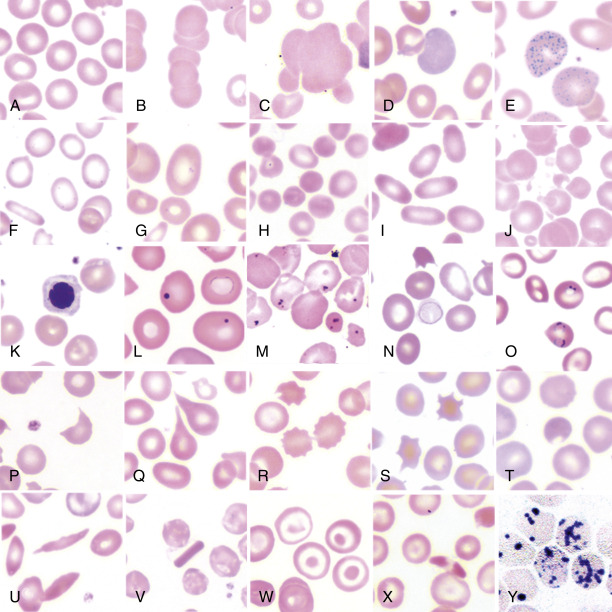
A, Normal red blood cells (RBCs). Note that the central pallor is one third the diameter of the entire cell. B, Rouleaux formation is indicative of increased plasma protein. C, Agglutination indicates an antibody-mediated process such as cold agglutinin disease. D, Polychromatophilic cell. The gray-blue color is attributable to RNA, and the cell is equivalent to a reticulocyte, which must be identified with a reticulocyte stain. E, Basophilic stippling. This also is attributable to increased RNA caused either by a left shift in erythroid cells or lead toxicity. F, Hypochromic microcytic cells typical of iron deficiency anemia. Note the widened central pallor and the “pencil” cell in the lower left. G, Macroovalocyte as can be seen in either megaloblastic anemia or myelodysplastic syndrome. H, Microspherocytes typical of hereditary spherocytosis. I, Elliptocytes (ovalocytes) from a patient with hereditary elliptocytosis. J, RBC fragments from thermal injury (burn patient). K, Nucleated RBC. L, Howell-Jolly bodies indicative of splenic dysfunction or absence. M, Pappenheimer bodies from a patient with sideroblastic anemia. N, Cabot ring, as can be seen in megaloblastic anemia or MDS. O, Malarial parasites (Plasmodium falciparum). P, Schistocyte typical of a microangiopathic hemolytic anemia. Q, Tear-drop form indicates marrow fibrosis and extramedullary hematopoiesis. R, Echinocyte (Burr cell) with rounded edges. S, Acanthocyte (spur cell) with more irregular pointed ends. This was from a patient with neuroacanthocytosis. They can also be seen in patients with liver disease and lipid abnormalities. T, “Bite” cell from a patient with glucose-6-phosphate dehydrogenase (G6PD) deficiency. U, Sickle cell, from a patient with homozygous sickle cell disease. V, Hemoglobin C crystal. W, Target cells. X, Hemoglobin C disease. Note that the RBC in center has condensed hemoglobin at each pole. Y, Heinz body preparation (supravital stain) from a patient with G6PD deficiency. Note that the cells to the right have increased precipitated hemoglobin.
From Hoffman R: Hematology: basic principles and practice, ed 7, Philadelphia, 2018, Elsevier.