section name header

Basic Information

AUTHORS: Sandeep Soman, MD and Snigdha t. Reddy, MD

Definition

Immunoglobulin A nephropathy (IgAN) is a proliferative glomerulonephritis associated with predominant mesangial IgA deposition. IgA deposition can occur in isolation or with IgG, IgM, or both, and often with complement protein C3.

Synonyms

Berger disease

IgAN

IgA vasculitis

ICD-10CM CODES
N02.1Recurrent and persistent hematuria with focal and segmental glomerular lesions
N02.3Recurrent and persistent hematuria with diffuse mesangial proliferative glomerulonephritis
N02.5Recurrent and persistent hematuria with diffuse mesangiocapillary glomerulonephritis
N02.7Recurrent and persistent hematuria with diffuse crescentic glomerulonephritis
N02.8Recurrent and persistent hematuria with other morphologic changes
Epidemiology & Demographics
Incidence

IgA nephropathy (IgAN) is the most common primary glomerulopathy worldwide. The annual incidence of IgAN is about 2.5 per 100,000 person-yr.1,2 There is a very low incidence in Africa relative to the U.S. and a higher incidence in East Asia. In Japan, the incidence is eight times as high as in the U.S.1 Because IgA is the primary enteric antibody, the increased incidence and prevalence in Asia has been postulated to relate to geographic differences in dietary exposure to antigens and genetic factors.

Prevalence

Autopsy studies reveal the prevalence of renal IgA deposits in 1.3% to 16% of cases in the general population.1 Many individuals remain undiagnosed, despite having histologic evidence of IgAN, because they do not have clinical signs of glomerular injury or renal dysfunction. Generally, prevalence rates are expressed as a percentage of cases of primary glomerulonephritis or percentage of total series of renal biopsies. The prevalence in kidney biopsies is 5% in the Middle East, 10% to 35% in Europe, 20% in the U.S., and 32% to 54% in Japan and China. The lower U.S. rates may be biased by a predilection of U.S. nephrologists not to perform kidney biopsy of asymptomatic patients with minimal renal abnormalities.

Predominant Sex & Age

IgAN is most prevalent in the second and third decades of life with a male:female ratio of 2:1 in the U.S. and 3:1 in Europe. In East Asia, the male:female ratio is closer to 1:1.

Risk Factors

Genetic and geographic background predisposes individuals to development of IgAN. The progression of IgAN is highly variable, but approximately 25% of patients develop end-stage renal disease (ESRD) within 10 to 25 yr. The most robust risk factors and predictors of poor renal outcomes at time of diagnosis include older age, hypertension, proteinuria >1 gram per day, and decreased glomerular filtration rate (GFR). Lower hemoglobin and albumin are additional risk factors of poor outcomes. Other remediable risk factors include obesity and smoking. Immunoglobulin levels, specifically IgA antibody, are not required for diagnosis, prognosis, or monitoring of patients.

Pathologic risk factors are based on the Oxford Classification of IgAN (MEST-C) and include increased mesangial cellularity, endocapillary proliferation, segmental glomerulosclerosis, tubular atrophy and interstitial fibrosis, and presence of crescents (see Box E1).3 The score predicts poor outcomes, but isolated endocapillary proliferation does not significantly worsen prognosis. Thrombotic microangiopathy, more commonly encountered in Asians, is associated with worse clinical outcomes.

Genetics and Pathogenesis

The primary defect is systemic and caused by an aberrant glycosylation that results in an abnormal antibody, galactose-deficient IgA1 (Gd-IgA1), a heritable trait in diverse racial and ethnic groups. Genetic predisposition is confirmed by elevated serum Gd-IgA1 levels in a quarter of relatives of patients with IgAN. The Gd-IgA1 binds IgG autoantibody and produces renal-inflammatory immune complexes.

Although IgAN is a sporadic disease in more than 90% of cases, recent genome-wide association studies have identified 6% to 8% of cases with at least 15 susceptibility loci, the strongest signal being from the major histocompatibility complex (MHC) region of chromosome 6.2 Certain loci are more prevalent in Asia than in the U.S. and Europe. In addition, Asian Americans have a fourfold higher ESRD incidence than European Americans and sevenfold higher ESRD incidence than African Americans.

Physical Findings & Clinical Presentation

  • Approximately 75% of children and young adults present with macroscopic hematuria often associated with upper respiratory infection or gastrointestinal illness. This is often referred to as “synpharyngitic hematuria.” Older adults present with microscopic hematuria, proteinuria, and hypertension.
  • Loin pain may be associated with macroscopic hematuria. This symptom should not be confused with the much rarer disorder, “loin pain hematuria syndrome.”
  • Physical findings are usually unremarkable. Hypertension is present in 20% to 30% of patients with chronic kidney disease, and edema is found in the 5% of patients with nephrotic-range proteinuria.
  • IgAN presents as acute kidney injury due to hemoglobin toxicity and intratubular obstruction due to red blood cell casts. It can present as macroscopic hematuria in 5% of patients and as chronic kidney disease in 10% to 20% of patients. Another cause for acute kidney injury can be rapidly progressive glomerulonephritis, frequently associated with the histologic finding of crescents in 50% of glomeruli.
Etiology

  • Most cases are idiopathic or primary. IgA vasculitis (formerly Henoch-Schönlein purpura) may represent a more systemic, vasculitic form of IgAN.
  • Secondary causes of IgAN (Table E1) include hepatitis B; alcoholic cirrhosis; celiac disease; inflammatory bowel disease (IBD); psoriasis; sarcoidosis; cystic fibrosis; cancer of the lungs, larynx, or pancreas; HIV infection; systemic lupus erythematosus; rheumatoid arthritis; diabetic nephropathy; Sjögren syndrome; and Reiter syndrome. Given the role of IgA in the gut, aside from IBD, infections in the gastrointestinal tract have been associated with IgAN or flares of the disease.
  • The four-hit pathogenesis model is depicted in Fig. E1. The first hit from disease heritability is abnormal IgA glycosylation that produces polymeric galactose-deficient antibody, Gd-IgA1. The second hit is when Gd-IgA1 becomes auto-antigenic and auto-antibodies and immune complexes form. The third hit occurs when the immune complexes of Gd-IgA1 and antiglycan antibodies deposit in the mesangium leading to enhanced production of growth factors such as PDGF and IGF-1 with mesangial expansion. The fourth hit is intrarenal complement activation with generation of inflammatory cytokines by activated leukocytes.

Figure E1 Pathogenesis of IgA nephropathy.

!!flowchart!!

IgA, Immunoglobulin A; IC, immune complex.

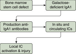
Table E1 Causes of Secondary IgAN

GroupDisease
Gastrointestinal and Liver DiseaseInflammatory bowel disease (Crohn disease, ulcerative colitis), celiac disease, cirrhosis
InfectionHBV, HCV, HIV, tuberculosis, leprosy
Autoimmune DiseasesAnkylosing spondylitis, rheumatoid arthritis, Sjogren syndrome
MalignancyLung cancer, renal cell carcinoma, non-Hodgkin and Hodgkin lymphoma, IgA myeloma
Respiratory tractSarcoidosis, bronchiolitis obliterans, pulmonary hemosiderosis, cystic fibrosis, pulmonary fibrosis
SkinDermatitis herpetiformis, psoriasis

HBV, hepatitis B virus; HCV, hepatitis C virus; HIV, human immunodeficiency virus; IgAN, immunoglobulin A nephropathy.

From Pattrapornpisut P et al: IgA nephropathy: core curriculum 2021, Am J Kidney Dis 78(3):429-441, 2021, https://doi.org/10.1053/j.ajkd.2021.01.024.

Diagnosis

Differential Diagnosis

The differential diagnosis is the group of glomerular disorders with hematuria.

  • IgA vasculitis (Henoch-Schönlein purpura)
  • Hereditary nephritis
  • Thin glomerular basement membrane disease
  • Lupus nephritis
  • Vasculitis
  • Postinfectious nephritis
  • IgA-dominant Staphylococcus-associated postinfectious glomerulonephritis
  • Secondary causes associated with IgAN are as discussed previously

While mesangial deposits in IgAN can be triggered by infection, Staphylococcus-associated glomerulonephritis is an immune-mediated glomerulonephritis with sole or dominant presence of IgA within mesangial and subepithelial deposits and hypocomplementemia.

Workup

  • Diagnosis is based on clinical history and laboratory data. Episodes of gross hematuria with upper respiratory infections and urinalysis with dysmorphic hematuria and/or red blood cell casts and negative serologic tests for other glomerulonephritides (e.g., lupus and vasculitis) are strongly representative of the diagnosis. The diagnosis is confirmed by kidney biopsy revealing predominant or codominant IgA mesangial deposits.
  • To standardize diagnosis and provide prognostication, the pathology is scored via the Oxford IgAN (MEST-C) classification that evaluates mesangial hypercellularity, endocapillary hypercellularity, segmental glomerulosclerosis, tubular atrophy/interstitial fibrosis, and crescent formation.
  • Prognosis is excellent with normal renal function and low proteinuria; accordingly, kidney biopsy is restricted to patients with sustained proteinuria >1 gram per day or worsening kidney function.
Laboratory Tests

  • Urinalysis shows positive blood reaction and variable degrees of proteinuria. Microscopy demonstrates protein, dysmorphic red blood cells, and red blood cell casts.
  • Serum creatinine may be elevated.
  • Urine protein-to-creatinine ratio may be elevated.
  • Serum IgA is elevated in only 50% of patients, but this has little clinical utility and not measured.
  • Measurement of Gd-IgA1 and Gd-IgA1-specific IgG antibodies have been proposed as diagnostic biomarkers, but neither has been validated for this purpose.

Treatment

Although initially considered a benign disease, IgAN is recognized as a common cause of kidney failure. Currently there is no definitive cure. Supportive care is the mainstay of IgAN management.

Nonpharmacologic Therapy

  • Weight reduction in overweight or obese patients
  • Smoking cessation
  • Low sodium diet
Acute & Chronic General Rx

  • Antihypertensive therapy, preferably with ACE inhibitors or angiotensin II type 1 receptor blockers (ARB). The goal blood pressure is <125/75 mm Hg if proteinuria is >1 gram per day and <130/80 mm Hg if <1 gram per day. Dual blockade of the renin-angiotensin-aldosterone system (RAAS) is not recommended due to risk of hyperkalemia and/or acute kidney injury.
  • Patients with recurrent gross hematuria, isolated microscopic hematuria, no or minimal proteinuria (<1 gram per day), normal blood pressure, and normal kidney function should be monitored every 6 to 12 mo to evaluate disease progression. Fish oil may be considered, but supporting data are limited.
  • If recurrent episodes of macroscopic hematuria are associated with tonsillitis, tonsillectomy may benefit these patients; however, this treatment is controversial.
  • Patients with persistent proteinuria >1 gram per day with or without hypertension are treated with ACE inhibitors or ARB.
  • A 6-mo regimen of glucocorticoid steroids has been recommended for patients with good kidney function and persistent proteinuria of >1 gram per day, despite ACE inhibitor or ARB administration and fish oil after 6 mo. However, a recent, prospective, randomized, controlled trial (STOP-IgAN) showed no difference in decline in GFR with corticosteroids in subnephrotic patients followed for 3 yr. Infections, glucose intolerance, and obesity were more frequent in the steroid-treatment group.4 Another recent, multicenter study (TESTING) of 262 subnephrotic patients randomized to steroids vs. placebo showed potential renal benefit of steroid treatment. The trial was terminated early because of excess infections in the steroid group (14.7% vs. 3.2%).5 Overall, definitive conclusions regarding steroid treatment are not established. Patients with nephrotic syndrome and relatively preserved kidney function on maximal RAAS blockade can be treated with steroids for a 6-mo course, especially if there is disease progression. Steroid treatment is not indicated if significant chronic disease is demonstrated on renal biopsy or a GFR <20 ml/min per 1.73 m2.
  • Rapidly progressive glomerulonephritis in the absence of severe fibrosis on kidney biopsy is treated with pulse steroids followed by oral steroids, combined with cyclophosphamide for the first 2 to 3 mo. This initial regimen is followed by oral steroids and azathioprine for 2 yr as a maintenance treatment.
  • Patients with acute kidney injury require a kidney biopsy to differentiate acute tubular necrosis due to pigment nephropathy, which requires only supportive therapy, from crescentic IgAN, which requires aggressive medical management.
  • Kidney transplantation is the treatment of choice for ESRD. Although the recurrence rate of IgAN in the transplanted kidney is 50%, allograft loss from recurrent IgAN is rare, at 5%.
  • Statins are indicated for all IgAN patients with dyslipidemia, hypertension, and other cardiovascular risks.
  • The roles of mycophenolate, rituximab, and plasmapheresis are not clearly defined.
  • Budesonide delayed release capsules (Tarpeyo) was recently approved for reduction of proteinuria in those at risk for rapid progression.
  • A phase 2b clinical trial (NEFIGAN) showed potential benefit of enteric budesonide added to conventional therapy.6
  • Another randomized phase 2 clinical trial showed antiproteinuric effect of combination therapy with hydroxychloroquine and RAAS inhibitors over 6 mo without significant adverse effects.7
  • In the DAPA-CKD trial, the SGLT2 inhibitor dapagliflozin was shown to reduce proteinuria and progression of kidney disease in addition to RAAS blockers for patients with eGFR between 25 and 75 ml/min/1.73 m2.8
  • Future treatments may include targeted immunosuppression of the gut and modification of abnormal complement activation.
Disposition/Prognosis

  • Complete remission occurs in <10% of patients.
  • ESRD develops in 12% of patients followed for 5 yr, in 15% to 20% within 10 yr, and in 20% to 40% within 20 yr. Prognostic markers for IgAN are described in Box E1.
  • Poor prognostic indicators include hypertension, degree of renal insufficiency, greater proteinuria, and particular histologic features on renal biopsy, including crescents, increased mesangial cells, glomerulosclerosis, and tubulointerstitial fibrosis or atrophy.
  • Potential future biomarkers include increased serum levels of glycan-specific IgG antibodies that predict progression to ESRD or death, elevated serum levels of galactose-deficient IgA1, factor H-related proteins, and selected urine microRNAs.

BOX E1 Prognostic Markers in IgA Nephropathy

Clinical
Poor prognosis

  • Increasing age
  • Severity of proteinuria
  • Hypertension (increased systolic blood pressure)
  • Kidney impairment (decreased GFR)
  • Decreased serum albumin
  • Decreased hemoglobin
  • Persistent microscopic hematuria
Good prognosis

  • Recurrent macroscopic hematuria
Histopathologic (Oxford Classification)
Poor prognosis

  • Increased mesangial cellularity
  • Segmental glomerulosclerosis
  • Tubular atrophy/interstitial fibrosis
  • Crescents
No impact on prognosis

  • Endocapillary hypercellularity

GFR, Glomerular filtration rate; IgA, immunoglobulin A.From Canetta PA et al: Glomerular diseases: emerging tests and therapies for IgA nephropathy, Clin J Am Soc Nephrol 9:617, 2014; and Lv J et al: Evaluation of the Oxford Classification of IgA nephropathy: a systematic review and meta-analysis, Am J Kidney Dis 62:891-899, 2013.

Complementary & Alternative Medicine

The role of fish oil is controversial. One large, randomized, placebo-controlled study showed benefit, although many small, randomized studies have shown no benefit. The recommendation for mild disease is that fish oil is optional for patients. International consensus guidelines recommend fish oil along with ACE inhibitors/ARB if proteinuria is >1 gram per day.

Referral

Patients with suspected IgAN are commonly referred to nephrologists for diagnosis and treatment, typically when presenting with gross hematuria and urinalysis with blood and proteinuria.

Pearls & Considerations

Comments

  • Gross hematuria (red urine) is uncommon with intrinsic kidney disease but commonly encountered in IgAN and in cyst rupture from autosomal dominant polycystic kidney disease.
  • IgAN is not an entirely benign condition, even when microhematuria is the only clinical presentation. Nearly 40% of patients reach ESRD after 20 yr of illness.
  • Episodic gross hematuria that occurs during upper respiratory infections is a classic presentation for IgAN. If present, an ACE inhibitor or ARB should be started.

Related Content

  1. Rodrigues J.C. : IgA nephropathyClin J Am Soc Nephro. ;12(4):677-686, 2017.doi:10.2215/cjn.07420716
  2. Lafayette R.A., Kelepouris E. : Immunoglobulin A nephropathy: advances in understanding of pathogenesis and treatmentAm J Nephro. ;47(Suppl. 1):43-52, 2018.doi:10.1159/000481636
  3. Canetta P.A. : Glomerular diseases: emerging tests and therapies for IgA nephropathyClin J Am Soc Nephrol. ;9(3):617-625, 2014.doi:10.2215/CJN.07260713
  4. Rauen T. : Intensive supportive care plus immunosuppression in IgA nephropathyNew Engl J Med. ;373(23):2225-2236, 2015.doi:10.1056/nejmoa1415463
  5. Lv J. : Effect of oral methylprednisolone on clinical outcomes in patients with IgA nephropathyJAMA. ;318(5), 2017.doi:10.1001/jama.2017.9362
  6. Fellström B.C. : Targeted-release budesonide versus placebo in patients with IgA nephropathy (NEFIGAN): a double-blind, randomised, placebo-controlled phase 2b trialLancet. ;389(10084):2117-2127, 2017.doi:10.1016/s0140-6736(17)30550-0
  7. Liu L.-J. : Effects of hydroxychloroquine on proteinuria in IgA nephropathy: a randomized controlled trialAm J Kidney Dis. ;74(1):15-22, 2019.doi:10.1053/j.ajkd.2019.01.026
  8. Wheeler D.C. : A pre-specified analysis of the DAPA-CKD trial demonstrates the effects of dapagliflozin on major adverse kidney events in patients with IgA nephropathyKidney Int. ;100(1):215-224, 2021.doi:10.1016/j.kint.2021.03.033