section name header

Basic Information

AUTHOR: Harlan G. Rich, MD, FACP, AGAF

Definition

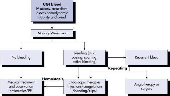
A Mallory-Weiss tear (MWT) is a longitudinal mucosal laceration in the region of the gastroesophageal junction and gastric cardia typically occurring after repeated episodes of severe vomiting or retching.

Synonyms

Mallory-Weiss syndrome

MWT

ICD-10CM CODE
K22.6Mallory-Weiss syndrome
Epidemiology & Demographics

  • Accounts for 5% to 15% of cases of upper GI bleeding1
  • Reported from early childhood to old age; historically the majority of patients are age 40 to 60 yr, though a recent international study demonstrated a median age of 612
  • More common in males
  • Alcohol use is present in 30% to 60%
Physical Findings & Clinical Presentation

  • Vomiting, retching, or vigorous coughing will often, but not always, precede hematemesis.
  • There are no specific physical exam findings. Patients may be clinically stable or present with tachycardia, hypotension, melena, hematochezia, epigastric pain, back pain, syncope, or hemorrhagic shock.
  • Bleeding may be self-limited or severe. Rebleeding is more common in patients with advanced alcoholic liver disease.3
  • Tears may be seen in association with other upper GI tract lesions, including hiatal hernia (present in as many as 90% of patients), ulcers, and esophageal varices, particularly in alcoholics.
Etiology

  • An acute increase in intragastric and intraabdominal pressure is transmitted to the gastroesophageal junction and esophagus, resulting in mucosal laceration.
  • Vomiting may be associated with alcohol use, cannabinoid use, ketoacidosis, ulcer disease, uremia, pancreatitis, chemotherapy, cholecystitis, pregnancy (in particular associated with hyperemesis gravidarum), bulimia, myocardial infarction, or the postoperative period.
  • Infrequently reported causes include chest wall trauma (including cardiopulmonary resuscitation), hiccups, coughing, seizures, lifting/straining, blunt abdominal trauma, acute severe asthma, labor and delivery, and even primal scream therapy.
  • Tears may be iatrogenic, related to routine endoscopy (especially in struggling, retching, or older patients, or in association with hiatal hernias or distal gastrectomy), enteroscopy with or without spiral or balloon overtubes, esophageal dilation, lower esophageal pneumatic disruption therapy for achalasia, endoscopic submucosal dissection, transesophageal echocardiography, bariatric intragastric balloons, or in association with polyethylene glycol electrolyte colonic lavage preparation.4
  • Tears are frequently found on the right lateral wall of the esophagus.

Diagnosis

Differential Diagnosis

  • Esophageal or gastric varices
  • Esophagitis or esophageal ulcers (peptic or pill-induced)
  • Gastric erosions
  • Gastric or duodenal ulcer
  • Dieulafoy lesion
  • Arteriovenous malformations
  • Neoplasms (usually esophageal or gastric)
  • Boerhaave syndrome
Workup

Endoscopy is the diagnostic method of choice.

Laboratory Tests

  • CBC, prothrombin time, partial thromboplastin time
  • Electrolytes, blood urea nitrogen, creatinine, liver function tests, pregnancy test, tests to evaluate for predisposing conditions
Imaging Studies

  • Upper GI series is usually not sensitive.
  • Patients with concurrent chest pain, dyspnea, shock, or physical examination findings of crepitus or pleural effusion should have a chest radiograph or computed tomography to exclude Boerhaave syndrome.

Treatment

Nonpharmacologic Therapy

  • Supportive care
  • Avoidance of aspirin, nonsteroidal antiinflammatory drugs, and anticoagulants
Acute General Rx

  • Patients with active bleeding or hemodynamic instability require large-bore intravenous lines, fluid resuscitation, transfusion of blood products (red blood cells to maintain hemoglobin >7 g/dl, fresh frozen plasma, platelets), and holding or reversal of anticoagulation as appropriate.
  • Nasogastric decompression and antiemetics may be considered. Electrolyte imbalances should be corrected.
    • Endoscopic therapy for patients with active or ongoing hemorrhage (Fig. E1). Therapeutic modalities include electrocoagulation, argon plasma photocoagulation, heater probe coagulation, injection (e.g., 1:10,000 epinephrine, polidocanol), sclerotherapy (for bleeding associated with esophageal varices), band ligation, endoscopic hemoclips (Fig. E2), over-the-scope clips, and hemostatic sprays. Therapies may be used alone or in combination (Fig. E3). Hemoclips combined with a nylon snare (“tulip-bundle” technique) have been successfully used for refractory bleeding.5
  • Arterial embolization in patients with active bleeding who are poor surgical candidates.
  • Laparotomy, with gastrotomy and oversewing of the tear or laparoscopic repair, is required in a small percentage of patients with uncontrolled bleeding.

Figure E1 Treatment Algorithm for Mallory-Weiss Syndrome. IV, Intravenous; Ppi, Proton-Pump Inhibitor; Ugi, Upper Gastrointestinal

From Cameron JL, Cameron AM: Current surgical therapy, ed 12, Philadelphia, 2017, Elsevier.

Figure E2 A, Endoscopic View of a Mallory-Weiss Tear (Blue Arrow) at the Gastroesophageal Junction

B, After Hemoclip Deployed at the Lesion with Good Hemostasis.

From Cameron JL, Cameron AM: Current surgical therapy, ed 12, Philadelphia, 2017, Elsevier.

Figure E3 Mallory-Weiss Tear

A and B, Endoscopic Views of Mallory-Weiss Tear Being Treated with Hemoclips.

Courtesy Isaac Raijman, MD, Associate Professor, Baylor College of Medicine. From Cameron JL, Cameron AM: Current surgical therapy, ed 10, Philadelphia, 2011, Saunders.

Chronic Rx

  • Healing will usually occur without specific therapy.
  • H2 blockers or proton pump inhibitors may be given to help facilitate healing but should not be used long term unless appropriate indications are present.
Disposition

  • Prognosis is good, with spontaneous cessation of bleeding in upward of 90% of patients. Endoscopic features can guide treatment. Blatchford score <6 suggests no need for transfusion or endoscopic intervention.
    1. Blatchford score is calculated by specific indices (sex, blood urea nitrogen, hemoglobin, systolic blood pressure, pulse, and the presence of melena, syncope, hepatic disease, or cardiac failure) and is used to determine which patients need clinical intervention for acute upper gastrointestinal bleeding.
  • Delayed rebleeding is described in patients with high-risk stigmata (shock at initial presentation, spurting, or oozing at initial endoscopy). The Forrest Classification may not be a good predictor of rebleeding.2
  • Death has been reported in 3% to 12%, often in association with severe bleeding and underlying comorbid conditions such as advanced age, coagulopathy, elevated transaminases, thrombocytopenia, alcohol use, presentation with a very low hemoglobin level or melena, and multisystem organ failure. Over the past decade, associated mortality appears to be significantly declining,6though it is similar to mortality related to peptic ulcer bleeding.2
Referral

  • Gastrointestinal referral for endoscopy
  • Surgical or interventional radiology referral for bleeding unresponsive to endoscopic treatment or surgical referral in the setting of coexistent perforation

Pearls & Considerations

Conditions predisposing to retching or vomiting should be identified and treated at presentation.

Related Content

Mallory-Weiss Tear (Patient Information)

Related Content

  1. Wuerth B.A., Rockey D.C. : Changing epidemiology of upper gastrointestinal hemorrhage in the last decade: a nationwide analysisDig Dis Sci. ;63:1286-1293, 2018.
  2. Tham J.E. : International multicenter study comparing demographics, therapy and outcomes in bleeding from Mallory Weiss tears and peptic ulcersEndosc Int Open. ;10:E653-E658, 2022.
  3. Nojkov B., Cappell M.S. : Distinctive aspects of peptic ulcer disease, Dieulafoy’s lesion, and Mallory-Weiss syndrome in patients with advanced alcoholic liver disease or cirrhosisWorld J Gastroenterol. ;22:446-466, 2016.
  4. Na S. : Risk factors for an iatrogenic Mallory-Weiss tear requiring bleeding control during a screening upper endoscopyGastroenterol Res Pract. ;1-6, 2017.
  5. Kim H.S. : Endoscopic management of Mallory-Weiss tearingClin Endosc. ;48:102-105, 2015.
  6. Ljubicic´ N. : Mortality in high-risk patients with bleeding Mallory-Weiss syndrome is similar to that of peptic ulcer bleeding. Results of a prospective database studyScand J Gastroenterol. ;49(458), 2014.