section name header

Basic Information

AUTHOR: Kapil S. Meleveedu, MD

Definition

Myelodysplastic syndromes (MDSs) are a group of acquired clonal disorders of hematopoietic stem cells characterized by altered differentiation and proliferation. Presenting features include peripheral blood cytopenias and bone marrow hypercellularity with morphologic abnormalities, which reflects underlying ineffective hematopoiesis with inadequate maturation.

Classification

  • Earlier classification systems for MDS have included the French-American-British (FAB) classification (1997), the 1997 World Health Organization (WHO) classification, and the 2008 modified WHO classification.
  • Currently, the revised 2016 WHO classification is in clinical use (Table 1).

TABLE 1 2016 World Health Organization Classification of the Adult Myelodysplastic Syndromes

NameDysplastic LineagesCytopeniasRing Sideroblasts as % of Marrow Erythroid ElementsBM and PB BlastsCytogenetics by Conventional Karyotype Analysis
MDS with single lineage dysplasia (MDS-SLD)11 or 2<15%/<5%BM <5%, PB <1%, no Auer rodsAny, unless fulfills all criteria for MDS with isolated del(5q)
MDS with multilineage dysplasia (MDS-MLD)2 or 31-3<15%/<5%BM <5%, PB <1%, no Auer rodsAny, unless fulfills all criteria for MDS with isolated del(5q)
MDS With Ring Sideroblasts (MDS-RS)
MDS-RS with single lineage dysplasia (MDS-RS-SLD)11 or 215%/5%BM <5%, PB <1%, no Auer rodsAny, unless fulfills all criteria for MDS with isolated del(5q)
MDS-RS with multilineage dysplasia (MDS-RS-MLD)2 or 31-315%/5%BM <5%, PB <1%, no Auer rodsAny, unless fulfills all criteria for MDS with isolated del(5q)
MDS with isolated del(5q)1-31-2None or anyBM <5%, PB <1%, no Auer rodsdel(5q) alone or with 1 additional abnormality except –7 or del(7q)
MDS With Excess Blasts (MDS-EB)
MDS-EB-10-31-3None or anyBM 5%-9% or PB 2%-4%, no Auer rodsAny
MDS-EB-20-31-3None or anyBM 10%-19% or PB 5%-19% or Auer rodsAny
MDS, Unclassifiable (MDS-U)
With 1% blood blasts1-31-3None or anyBM <5%, PB = 1%, no Auer rodsAny
With single lineage dysplasia and pancytopenia13None or anyBM <5%, PB <1%, no Auer rodsAny
Based on defining cytogenetic abnormality01-3<15%§BM <5%, PB <1%, no Auer rodsMDS-defining abnormality
Refractory cytopenia of childhood1-31-3NoneBM <5%, PB <2%Any

Cytopenias defined as hemoglobin <10 g/dl; platelet count <100 × 109/L; and absolute neutrophil count <1.8 × 10⁹/L. Rarely, MDS may present with mild anemia or thrombocytopenia above these levels. PB monocytes must be <1 × 10⁹/L.

If SF3B1 mutation is present.

1% PB blasts must be recorded on at least 2 separate occasions.

§ Cases with 15% ring sideroblasts by definition have significant erythroid dysplasia and are classified as MDS-RS-SLD.

From Arber DA et al: The 2016 revision to the World Health Organization classification of myeloid neoplasms and acute leukemia, Blood 127(20):2391-2405, 2016.

Synonyms

MDS

Preleukemia

ICD-10CM CODES
D46.9Myelodysplastic syndrome, unspecified
D46.CMyelodysplastic syndrome with isolated del(5q) chromosomal abnormality
D46.ZOther myelodysplastic syndromes
Epidemiology & Demographics
Incidence (In U.S.)

Approximately 4 new cases/100,000 persons per yr. An estimated 30,000 new cases are diagnosed annually in the U.S. The true incidence is likely to be higher because of incomplete case assessment and underreporting of MDS in cancer registries, and it may be close to 75 per 100,000 among persons over the age of 70 years.1

Predominant Age

More common in elderly patients; median age about 70 years

Physical Findings & Clinical Presentation

  • Patients often present with fatigue due to anemia.
  • Patients can present with thrombocytopenia and leukopenia.
  • Skin pallor, mucosal bleeding, and ecchymosis may be present.
  • Fever, infection, and dyspnea are common.
Etiology

Normal human aging is the most important risk factor for development of MDS due to progressive acquisition of somatic mutations by the hematopoietic stem cells throughout the human life span. Exposure to radiation, chemotherapeutic agents, benzene, or other organic compounds is also associated with myelodysplasia. Table 2 describes predisposing factors and epidemiologic associations of patients with MDS. MDS is extremely rare in pediatric age group but if does arise in patients <18 years of age, work up for inherited or congenital disorders should be sought. With recent advances in genomic sequencing technologies, some individuals with or without cytopenias are found to have somatic clonal mutations which in some cases go on to develop MDS or AML. The understanding regarding these potential premalignant conditions (ICUS, CHIP, CCUS) including factors that determine progression continues to evolve. Up to 40 genes that affect specific functional pathways are mutated in MDS, with 90% of patients having at least one mutation and a median of two to three mutations detected per patient. The most common mutations occur in genes involved in RNA splicing (SF3B1, SRSF2, U2AF1, and ZRSR2), epigenetic modification (TET2, ASXL1, and DNMT3A), regulators of signal transduction (NRAS and JAK2), and transcription factors (RUNX1 and TP53). An increasing number of germline mutations (RUNX1, GATA2, DDX41, etc) are also being identified which are shown to be associated with a familial syndrome with inherited predisposition of developing MDS.

TABLE 2 Predisposing Factors and Epidemiologic Associations of Patients With Myelodysplastic Syndrome

Heritable
Constitutional Genetic Disorders
Trisomy 8 mosaicism
Familial monosomy 7
Down syndrome (trisomy 21)
Neurofibromatosis 1
Germ cell tumors [embryonal dysgenesis del(12p)]
Congenital Neutropenia
Kostmann syndrome
Shwachman-Diamond syndrome
DNA Repair Deficiencies
Fanconi anemia
Ataxia-telangiectasia
Bloom syndrome
Xeroderma pigmentosum
Pharmacogenomic polymorphisms (GSTq1-null)
Acquired
Senescence
Mutagen Exposure
Alkylator therapy (chlorambucil, cyclophosphamide, melphalan, N-mustards)
Topoisomerase II inhibitors (anthracyclines)
βEmitters (32p)
Autologous stem cell transplantation
Environmental/occupational (benzene)
Tobacco
Aplastic anemia
Paroxysmal nocturnal hemoglobinuria

DNA, Deoxyribonucleic acid.

From Hoffman R et al: Hematology, basic principles and practice, ed 7, Philadelphia, 2018, Churchill Livingstone.
Diagnosis

Differential Diagnosis

  • Hereditary dysplasias (e.g., Fanconi anemia, Diamond-Blackfan syndrome)
  • Vitamin B12/folate deficiency
  • Copper deficiency
  • Exposure to toxins (drugs, alcohol, chemotherapy)
  • Renal failure
  • Irradiation
  • Autoimmune disease
  • Paroxysmal nocturnal hemoglobinuria
Workup

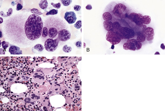
Diagnostic workup includes laboratory evaluation and bone marrow examination (Fig. E1). Cytogenetic analysis (Box E1) by conventional metaphase karyotyping or by MDS FISH assessment should be performed in patients with MDS. Next-generation sequencing (NGS) can be performed on bone marrow to provide additional information on mutations. Genes recurrently mutated in myelodysplastic syndrome are summarized in Table 3. Physical examination, medical history, and laboratory tests aiding in diagnosis of MDS are described in Table 4. Key features of the major myelodysplastic syndromes are summarized in Table 5.

TABLE 5 Diagnostic Criteria for Myelodysplastic Syndrome

A. Presence of at Least One Unexplained Cytopenia for at Least 6 Moa
Hemoglobin <11 g/dl, or
Absolute neutrophil count <1.5 × 10⁹/L, or
Platelet count <100 × 109/L
plus B. Presence of One or More MDS-Qualifying Criteria:
>10% dysplasia in one or more hematopoietic lineage, or
5%-19% blasts in bone marrow, or
MDS-defining cytogenetic abnormality, such as:
t(1;3)(p36.3;q21.1)t(2;11)(p21;q23)inv(3)(q21;q26.2)
t(3;21)(q26.2;q22.1)–5 or del(5q)t(6;9)(p23;q34)
–7 or del(7q)del(9q)
del(11q)t(11;16)(q23;p13.3)del(12p) or t(12p)
–13 or del(13q)i(17q) or del(17p)idic(X)(q13)
plus C. Exclusion of Alternative Diagnoses
AML (i.e., <20% blasts, and no t(8;21), inv(16), t(16;16), t(15;17), or erythroleukemia) or ALL
Other hematologic diseases (aplastic anemia, PNH, LGL, lymphoma, myelofibrosis, and other MPN)
Viral infections (HIV, EBV, parvovirus)
Nutritional deficiencies (iron, copper, B12, folate)
Medications (methotrexate, azathioprine, isoniazid, cytotoxic chemotherapy)
Alcohol or other toxins
Autoimmune diseases (SLE, Felty syndrome, ITP, autoimmune hemolytic anemia)
Congenital disorders (Diamond-Blackfan anemia, Shwachman-Diamond syndrome, Fanconi anemia, and others)

ALL, Acute lymphoblastic leukemia; AML, acute myeloid leukemia; EBV, Epstein-Barr virus; HIV, human immunodeficiency virus; ITP, immune thrombocytopenic purpura; LGL, large granular lymphocyte leukemia; MDS, myelodysplastic syndrome; MPN, myeloproliferative neoplasm; PNH, paroxysmal nocturnal hemoglobinuria; SLE, systemic lupus erythematosus.

a Diagnosis can be made earlier than 6 mo if no other cause is apparent for cytopenias, or there are excess blasts or an MDS-defining cytogenetic abnormality.

From Hoffman R et al: Hematology, basic principles and practice, ed 7, Philadelphia, 2018, Elsevier.

TABLE 4 Physical Examination, Medical History, and Laboratory Tests Aiding in Diagnosis of Myelodysplastic Syndrome

Medical History
  • Duration of symptoms
  • History of blood disease
  • History of exposure to occupational toxins or cytotoxic agents
  • Medication history
  • Alcohol intake
  • Comorbid conditions
  • Family history of hematological disorders
Physical Examination
  • Pallor
  • Petechiae
  • Purpura
  • Bruising
  • Tachypnea
  • Signs of infection
  • Splenomegaly
Laboratory Testing
  • Complete blood count with a manual differential
  • Reticulocyte count
  • Vitamin B12 and folate levels
  • Consider methylmalonic acid and red blood cell folate levels
  • Iron, total iron-binding capacity, and ferritin level
  • Thyroid-stimulating hormone level
  • Lactate dehydrogenase
  • Antinuclear antibody
  • Coombs test and haptoglobin
  • Serum erythropoietin level
  • Human leukocyte antigen (histocompatibility antigens) typing in appropriate patients
  • Paroxysmal nocturnal hemoglobinuria screen
Bone Marrow Testing
  • Hematopathology
  • Percentage of blasts on 200 cell aspirate differential
  • Presence or absence of Auer rods
  • Percentage of cellularity of bone marrow biopsy
  • Iron stain on aspirate (ringed sideroblasts)
  • Iron stain on biopsy (storage)
  • Dysplastic features (% and number of dysplastic lineages)
  • Cytogenetics (karyotype of 20 metaphase cells)
  • Fluorescent in situ hybridization
  • Flow cytometry (not useful for quantitation)

From Hoffman R et al: Hematology, basic principles and practice, ed 7, Philadelphia, 2018, Churchill Livingstone.
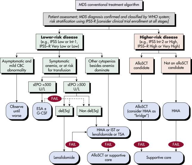
TABLE 3 Genes Recurrently Mutated in Myelodysplastic Syndrome

GeneFrequency (%)Notes
Splicing Factors
SF3B120-30Strong association with RARS
SRSF210-15 (MDS)
40 (CMML)
Enriched in CMML
U2AF15-12Association with del(20q)
Epigenetic Modifiers
TET220-30 (MDS)
40-50 (CMML)
Enriched in CMML
Mutually exclusive with IDH
DNMT3A8-13
ASXL110-20 (MDS)
30-40 (CMML)
Enriched in CMML
EZH25-10 (MDS)
20-30 (CMML)
Enriched in CMML
May be functionally involved in 7q–
IDH1/2<5More frequent in AML
ATRXRareAssociated with acquired thalassemia
Transcription Factors
RUNX110-15Can be somatic or germline
GATA2RareMostly germline
ETV6<5Can be somatic or germline
TP5310-12Association with complex karyotype, therapy-related disease
Kinases and Receptors
JAK2<5Enriched in RARS-T
NRAS5-10Seen in progression to AML
CBL<5Enriched in JMML
PTPN11<5More common in JMML
BRAFRareAlso seen in hairy cell leukemia
Cohesin Complex
STAG25-10Cohesin class mutations enriched in high-risk MDS and secondary AML
RAD21<5
SMC3<2
SMC1A<2
GCPR Complex
GNASRareMutations recently described in wide range of hematologic malignancies, including MDS
GNB1Rare

AML, Acute myeloid leukemia; CMML, chronic myelomonocytic leukemia; GCPR, G-coupled protein receptor; IDH, isocitrate dehydrogenase; JMML, juvenile myelomonocytic leukemia; MDS, myelodysplastic syndrome; RARS, refractory anemia with ring sideroblasts; RARS-T, RARS with thrombocytosis.

From Hoffman R et al: Hematology, basic principles and practice, ed 7, Philadelphia, 2018, Elsevier.

The 2016 WHO classification of myeloid neoplasms is now used to classify MDS and is broadly categorized into the following subtypes: MDS with dysplasia, MDS with ring sideroblasts (MDS-RS), MDS with excess blasts (MDS-EB), and MDS unclassifiable (MDS-U).

Figure E1 Megakaryocytic Dysplasia in Bone Marrow Aspirate Smears and Biopsies in Myelodysplastic Syndrome

(A) Abnormal nuclear features in bone marrow aspirate smears include monolobate and multiple separate small nuclei. (B) Hyperlobation of megakaryocytic nuclei. (C) Clustered dysplastic megakaryocytes in a bone marrow biopsy.

From Jaffe ES et al: Hematopathology, Philadelphia, 2001, Saunders.

BOX E1 Cytogenetic Abnormalities in Myelodysplastic Syndrome

Gain or loss of chromosomal material

  • -7, 7q-
  • 5q-, -5
  • +8
  • +21, -21
  • 17p-, -17
  • -20, 20q-
  • 11q-, +11
  • -Y
  • 9q-
  • +6
  • 12p-
  • 13q-

Uncommon translocations and inversions

  • t(3;3)(q21;q26), inv3(q21q26), t(3;21)(q26;q22), and other 3q21 and 3q26 translocations
  • t(1;7)(p11;p11)
  • t(2;11)(p21;q23)
  • t(11;16)(q23;p13)
  • i(17)q, unbalanced and dicentric translocations at 17p

Any acquired clonal cytogenetic abnormality in hematopoietic cells, except characteristic de novo acute myeloid leukemia (AML) translocations.

Complex abnormalities (multiple cytogenetic abnormalities, excluding those characteristic of de novo AML).

From Jaffe ES et al: Hematopathology, Philadelphia, 2011, Saunders.

Common de novo AML translocations: t(15;17), t(8;21), inv(16) or t(16;16), t(9;11), t(11;19), t(11;17), t(8;16), t(1:22).

Treatment

Nonpharmacologic Therapy

  • Packed red blood cell (PRBC) transfusions in patients with severe symptomatic anemia
  • Platelet transfusions in patients with severe thrombocytopenia or those with bleeding episodes
Acute General Rx

  • The initial focus of MDS therapy involves stratification of patients into very-low-, low-, intermediate-, high-, and very-high-risk states using the well-defined and validated revised International Prognostic Scoring System (IPSS-R). This was developed from an evaluation of 7012 patients and utilizes five major variables for evaluating clinical outcomes: Cytogenetic risk groups, marrow blast percentage, and depth of cytopenias (hemoglobin, platelet, and absolute neutrophil count levels, respectively). New prognostic systems incorporating molecular mutations into the IPSS (IPSS-Molecular or IPSS-M) are available (Published June 12, 2022, NEJM Evid 2022; 1 (7) although it is not widely adopted yet. The WHO classification has also recently been updated to distinguish patients with defining genetic abnormalities (SF3B1, deletion 5q or TP53) from morphologically defined subtypes.
  • Although the current WHO diagnostic classification (5th edition) distinguishes MDS from AML based on the percentage of blasts (MDS, <20% blasts and AML 20% blasts), MDS with >10% blasts could be treated similarly to AML.

Fig. 2 and Table 6 summarize a treatment approach for MDS.

  • Very-low- and low-risk patients are treated with supportive care or growth factors.
  • Intermediate-, high-, and very-high-risk patients are offered treatment with hypomethylating agents (HMAs) along with supportive care.
  • The only potentially curative treatment for MDS is hematopoietic cell transplantation (HCT). All patients with higher risk MDS (and selected low risk patients with poor risk molecular features) should be referred for consideration of HCT soon after diagnosis. Older age should not preclude HCT as a reduced intensity conditioning HCT has been shown to offer better survival in patients with higher risk MDS compared to HMAs or best supportive care.
  • Erythropoietin (10,000 to 60,000 units/wk) or pegylated erythropoietin (200 to 500 mg every 1 to 3 wk) is used in patients with symptomatic anemia. Responses with increase in hemoglobin and decreased transfusion requirements are achieved typically in patients who have serum erythropoietin levels <500 U/L and adequate iron stores.
  • Luspatercept is a recombinant fusion protein that binds transforming growth factor β superfamily ligands to reduce SMAD2 and SMAD3 signaling. It was evaluated in patients with very-low-risk to intermediate-risk MDS with ring sideroblasts (often associated with SF3B1variant) who had been receiving regular PRBC transfusions and was found to achieve transfusion independence in 38% patients versus 13% receiving placebo. It is particularly useful for anemia either refractory to erythropoietin stimulating agents (ESA) or unlikely to respond to ESA.
  • DNA methyltransferase inhibitors: Azacitidine, a pyrimidine nucleoside analogue of cytidine, has been shown to improve the quality of life for patients and to prolong overall survival. Decitabine, another nucleoside analogue, has also been FDA approved for patients with MDS. These agents may also be useful in preventing the transition of MDS to AML.
  • The fixed-dose oral combination of decitabine and cedazuridine was approved by FDA on July 2020 for adult patients with intermediate -1, intermediate-2, and high risk MDS, including those with previously treated and untreated, de novo, and secondary MDS. This was based on two open-label randomized cross over trials which demonstrated a CR rate of 18-21% and median duration of CR of 7.5 - 8.7 months.
  • Immunomodulators: Lenalidomide, an immunomodulatory agent, reduces PRBC transfusion dependence in low-risk, non-5q deletion MDS patients but with increased hematologic adverse events. It can also reduce transfusion requirements and reverse cytologic and cytogenetic abnormalities in patients who have 5q deletion MDS.
  • Additionally, lenalidomide augments erythropoietin receptor signaling in vitro and improves hemoglobin response significantly when used in combination with erythropoietin in patients with lower-risk, non-del(5q) MDS.
  • Thrombopoietin receptor agonists are often avoided in MDS patients with excess blasts (except in palliative settings) in view higher rates of transformation to AML in clinical trials.
  • Results of chemotherapy are generally disappointing. AML-style combination chemotherapy regimens generally induce a complete response in only a minority of patients, and the average duration of response is <1 yr.
  • Myeloid growth factors (granulocyte colony-stimulating factor [G-CSF], granulocyte-macrophage colony-stimulating factor [GM-CSF]) are used in patients with severe neutropenias and high infection risk. Additionally, these provide a synergistic effect when used in combination with erythropoietin in terms of improvement in the hemoglobin levels.

TABLE 6 Available Therapies for Myelodysplastic Syndromes in the U.S. and Preferred Dosage Regimena

  1. Erythropoiesis-stimulating agents (should hold if hemoglobin 10 g/dl)
    1. Epoetin alfa 20-60,000 mIU/wk subcutaneously; dose can be adjusted every 1-2 mo depending on response; or
    2. Darbepoetin alfa 200-300 μg subcutaneously every 1-2 wk or 500 μg subcutaneously every 3 wk; or
    3. Epoetin alfa as per (a) plus G-CSF (filgrastim) 0.5 μg /kg subcutaneously 1-3 times per wk (pegfilgrastim is commonly substituted in the U.S., but data are absent, and splenic rupture and leukemoid reactions are risks)
  2. Iron chelation therapy
    1. Deferoxamine (starting dose 20 mg/kg/day; a typical effective dose for an adult is 6 g/day subcutaneously or intravenously, administered by infusion pump over 8-16 h daily); or
    2. Deferasirox, tablet for oral suspension 20-30 mg/kg/day orally once daily oral tablet, 14-28 mg/kg/day, adjust as tolerated and according to efficacy.
    3. Deferiprone (L1) is available outside the U.S., but is only approved for thalassemia in the U.S. and is a weaker chelator than the other two and cytopenias can be problematic
  3. Lenalidomide: 10 mg orally once daily for 21-28 days per 28-day cycle (dose reduction due to cytopenias is commonly required)
  4. Azacitidine: 75 mg/m2/day subcutaneously or intravenously for 7 days, repeat every 28 days; “weekend-sparing” schedules may also be effective
  5. Decitabine: 15 mg/m2/dose over 3-4 h intravenously every 8 h × 9 doses (= 3 days); or 20 mg/m2/day intravenously over 1-2 h daily × 5 consecutive days; repeat every 4-6 wk
  6. Immunosuppression: Equine ATG per aplastic anemia regimens (e.g., 40 mg/kg/day × 4 days intravenously, after test dose), with corticosteroids to prevent serum sickness, with or without cyclosporine A or tacrolimus
  7. Miscellaneous therapies such as antifibrinolytics (aminocaproic acid, tranexamic acid) and androgens (danazol) can be useful in some cases.

ATG, Antithymocyte globulin; G-CSF, granulocyte colony-stimulating factor.

a Consult the package insert in each case. Only azacitidine, lenalidomide, and decitabine are approved by the U.S. Food and Drug Administration for myelodysplastic syndrome-related indications in the U.S.

From Niederhuber JE: Abeloff’s clinical oncology, ed 6, Philadelphia, 2020, Elsevier.

Figure 2 A Suggested Treatment Algorithm for Myelodysplastic Syndrome (MDS), Described Further in the Text and in Table 6

!!flowchart!!

Risk assessment has traditionally been based on the International Prognostic Scoring System (IPSS) (and more recently the IPSS-R), but that is likely to change with the advent of new prognostic scoring systems and molecular markers. IPSS-R intermediate-risk disease may follow the track for lower- or higher-risk disease. Disease classification should use the 2016 World Health Organization (WHO) system. Clinical trial enrollment should be considered at each step. Some clinicians would proceed to lenalidomide without a trial of an ESA if del(5q) is present, even if the serum erythropoietin level is less than 500 U/L. alloSCT, Allogeneic stem cell transplantation; CBC, complete blood count; ESA, erythropoiesis-stimulating agent (epoetin or darbepoetin-off-label uses); G-CSF, granulocyte colony-stimulating factor (e.g., filgrastim or tbo-filgrastim); HMA, hypomethylating agent (azacitidine or decitabine); IPSS-R, revised International Prognostic Scoring System; IST, immunosuppressive therapy (antithymocyte globulin with cyclosporine A or tacrolimus-off-label uses); SCT, stem cell transplantation; sEPO, serum erythropoietin level; TSA, thrombopoiesis-stimulating agent (e.g., eltrombopag, romiplostim-off-label uses).

From Niederhuber JE: Abeloff’s clinical oncology, ed 6, Philadelphia, 2020, Elsevier.

Chronic Rx

  • Monitor for infections, bleeding, and complications of anemia.
  • Supportive measures include PRBC transfusions and erythropoietin for anemia, platelet transfusions for severe thrombocytopenia, and antimicrobials to treat opportunistic infections.
  • Iron overload from frequent PRBC transfusions (>50 to 100 PRBC units) should prompt the use of iron chelation therapy with a view to improve survival.
Disposition

  • The 5-yr overall survival is approximately 30%. Median overall survival rates are best for patients with refractory anemia (56 mo), refractory anemia with ring sideroblasts (48 mo), and deletion 5q subtype (32 mo). The International Prognostic Scoring System (IPSS) for MDS is summarized in Tables 7 and E8. Table 9 describes survival based on IPSS.
  • Long-term remission rates in young patients with allogeneic stem cell transplantation approach 40% to 50%.
  • The risk of transformation to AML varies with cytogenetics and bone marrow blast percentage.

TABLE 7 1997 International Prognostic Scoring System for Myelodysplastic Syndromes (IPSS)

VariableScore
00.511.5
Marrow blasts (%)<55-10-11-20
KaryotypeGoodIntermediatePoor-
Cytopenias0-12-3--

From Hoffman R et al: Hematology, basic principles and practice, ed 7, Philadelphia, 2018, Elsevier.

TABLE 9 Survival Based on International Prognostic Scoring System for Myelodysplastic Syndromes (Percent)

IPSS Risk Group# Patients2 Yr5 Yr10 Yr15 Yr
Low267 (33%)85552820
Intermediate-1314 (38%70351712
Intermediate-2179 (22%)3080-
High56 (7%)50--

IPSS, International Prognostic Scoring System.

From Hoffman R et al: Hematology, basic principles and practice, ed 7, Philadelphia, 2018, Elsevier.

TABLE E8 2012 Revised International Prognostic Scoring System for Myelodysplastic Syndrome (IPSS-R)

CYTOGENETIC RISKINCLUDED KARYOTYPIC ABNORMALITIES
Very gooddel(11q), –Y
GoodNormal, del(20q), del(5q) alone or +1 other abnormality, del(12p)
Intermediate+8, del(7q), i(17q), +19, +21
Any other single or double abnormality
Two or more independent clones
Poorder(3q), –7, double with del(7q), complex with exactly 3 abnormalities
Very poorComplex with >3 abnormalities
Scoring Table
ParameterCategory/Score
Cytogenetic riskVery good
0
Good
1
Intermediate
2
Poor
3
Very poor
4
Marrow blasts (%)2
0
3-4
1
5-10
2
>10
3
Hemoglobin (g/dl)10
0
8-9.9
1
<8
1.5
Platelet count (×109/L)100
0
50-99
0.5
<50
1
Neutrophil count (×109/L)0.8
0
<0.8
0.5
IPSS-R Risk GroupTotal Score% of PatientsMedian Survival (Years)25% With AML (Years)
Very low1.5198.8NR
Low2-3385.310.8
Intermediate3.5-4.52033.2
High5-6131.61.4
Very high>6100.80.73

AML, Acute myeloid leukemia; NR, not reached.

From Hoffman R et al: Hematology, basic principles and practice, ed 7, Philadelphia, 2018, Elsevier.

Referral

  • Hematology referral for all patients
  • Bone marrow transplant evaluation for stem cell transplant eligibility as a potentially curative modality

Pearls & Considerations

Comments

  • Somatic point mutations in TP53, EZH2, ETV6, RUNX1, and ASXL1 are predictors of poor overall survival in patients with MDS independent of established risk factors. Patients with cytogenetic abnormalities associated with poor prognosis should be considered for upfront allogeneic stem cell transplantation.
  • Many younger patients who respond to immunosuppressive therapy with drugs such as antithymocyte globulin and cyclosporine have clonal expansions of cytotoxic CD8+ T cells that suppress normal hematopoiesis, as well as expansion of CD4+ helper T cell subsets that promote and sustain autoimmunity.
  • Nearly 50% of the deaths that result from MDS are the result of cytopenias associated with bone marrow failure.
Related Content

Myelodysplastic Syndrome (Patient Information)

Suggested Readings

    1. Arber D.A. : The 2016 revision to the World Health Organization classification of myeloid neoplasms and acute leukemiaBlood. ;127(20):2391-2405, 2016.
    2. Cazzoza M. : Myelodysplastic syndromesN Engl J Med. ;383(14), 2020.
    3. Fenaux P. : How we manage adults with myelodysplastic syndromeBr J Haematol. ;189(6):1016-1027, 2020.
    4. Fenaux P. : Luspatercept in patients with lower-risk myelodysplastic syndromesN Engl J Med. ;382:140-151, 2020.
    5. Greenberg P.L. : Revised international prognostic scoring system for myelodysplastic syndromesBlood. ;120(12):2454-2465, 2012.
    6. Zeidan A.M. : Epidemiology of myelodysplastic syndromes: why characterizing the beast is a prerequisite to taming itBlood Rev. ;34:1-15, 2019.

Related Content

  1. Cazzola M. : Myelodysplastic syndromesN Engl J Med. ;383(14):1358-1374, 2020.