section name header

Basic Information

AUTHOR: Glenn G. Fort, MD, MPH

Definition

Necrotizing fasciitis (NF) is a rapidly spreading bacterial infection of the deep fascia, with associated inflammation, leading to necrosis of subcutaneous tissue planes. This infection can occur in wounds from trauma or surgical wounds or can be spontaneous or idiopathic. There are two clinical types, both of which carry a high rate of morbidity and mortality.

Synonyms

NF

Soft tissue gangrene

Flesh-eating bacteria

Fournier gangrene

Hemolytic streptococcal gangrene

ICD-10CM CODE
M72.6Necrotizing fasciitis
Epidemiology & Demographics
Incidence

Invasive group A Streptococcus infection occurs at a rate of 3.5 cases per 100,000 persons, with a case fatality rate of around 24%.

Predominant Sex

Male >female.

Predominant Age

  • 6 to 50 yr; less common in children.
Physical Findings & Clinical Presentation
Clinical Types of Necrotizing Fasciitis

  • Type I: Necrotizing fasciitis: At least one anaerobic species is isolated in conjunction with one or more facultative anaerobic species, such as streptococci (not group A), and members of the Enterobacteriaceae (gram-negative rods)
  • Anaerobic bacteria, most commonly Bacteroides or Peptostreptococcus spp.
  • Enterobacteriaceae: Escherichia coli, Klebsiella spp., Proteus spp., Enterobacter spp.
  • Usually associated with diabetes or peripheral vascular disease
  • Example of type I: Fournier gangrene of the perineum
  • Type II: Necrotizing fasciitis: Group A Streptococcus is isolated alone or in combination with other bacteria, most likely Staphylococcus aureus. Also known as hemolytic streptococcal gangrene
    1. Example of type II: Invasive group A Streptococcus, associated with virulence factors type 1 and type 3 M protein
Examples of Necrotizing Fasciitis

  • Fournier gangrene: Aggressive type I infection of the perineum usually caused by penetration of the gastrointestinal or urethral mucosa by enteric organisms. It can rapidly spread to involve the scrotum, penis, and abdominal wall or gluteal muscles, causing gangrene. Use of sodium-glucose cotransporter-2 (SGLT2) inhibitors in diabetics has been associated with Fournier gangrene.
  • Clostridial cellulitis: Caused by Clostridium perfringens associated with local trauma or surgery and crepitus caused by gas production; generally noted in the skin, with deeper tissues generally spared.
Physical Findings

Minor skin trauma, toxic-appearing patient:

  • Open skin wound
  • Severe pain at injury or surgical site
  • Fever, confusion, weakness, diarrhea
  • Early skin erythema, quickly spreading in hours to days
  • Skin redness changes to purple discoloration
  • Gangrenous skin changes may develop
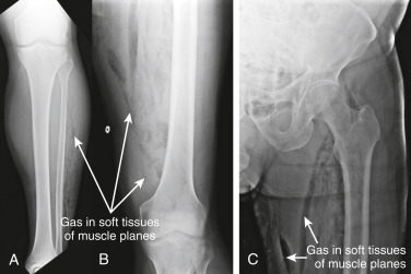
  • Loosening of skin and subcutaneous skin in association with deep fascial necrosis (Fig. E1). “Woody” induration and crepitus of involved area are characteristics
  • Muscle involvement, thrombosis of blood vessels, and myonecrosis may develop
  • Bullae and gas formation at site

Figure E1 Necrotizing fasciitis.

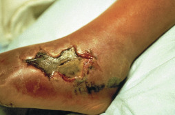
The so-called flesh-eating bacteria, group A β-hemolytic Streptococcus, can cause significant tissue destruction rapidly. This 32-yr-old woman had pain, erythema, and swelling of the foot followed by necrotic ulceration over a week. There was no history of trauma.

Courtesy Roger Bitar, MD. From White GM, Cox NH [eds]: Diseases of the skin, a color atlas and text, ed 2, St Louis, 2006, Mosby.

Etiology

  • NF usually arises from skin damage or trauma. Risk is increased with presence of comorbidities (diabetes mellitus, cancer, liver disease, immunosuppression, use of SGLT2 inhibitors)
  • Polymicrobial: Mixture of anaerobes and aerobic enteric gram-negative rods
  • Group A streptococci (S. pyogenes)
  • S. aureus
  • C. perfringens
  • Bacteroides fragilis
  • Vibrio vulnificus
  • Methicillin-resistant S. aureus (MRSA), especially community-acquired MRSA

Diagnosis

Differential Diagnosis

  • Cellulitis
  • Pyomyositis
  • Gas gangrene
  • A classification of necrotizing skin, soft tissue, and muscle infections is described in Table 1

TABLE 1 Classification of Necrotizing Skin, Soft-Tissue, and Muscle Infections

DiseaseBacteriologyComments
Necrotizing Cellulitis
Clostridial cellulitisClostridium perfringensLocal trauma, recent surgery; fascial/deep muscle spared
Nonclostridial cellulitisMixed: Escherichia coli, Enterobacter, Peptostreptococcus spp., Bacteroides fragilisDiabetes mellitus predisposes; produces foul odor
Meleney synergistic gangreneStaphylococcus aureus, microaerophilic streptococciRare infection; postoperative; slowly expanding, indolent, ulceration in superficial fascia
Synergistic necrotizing cellulitisMixed aerobic and anaerobic, including B. fragilis, Peptostreptococcus spp.Diabetes mellitus predisposes; variant of necrotizing fasciitis type I; involves skin, muscle, fat, and fascia
Necrotizing Fasciitis
Type IMixed aerobic and anaerobic; staphylococci, B. fragilis, E. coli, group A streptococci, Peptostreptococcus spp., Prevotella, Porphyromonas spp., Clostridium spp.Usually requires a breach in the mucous membrane layer either through surgery or penetrating injuries or from chronic medical conditions such as diabetes, peripheral vascular disease, malignancy, and anal fissures
Type IIGroup A streptococciIncreasing in frequency and severity since 1985; very high mortality; often begins at site of nonpenetrating minor trauma such as a bruise or muscle strain but often no identified precursor
Predisposing factors: Blunt/penetrating trauma, varicella (chickenpox), intravenous drug abuse, surgical procedures, childbirth, nonsteroidal antiinflammatory drug use
Myonecrosis
Clostridial myonecrosisClostridium spp.Predisposing factors: Deep/penetrating injury, bowel and biliary tract surgery, improperly performed abortion and retained placenta, prolonged rupture of the membranes, and intrauterine fetal demise or missed abortion in postpartum patients. Recurrent gas gangrene occurs at sites of previous gas gangrene
Streptococcal myonecrosisStreptococci
Special Type of Necrotizing Soft-Tissue Infection
Fournier gangrenePolymicrobial, with E. coli the predominant aerobe and Bacteroides the predominant anaerobe. Other microflora: Proteus, Staphylococcus, Enterococcus, aerobic and anaerobic Streptococcus, Pseudomonas, Klebsiella, and ClostridiumNecrosis of the scrotum or perineum that starts with scrotal pain and erythema and rapidly spreads onto anterior abdominal wall and gluteal muscle. It is more often seen in diabetics and can be associated with trauma

From Vincent JL et al: Textbook of critical care, ed 6, Philadelphia, 2011, Saunders.

Workup

  • Diagnosis of necrotizing fasciitis generally requires incision and probing. In patients with necrotizing fasciitis, there is no resistance to probing subcutaneously, and there is fascial plane involvement. Fig. E2 is an algorithm for assessment and treatment of suspected necrotizing fasciitis.
  • Laboratory tests:
    1. The laboratory risk indicator for necrotizing fasciitis (LRINEC) consists of the following six variables (Table 2). When present, the reported positive predictive value is 92%: CBC with differential (leukocytosis [white blood count >15,000], anemia [Hb <13.5]), elevated C-reactive protein (15 mg/dl), hyponatremia (sodium <135 mEq/L), elevated creatinine (>1.6 mg/dl), hyperglycemia (glucose >180 mg/dl).
    2. Cultures of skin, soft tissue, or debrided tissue, aerobically and anaerobically. Blood cultures are positive in 60% of patients with type II infections and 20% with type I infections.
  • Imaging:
    1. Radiographs may show subcutaneous gas in fascial planes.
    2. Computed tomography (CT) or MRI may be helpful because they can detect gas in the tissues. MRI with contrast is more sensitive than CT.

TABLE 2 Laboratory Risk Indicator for Necrotizing Fasciitis (LRINEC)a

VariablePoints
CRP >150 mg/L4
Leukocytosis
WBC count between 15,000 and 25,000/mm3
WBC count >25,000/mm3
1
2
Anemia
Hgb between 11 and 13.5 g/dl
Hgb <11 g/dl
1
2
Hyponatremia
Na <135 mmol/L
2
Renal insufficiency
Cr >1.6 mg/dl
2
Serum glucose >180 mg/dl1

Cr, Creatinine; CRP, C-reactive protein; Hgb, hemoglobin; WBC, white blood cell.

a LRINEC score <6 points is considered low risk for necrotizing soft tissue infection but does not rule out the diagnosis. High clinical suspicion for necrotizing soft tissue infection warrants surgical debridement, irrespective of LRINEC score.

From Spec A et al: Comprehensive review of infectious diseases, Philadelphia, 2019, Elsevier.

Figure E2 Necrotizing fasciitis.

This 71-yr-old man with aplastic anemia presented with fevers to 38.9° C (102.02° F), leg weakness, and extreme leg pain. Initially, the patient was thought to have neuropathic pain and weakness, possibly indicating spinal disease such as epidural abscess. He rapidly developed crepitus of his legs. X-rays of the patient ’s legs were obtained, followed by noncontrast computed tomography. (A) Anterior-posterior (AP) tibia and fibula. (B) AP femur. (C) AP hip. Air is seen dissecting in muscle planes of the legs. On X-ray, air appears black. Given the wide distribution of air, a focal abscess is unlikely, and necrotizing fasciitis with gas-producing organisms should be suspected.

From Broder JS: Diagnostic imaging for the emergency physician, Philadelphia, 2011, Saunders.

Treatment

Suggested Readings

    1. Bersoff-Matcha S.J. : Fournier gangrene associated with sodium-glucose cotransporter-2 inhibitors: a review of spontaneous postmarketing casesAnn Intern Med. ;170(11):764-769, 2019.
    2. Bonne S.L., Kadri S.S. : Evaluation and management of necrotizing soft tissue infectionsInfect Dis Clin North Am. ;31:497-511, 2017.
    3. Stevens D.L., Bryant A.E. : Necrotizing soft-tissue infectionsN Engl J Med. ;377:2253-2265, 2017.