section name header

Basic Information

AUTHORS: Benjamin J. Ahn, BS and John D. Milner, MD, and Manuel F. DaSilva, MD

Definition

Ankylosing spondylitis (AS) is a type of inflammatory arthritis involving the sacroiliac joints and axial skeleton characterized by ankylosis (abnormal stiffening of a joint) and enthesitis (inflammation at tendon insertions). It is part of a family of overlapping syndromes called seronegative spondyloarthropathies (SpA) that includes reactive arthritis (formerly Reiter syndrome), psoriatic spondylitis, and enteropathic arthritis.1

Synonyms

Marie-Strümpell disease

Bechterew disease

ICD-10CM CODES
M45.9Ankylosing spondylitis of unspecified sites in spine
M08.1Juvenile ankylosing spondylitis
M45.0Ankylosing spondylitis of multiple sites in spine
M45.1Ankylosing spondylitis of occipito-atlanto-axial region
M45.2Ankylosing spondylitis of cervical region
M45.3Ankylosing spondylitis of cervicothoracic region
M45.4Ankylosing spondylitis of thoracic region
M45.5Ankylosing spondylitis of thoracolumbar region
M45.6Ankylosing spondylitis lumbar region
M45.7Ankylosing spondylitis of lumbosacral region
M45.8Ankylosing spondylitis sacral and sacrococcygeal region
Epidemiology & Demographics
Prevalence

Between 0.1% and 1% of the population. Varies with prevalence of HLA-B27 and ethnicity. The U.S. prevalence of AS has been reported between 5% and 6% in HLA-B27 populations. Much higher in those with positive family history of spondyloarthropathy.2

Predominant Sex

70.4% male predominance

Predominant Age

15 to 35 yr, usually symptoms present before age 45

Physical Findings & Clinical Presentation (

  • Prolonged morning back stiffness of insidious onset lasting more than 3 mo
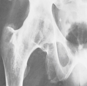
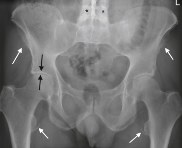
  • Bilateral sacroiliac joint tenderness (sacroiliitis)
  • Inflammatory back pain; often improves with exercise and is worse with rest
  • Limited lumbar spine motion
  • Tenderness at tendon insertion sites, especially the Achilles tendons and plantar fascia
  • Loss of chest expansion reflecting rib cage involvement
  • Possible to have peripheral joint arthritis, usually involving lower extremities
  • In advanced cases the typical posture consists of compensatory hyperextension of neck, fixed flexion of hips, and compensatory flexion of knees (Fig. 1)
  • There is an increased incidence of iritis and uveitis (30% to 40% lifetime prevalence)
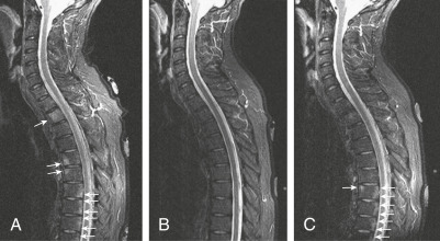
  • Other extra-skeletal manifestations include effects on the cardiovascular system (aortic insufficiency, cardiovascular disease), cerebrovascular system, and lungs (pulmonary fibrosis). There is also an increased risk for osteoporosis.3,4
FIG 1 Typical Posture of Patient with Ankylosing Spondylitis; Note the Flexed Neck, Protuberant Abdomen, and Loss of Lumbar Lordosis

From Talley NJ et al: Essentials of internal medicine, ed 4, Chatswood, NSW, 2021, Elsevier Australia.

TABLE E1 Aspects of Inflammatory Back Pain in Ankylosing Spondylitis and Axial Spondyloarthritis

Onset of symptoms before age 45 yr
Duration of symptoms more than 3 mo (chronic pain)
Located at the lower back
Alternating buttock pain
Awaking due to back pain during the second half of the night
Morning stiffness for at least 30 min
Insidious onset of complaints
Improvement with exercises
No improvement of back pain with rest
Improvement with use of nonsteroidal agents

From Firestein GS et al: Firestein & Kelley’s textbook of rheumatology, ed 11, Philadelphia, 2021, Elsevier.

TABLE E2 Characteristic Features of Ankylosing Spondylitis

Chronic inflammatory spinal pain
Chest pain
Alternate buttock pain
Acute anterior uveitis
Synovitis (predominantly of lower limbs, asymmetric)
Enthesitis (heel, plantar)
Radiographic sacroiliitis
Positive family history of ankylosing spondylitis
Chronic inflammatory bowel disease
Psoriasis

From Firestein GS et al: Firestein & Kelley’s textbook of rheumatology, ed 11, Philadelphia, 2021, Elsevier.

Etiology

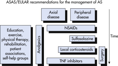
Although significant progress has been made, the etiology of AS still remains unclear to an extent. To date, it is believed that genetic background, immune reactions, infection, and endocrinal abnormities play a role in susceptibility to the spondyloarthropathies. Major histocompatibility complex class I allele HLA-B27 has been described as the predominant genetic predisposing factor. Infections such as Klebsiella pneumonia, an opportunistic pathogen that makes up part of the normal gut microflora, may be an exacerbating agent in the autoimmune process of AS. Tumor necrosis factor is important in the inflammatory response.5

Diagnosis

Differential Diagnosis

  • Diffuse idiopathic skeletal hyperostosis (Forestier disease)
  • Noninflammatory back pain (a clinical algorithm for the evaluation of back pain is described in Section III)
  • Table 3 compares ankylosing spondylitis and related disorders

TABLE 3 Comparison of Ankylosing Spondylitis and Related Disorders

FeatureAnkylosing SpondylitisPsoriatic ArthritisReactive ArthritisEnteropathic Arthropathy
Gender (male:female)2-3:11:11:11:1
Age of onset<40 yr35-55 yr20-40 yrAny age
Sacroiliitis or spondylitis (%)1002040<20
Symmetry of sacroiliitisSymmetricAsymmetricAsymmetricSymmetric
Peripheral arthritis (%)2595905-20
DistributionAxial and lower limbsVariableLower limbsVariable
HLA-B27 positivity (%)85-9525-6030-707-70
Uveitis (%)0-402050<15

, approximately.

60% when spondylitis is present.

60% when spondylitis is present.

From Hochberg MC: Rheumatology, ed 7, Philadelphia, 2019, Elsevier.

Laboratory Tests

  • Elevated sedimentation rate, C-reactive protein
  • Mild hyperchromic anemia
  • Demonstration of inflammatory sacroiliitis by radiography or MRI is diagnostic for most patients, although some patients may meet criteria for “nonradiographic spondyloarthropathy” based on compelling clinical evaluation6
  • HLA-B27 antigen is not useful in the evaluation of noninflammatory back pain because it is present in up to 10% of the normal population (varies based on ethnicity)
Imaging Studies

  • Classic feature is bilateral sacroiliitis on pelvic x-rays (modified NY criteria).
  • Vertebral bodies lose anterior concave shape and become square.
  • With progression, calcification of the annulus fibrosus and paravertebral ligaments develops, giving rise to the so-called bamboo spine (Figs. E2 through Fig. E6).
  • Severe diskovertebral erosions and destruction may occur (Andersson lesion, Fig. 7).
  • MRI (Fig. E8) may be useful in detecting early inflammatory lesions and is especially helpful when the history is suggestive but x-rays are equivocal; can see bone marrow edema, fatty metaplasia, sacroiliitis, erosions, shiny corners.7
Figure 7 Severe Andersson Lesion (Diskovertebral Erosions and Destruction) (A, Arrow) of the Thoracic Spine in a Patient with Ankylosing Spondylitis Resulting in Instability and Severe Pain, Which was Treated with a Fusion Operation (B)

From Hochberg MC: Rheumatology, ed 7, Philadelphia, 2019, Elsevier.

Figure E2 A, Ankylosing Spondylitis (AS) with Sclerosis of the Vertebral Corners (Romanus Lesions)

The x-Ray Shows Advanced Lesions () and an Early Lesion (Arrow). B, Stir Sagittal Magnetic Resonance Image of a Different AS Patient with Edema of the Vertebral Corners (Arrows) Termed MR Romanus Lesions.

From Grant LA: Grainger & Allison’s diagnostic radiology essentials, ed 2, Philadelphia, 2019, Elsevier.

Figure E3 T1-Weighted (A) and T2 Fat-Suppressed (B) Coronal Magnetic Resonance Images of the Sacroiliac Joints (Sijs) in Ankylosing Spondylitis

The T1-Weighted Image Readily Demonstrates Erosion (Arrows) in the Right Sij with Joint Space Loss on the Left (), All Indicative of Damage. Note that the T2-Weighted Image Poorly Demonstrates Erosion; However, It Shows Subchondral Edema (Arrows) More Reflective of Disease Activity.

From Grant LA: Grainger & Allison’s diagnostic radiology essentials, ed 2, Philadelphia, 2019, Elsevier.

Figure E4 T1-Weighted (A) and T2 Fat-Supressed (B) Coronal MR Images of the Sacroiliac Joints (Sij) in Ankylosing Spondylitis

The T1-Weighted Image Readily Demonstrates Erosion (Arrows) in the Right Sij with Joint Space Loss on the Left (), All Indicative of Damage. Note How Poor the T2-Weighted Image is at Demonstrating Erosion; However, It Shows Subchondral Edema (Arrows) More Reflective of Disease Activity.

From Grant LA: Grainger & Allison’s diagnostic radiology essentials, ed 2, 2019, Elsevier.

Figure E5 Ankylosing spondylitis. Bone ankylosis across the joint cartilage.

From Grant LA: Grainger & Allison’s diagnostic radiology essentials, ed 2, Philadelphia, 2019, Elsevier.

Figure E6 Patient with Ankylosing Spondylitis with Spinal Fusion (Black) and Sij Fusion (White)

There is Hip Arthropathy with Diffuse Loss of Joint Space (Black Arrows) and Flattened Configuration of the Femoral Heads. Widespread Entheseal New Bone Formation is Noted around the Pelvis (White Arrows).

From Grant LA: Grainger & Allison’s diagnostic radiology essentials, ed 2, Philadelphia, 2019, Elsevier.

Figure E8 Spine Inflammation in Ankylosing Spondylitis (Magnetic Resonance Imaging [MRI])

A 43-Yr-Old Man with HLA-B27-Positive Ankylosing Spondylitis with Deteriorating Symptoms, Including Inflammatory Back Pain, Had an MRI Scan Before Starting Biologic Therapy. Baseline Sagittal Short Tau Inversion Recovery (Stir) MRI (A) Shows Diffuse Increased Signal (Edema) in the T2 Vertebral Body and Multiple Foci of Corner Inflammation Anteriorly at T5 and T6, and Posteriorly at T7, T8, T9, and T10 (Arrows). Other Images Confirmed Extensive Active Inflammation in the Spine. The Patient Responded Very Well, and after 6 Mo of Therapy, a Repeat Stir MRI (B) Showed Complete Resolution of Bone Marrow Inflammation. Subsequently, the Patient Experienced Recurrence of Symptoms, and a Third MRI (C) was Performed (2 Mo after Anti-Tumor Necrosis Factor Therapy was Stopped). This MRI Shows No Edema at T5 to T6, a Conspicuous New Lesion Anteriorly at T7, and Recurrent Inflammation Posteriorly in the Lower Thoracic Spine (Arrow).

From Firestein GS et al: Kelley’s textbook of rheumatology, ed 9, Philadelphia, 2013, Saunders.

Treatment

Nonpharmacologic Therapy

  • Exercises primarily to maintain flexibility and aerobic activity are important (Fig. E9).
  • Postural training:
    1. Patients must be instructed on spinal extension exercises to avoid fusion in a flexed position.
    2. Sleeping should be in the supine position on a firm mattress; pillows should not be placed under the head or knees.

Figure E9 An Exercise Sequence Used in Patients with Ankylosing Spondylitis

Cervical Spine Exercises Include Full Extension (A) and Rotation (B). A Sequence of Back Extension (C and D) is Followed by Rotation in the Lying (E and F) and Upright Kneeling (G) Positions. Finally, Breathing with the Thoracic Muscles is Practiced (Not Shown). Nsaid, Nonsteroidal Antiinflammatory Drug; Tnf, Tumor Necrosis Factor.

From Hochberg MC: Rheumatology, ed 7, Philadelphia, 2019, Elsevier.

Pharmacologic Therapy

  • NSAIDs: Patients with ankylosing spondylitis should be trialed on full-dose continuous NSAID therapy. There is anecdotal evidence suggesting that indomethacin may be more effective than other NSAIDs, but other NSAIDs are also efficacious and may be better tolerated. One study suggested that continuous NSAID therapy may retard the radiographic progression of ankylosing spondylitis, but data are conflicting.5
  • Sulfasalazine may be efficacious in patients with peripheral arthritis.5
  • Numerous controlled trials have shown that tumor necrosis factor (TNF) antagonists such as etanercept, infliximab, and adalimumab are very effective for relieving symptoms of spinal inflammatory arthritis. Anti-TNF therapy should be recommended for patients whose symptoms are not completely controlled with NSAIDs, and it can result in dramatic improvement in symptoms, range of motion of the spine, and quality of life for these patients. There is evidence suggesting that anti-TNF therapy slows the radiographic progression of the disease. Etanercept should generally be avoided if history of uveitis.5
  • Secukinumab, an antiinterleukin-17A monoclonal antibody, has been approved for treating AS. Ixekizumab, another interleukin-17A antibody, was also approved in 2019.8
  • Fig. 10 shows American College of Rheumatology (ACR)/The European League Against Rheumatism (EULAR) recommendations for management.
Figure 10 Recommended Management of Ankylosing Spondylitis (AS), Based on Clinical Expertise and Research Evidence

The Disease Progression with Time Moves Vertically from Top to Bottom. These Recommendations Were Developed Before the Availability of Secukinumab. Secukinumab is Approved but was Not Available at the Time of These Recommendations. Asas/Eular, Assessment in Ankylosing Spondylitis/European League Against Rheumatism; NSAIDs, Nonsteroidal Antiinflammatory Drugs; Tnf, Tumor Necrosis Factor.

From Firestein GS et al: Firestein & Kelley’s textbook of rheumatology, ed 11, Philadelphia 2021, Elsevier.

Disposition

Most patients have a normal life span, but many can suffer significant disability from loss of spinal mobility.

Referral

All patients with seronegative spondyloarthropathy should be referred to a rheumatologist for further evaluation and treatment.

Pearls & Considerations

A family history of seronegative spondyloarthropathy increases the specificity of testing for HLA-B27. Surgical osteotomy may benefit selected patients with severe spinal deformity. Recent data suggest that men with AS have increased risk of vascular mortality.

Related Content

Ankylosing Spondylitis (Patient Information)

Related Content

.

    1. Taurog J.D. : Ankylosing spondylitis and axial spondyloarthritisN Engl J Med. ;374:2563-2574, 2016.doi:10.1056/NEJMra1406182
    2. Reveille J.D., Weisman M.H. : The epidemiology of back pain, axial spondyloarthritis and HLA-B27 in the United StatesAm J Med Sci. ;345(6):431-436, 2013.doi:10.1097/maj.0b013e318294457f
    3. Bengtsson K. : Are ankylosing spondylitis, psoriatic arthritis and undifferentiated spondyloarthritis associated with an increased risk of cardiovascular events? A prospective nationwide population-based cohort studyArthritis Res Ther. ;19(1), 2017.doi:10.1186/s13075-017-1315-z
    4. Haroon N. : Patients with ankylosing spondylitis have increased cardiovascular and cerebrovascular mortalityAnn Intern Med. ;163:409-416, 2015.doi:10.7326/M14-2470
    5. Zhu W. : Ankylosing spondylitis: etiology, pathogenesis, and treatmentsBone Res. ;7(22), 2019.doi:10.1038/s41413-019-0057-8
    6. de Winter J.J. : Prevalence of peripheral and extra-articular disease in ankylosing spondylitis versus non-radiographic axial spondyloarthritis: a meta-analysisArthritis Res Ther. ;18(1), 2016.doi:10.1186/s13075-016-1093-z
    7. Griffith J.F. : Computer-aided assessment of spinal inflammation on magnetic resonance images in patients with spondyloarthritisArthritis Rheum. ;67:1789-1797, 2015.doi:10.1002/art.39126
    8. Marzo-Ortega H. : Secukinumab and sustained improvement in signs and symptoms of patients with active ankylosing spondylitis through two years: results from a Phase III studyArthritis Care Res. ;69(7):1020-1029, 2017.doi:10.1002/acr.23233