section name header

Basic Information

AUTHORS: Dianelys Mendez, MD and Vicky Cheng, MD

Definition

Hyperparathyroidism is an endocrine disorder caused by excessive secretion of parathyroid hormone (PTH) from the parathyroid glands. Autonomous production of PTH resulting in hypercalcemia defines primary hyperparathyroidism. Secondary hyperparathyroidism occurs when the parathyroid glands appropriately increase PTH production in response to low calcium or vitamin D states. Primary hyperparathyroidism is the focus of this section.

ICD-10CM CODES
E21.0Primary hyperparathyroidism
E21.1Secondary hyperparathyroidism, not elsewhere classified
E21.2Other hyperparathyroidism
E21.3Hyperparathyroidism, unspecified
N25.81Secondary hyperparathyroidism of renal origin
Epidemiology & Demographics
Incidence

66 cases/100,000 person-year in women and 25 cases/100,000 person-year in men in the United States.1 Although malignancy is the most common cause of hypercalcemia in hospitalized patients, primary hyperparath-yroidism is the most common cause of hypercalcemia in the outpatient setting.2

Prevalence

Varies by country and race with 23 cases/10,000 women and 8.5 cases/10,000 men in the United States.1

Predominant Sex & Age

Higher prevalence in women (female:male ratio 4:1)3 and peaks in the early postmenopausal year.4

Physical Findings & Clinical Presentation

The majority of patients with primary hyperparathyroidism are asymptomatic. Diagnosis is usually considered in patients after an incidental discovery of elevated calcium or PTH levels on biochemical screening or during the evaluation for decreased bone mass.

The development of symptoms varies with severity and rapidity of disease progression and reflects both the hypercalcemia and hyperparathyroid components of the disease process. Symptoms associated with hypercalcemia (Table 1) are as follows2,3,5:

  • Cardiovascular: Hypertension, shortened QT interval, bradycardia, arrhythmia, valvular calcification, left ventricular hypertrophy, and increased mean carotid intima-media thickness
  • GI: Anorexia, nausea, vomiting, constipation, abdominal pain, peptic ulcer disease, and pancreatitis
  • Genitourinary (GU): Nephrolithiasis (15% to 20%), nephrocalcinosis, renal insufficiency, polydipsia, polyuria, nocturia, nephrogenic diabetes insipidus, and renal tubular acidosis
  • Musculoskeletal: Weakness, myopathy, bone pain, osteopenia, osteoporosis, gout, pseudogout, chondrocalcinosis, osteitis fibrosa cystica, and subperiosteal bone resorption
  • Central nervous system: Confusion, anxiety, fatigue, difficulty concentrating, lethargy, obtundation, depression, and coma
  • Other: Pruritus, metastatic calcifications, band keratopathy

TABLE 1 Signs and Symptoms of Primary Hyperparathyroidism

Target Organ or SystemSymptomsComments
RenalNephrolithiasis, nephrocalcinosis, polyuria, polydipsia, renal insufficiency15%-20% of patients have kidney stones
SkeletalFragility fracturesUnrelated to significant trauma
Osteopenia/osteoporosisCortical bone >trabecular bone (distal third of radius most affected)
Bone painCommon
Osteitis fibrosa cysticaRare, but may occur with advanced disease, characterized by bone pain and multiple skeletal deformities, including salt and pepper appearance of the skull, bone cysts, and brown tumors of bone
NeuromuscularProximal muscle weakness, muscular atrophy, gait disturbanceRare
Easy fatigability, generalized weaknessCommon
GastrointestinalGastroesophageal reflux, constipation, abdominal pain, peptic ulcer diseaseCommon
Rare
Nausea, vomiting, acute pancreatitisRare, can be seen in cases of severe hypercalcemia
NeuropsychiatricFatigue, depression, anxiety, emotional lability, sleep disturbances, lethargy, memory loss, inability to concentrate, mental status change, psychosis, obtundation, and comaOften reported
Rare, can be seen with severe hypercalcemia
CardiovascularExacerbation of hypertension, valvular disease, myocardial calcifications, premature atherosclerosis, left ventricular hypertrophy, shortened QT interval, conduction abnormalities, and heart blockConflicting data on improvement of cardiac parameters after parathyroidectomy

From Townsend CM et al: Sabiston textbook of surgery, ed 21, St Louis, 2022, Elsevier.

Etiology

  • Regulators of PTH secretion are illustrated in Fig. 1. Most cases of primary hyperparathyroidism are sporadic.3 Hyperparathyroidism can be associated with rare familial conditions in <10%, such as multiple endocrine neoplasia (MEN-1 and MEN-2), familial hyperparathyroidism-jaw tumor syndrome, familial isolated hyperparathyroidism, and neonatal severe hyperparathyroidism.2 Higher prevalence of hyperparathyroidism is noted with head and neck irradiation, chronic low calcium or vitamin D status, and lithium therapy.6
  • Pathologic characteristics include adenoma (80%),3 hyperplasia (15% to 20%),3 or carcinomas (<1%).7
Figure 1 Regulators of Parathyroid Hormone (PTH) Secretion and PTH Effects on Calcium Homeostasis

FGF23, Fibroblast growth factor 23; 25(OH)D, 25-hydroxyvitamin D; 1,25(OH)2D, 1,25-dihydroxyvitamin D.

From Townsend CM et al: Sabiston textbook of surgery, ed 21, St Louis, 2022, Elsevier.

Diagnosis

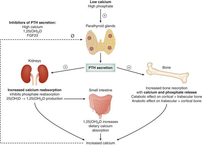
Differential Diagnosis

  • Primary hyperparathyroidism:
    1. Adenoma (80%)
    2. Hyperplasia (15% to 20%)
    3. Carcinomas (<1%)
  • Secondary hyperparathyroidism (Box 1) precipitated by conditions that result in hypocalcemia:
    1. Renal calcium loss (i.e., medication: Loop diuretics and hypercalciuria)
    2. Calcium deficiency
    3. Vitamin D deficiency
    4. Malabsorption
    5. Chronic kidney disease (most common)2
    6. Pseudohypoparathyroidism (PTH resistance)
    7. Inhibition of bone resorption (i.e., bisphosphonates and denosumab)
  • Other causes of hypercalcemia (Table 2) include:
    1. Medications: Thiazide diuretics, lithium therapy
    2. Vitamin D intoxication, milk-alkali syndrome
    3. Vitamin A toxicity
    4. Familial hypocalciuric hypercalcemia (FHH)
    5. Renal failure (tertiary hyperparathyroidism)
    6. Thyrotoxicosis
    7. Granulomatous disorders (e.g., sarcoidosis)
    8. Malignancy (e.g., lung cancer, lymphoma, multiple myeloma, and bone metastasis)
    9. Prolonged immobilization

TABLE 2 Causes of Hypercalcemia

EndocrinePrimary hyperparathyroidism
Tertiary hyperparathyroidism
Familial hypocalciuric hypercalcemia
Hyperthyroidism
MalignancyTumors producing PTHrP (SCC of lung, bladder cancer, renal cell cancer)
Osteolytic bone metastasis
Hematologic malignancies (lymphoma, leukemia, multiple myeloma)
Granulomatous diseaseSarcoidosis
Tuberculosis
Fungal infection
MedicationsCalcium
Thiazide diuretics
Lithium
Vitamin A and D intoxication
Milk alkali syndrome
MiscellaneousPaget and other bone diseases with prolonged immobilization

PTHrP, Parathyroid hormone-related peptide; SCC, squamous cell carcinoma.

From Townsend CM et al: Sabiston textbook of surgery, ed 21, St Louis, 2022, Elsevier.

BOX 1 Causes of Secondary Hyperparathyroidism

  • Chronic renal failure
  • 25-Hydroxyvitamin D deficiency
  • Malabsorption syndromes
    • Celiac disease
    • Cystic fibrosis
    • Short gut syndrome
    • Bariatric procedures
  • Medications
  • Metabolic abnormalities
    • Hypermagnesemia
    • Hyperphosphatemia
  • Congenital disorders
    • Transient neonatal hyperparathyroidism
    • DiGeorge syndrome

From Townsend CM et al: Sabiston textbook of surgery, ed 21, St Louis, 2022, Elsevier.

Workup (Table 3

  • Typically, primary hyperparathyroidism is confirmed with an elevated serum calcium and PTH level.
    1. Two measurements of serum calcium are required for the confirmation of hypercalcemia. Total calcium should be corrected for low albumin using the formula: Corrected calcium = serum calcium + 0.8 × 4 - serum albumin. If a reliable laboratory is available, ionized calcium should be considered, especially in conditions associated with acid-base disturbances or low albumin states. Patients with primary hyperparathyroidism can also present with normal calcium levels (normocalcemic primary hyperparathyroidism).
    2. The serum intact PTH level is the best test to evaluate the etiology of hypercalcemia. PTH is elevated or in the high normal range (i.e., inappropriately normal for an elevated calcium state) in primary hyperparathyroidism.
  • Other causes of hypercalcemia should be ruled out. These are typically associated with low PTH levels. Exceptions include lithium use and FHH.
    1. Review medication history to determine lithium, thiazide, vitamin D, or calcium intake.
    2. Check 24-h urine calcium: Creatinine to rule out FHH. Urine calcium is very low (<100 mg), and calcium clearance/creatinine clearance ratio is <0.01 in FHH. PTH can be normal or high in FHH.
    3. Consider PTH-related peptide (PTHrP) to evaluate hypercalcemia related to malignancies and 1,25-hydroxyvitamin D to assess hypercalcemia secondary to granulomatous diseases or lymphomas.
    4. Multiple myeloma and bone metastasis can also result in a high calcium state and therefore must be appropriately evaluated.
  • Rule out other causes of elevated PTH (i.e., secondary hyperparathyroidism). Serum calcium is typically low or low-normal in secondary hyperparathyroidism.
    1. Check calcium and 25-hydroxyvitamin D to rule out deficiency states.
    2. Check serum creatinine to assess renal function and 24-h urine calcium and creatinine to evaluate renal calcium loss.

TABLE 3 Evaluation of Patients With Suspected or Confirmed Primary HPT

TestsComments
Laboratory Tests
Serum total calciumBaseline laboratory tests for diagnosis of primary HPT and to rule out the most common causes of secondary HPT
Intact PTH
Creatinine, GFR
25-Hydroxyvitamin D
Ionized calciumFor patients with normocalcemic primary HPT
AlbuminIf low, calculate corrected calcium (mg/dl) = (0.8 [4.0-patient’s albumin (g/dl)] + total calcium (mg/dl)
Serum phosphateLow in approximately 50% of patients with primary HPT
Alkaline phosphataseMarker of bone turnover, indicates extent of bone disease
Urine Tests
24-H urine calcium and creatinineScreen for increased risk of kidney stones and for familial hypocalciuric hypercalcemia
If urine calcium <100 mg/24 h, calculate CCCRCCCR = (24-h calcium urine/calcium serum)/(24-h creatinine urine/creatinine serum)
Imaging
DXAMeasurements of bone mineral density at the lumbar spine, hip femoral neck, and distal radius
Abdominal imaging for kidney stones or nephrocalcinosisPlain abdominal x-ray, abdominal ultrasound, or noncontrast CT
Vertebral spine assessmentPlain x-ray, CT, or DXA
Genetic Testing
IndicationsPatients with pHPT less than 40 yr with multigland disease and patients with a family history of pHPT or syndromes associated with pHPT

CCCR, Calcium creatinine clearance ratio; CT, computed tomography; DXA, dual-energy x-ray absorptiometry; GFR, glomerular filtration rate; HPT, hyperparathyroidism; pHPT, primary hyperparathyroidism; PTH, parathyroid hormone.

Laboratory Tests

  • Serum calcium (ionized or corrected calcium): Normal or elevated in primary hyperparathyroidism
  • Serum phosphorus: Low or low-normal in primary hyperparathyroidism
  • PTH: Elevated or high-normal in primary hyperparathyroidism
  • Serum creatinine and estimated glomerular filtration rate (GFR)
  • 24-h urine calcium and creatinine
  • 25-hydroxyvitamin D, 1,25-hydroxyvitamin D, PTHrP levels
  • ECG may reveal shortening of the QT interval secondary to severe hypercalcemia (>12 mg/dl)
Imaging Studies (Table 4

  • Parathyroid localization with technetium-99m sestamibi (Fig. E2 and Fig E3) can identify potential adenomas to help with surgical planning.8 The inferior parathyroid glands are more commonly ectopic and can be found anywhere from the angle of the mandible to the pericardium. The most common ectopic location for an inferior gland is within the thymus (Fig. 4)
  • Parathyroid ultrasound (Fig. 5, Fig E6), dynamic (4D) CT, and MRI are also used to localize the parathyroid adenoma.8
  • Bone mineral density (BMD) imaging of the spine, hip, and forearm (distal third of radius) is recommended for all patients with hyperparathyroidism to assess the risk for osteoporosis and fragility fractures. Plain radiographic abnormalities are illustrated in Fig. E7 and Fig. E8. Cortical bone loss (i.e., distal one third radius) is greater than trabecular bone loss (i.e., hip and spine) in hyperparathyroidism.9
  • Renal ultrasound can be considered to assess asymptomatic renal stones.
Figure 5 High-Resolution Ultrasonography of the Left Lobe of the Thyroid (Llt) Anterior to a Parathyroid Adenoma (White Arrows)

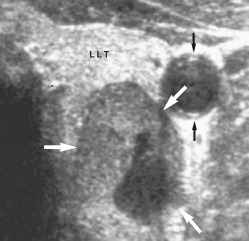
There is a small area of cystic degeneration within the posterior aspect of the adenoma. Carotid artery (black arrows).

From Adam A et al: Grainger & Allison’s diagnostic radiology, ed 5, London, 2007, Churchill Livingstone. In Grant LA: Grainger & Allison’s diagnostic radiology essentials, ed 2, London, 2019, Elsevier.

TABLE 4 Characteristics of Preoperative Imaging Studies in Patients With Hyperparathyroidism

ImagingImaging FindingsSensitivity, PpvAdvantagesLimitationsUpdates
USHypoechoic nodule with well-defined hypervascular echogenic capsule76%, 93%Noninvasive, inexpensive, can be performed rapidly by the surgeon with simultaneous evaluation of the thyroidUnable to see ectopic mediastinal, retroesophageal, or retropharyngeal glands; decreased sensitivity in multigland disease and small glands; thyroid nodules and lymph nodes can cause false-positivesUS elastography
Sestamibi-SPECTIncreased focal uptake and prolonged retention of the technetium-99m sestamibi79%, 90%Detects ectopic and posterior glands; lower radiation than 4D-CT, operator-independentLong duration of time for the exam; more expensive than US and 4D-CT, radiation exposure; decreased sensitivity for multigland disease and small glands; false-positives (lymph nodes, thyroid tissue, granulomatous disease)PET scanning with various radiopharmaceuticals under investigation
4D-CTSoft tissue nodule with peaked enhancement in arterial phase and washout in venous phase with polar vessel81%-89%, 93%Rapid acquisition time, superior anatomic information, superior sensitivity than other techniques, more successful in localizing small adenomas and multigland diseaseHigh radiation dose, intravenous contrast use, some contrast artifact in neck veins can occurProtocols with fewer phases or less contrast, to diminish radiation and contrast dose
MRIHomogeneous or marbled appearance with high intensity on T2-weighted images, intermediate to low intensity on T1-weighted images43%-94%No radiation, contrast not necessary, superior anatomic informationExpensive, long duration of study acquisition, cannot use in patients with metal implants, low specificityDixon fat suppression method
18F-fluorocholine PET/CTFocal tracer uptake93%, 90%Decreased radiation and acquisition time compared to some sestamibi protocols, higher sensitivityLimited data on new tracer, limited availabilityAbility to differentiate between parathyroid adenoma and hyperplasia preoperatively based on maximal standardized uptake values

CT, Computed tomography; 4D-CT, four-dimensional CT; MRI, magnetic resonance imaging; PET/CT, positron emission tomography/CT; SPECT, single photon emission CT; US, ultrasound.

From Townsend CM et al: Sabiston textbook of surgery, ed 21, St Louis, 2022, Elsevier.

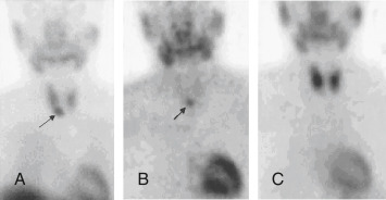
Figure E2 Role of scintigraphy in detecting parathyroid adenomas.

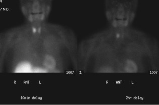
A 66-yr-old woman with hypercalcemia. CT (not shown) did not reveal a parathyroid adenoma. 99mTc-sestamibi radionuclide imaging demonstrates uptake in both thyroid and parathyroid parenchyma in the 10-min delayed image (left); however, at 2-h delay, imaging (right) demonstrates persistent uptake in the right lobe of the thyroid gland, representing the parathyroid adenoma.

From Adam A et al: Grainger and Allison’s diagnostic radiology, ed 6, London, 2015, Elsevier. In Grant LA: Grainger & Allison’s diagnostic radiology essentials, ed 2, London, 2019, Elsevier.

Figure E3 Ectopic intrathymic parathyroid adenoma.

From Townsend CM et al: Sabiston textbook of surgery, ed 21, St Louis, 2022, Elsevier.

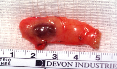
Figure E4 Parathyroid adenoma.

99mTc-MIBI images at 10 min (A) and 3 h (B), showing a persistent focus of activity inferior to the right lobe of the thyroid; 99mTcO 4 image (C) shows normal thyroid uptake but the adenoma is not visualized.

From Grant LA et al: Grainger & Allison’s diagnostic radiology essentials, ed 2, Philadelphia, 2019, Elsevier.

Figure E6 High-Resolution Ultrasound Images Demonstrating a 1

57 × 1.03 × 1.13 cm Hypoechoic Mass Posterior to the Superior Pole of the Left Lobe of the Thyroid Gland in Longitudinal and Axial Views, Which Corresponded to a Left Superior Parathyroid Adenoma.

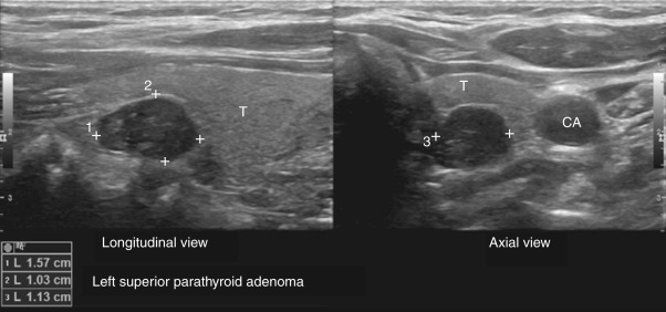
Parathyroid gland marked by measuring points. CA, Carotid artery; T, thyroid.

From Townsend CM et al: Sabiston textbook of surgery, ed 21, St Louis, 2022, Elsevier.

Figure E7 Hyperparathyroidism.

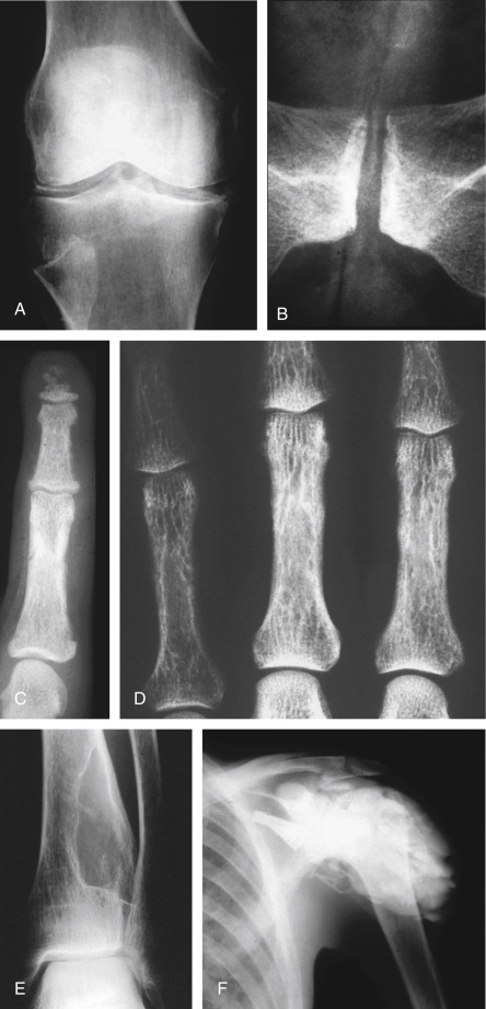
Chondrocalcinosis in the knee menisci (A) and (B) the symphysis pubis. (C) Subperiosteal erosions along the radial side of the middle phalanx of the 2nd finger. Acroosteolysis is also present. Metastatic calcification of the digital artery confirms this is secondary hyperparathyroidism. (D) Cortical “tunneling” in the proximal phalanges. (E) Brown tumor within the distal tibia. (F) Precipitation of amorphous calcium phosphate in the soft tissues of the shoulder (hyperparathyroidism secondary to chronic renal disease).

From Adam A et al: Grainger & Allison’s diagnostic radiology, ed 5, London, 2007, Churchill Livingstone. In Grant LA: Grainger & Allison’s diagnostic radiology essentials, ed 2, London, 2019, Elsevier.

Figure E8 Secondary hyperparathyroidism.

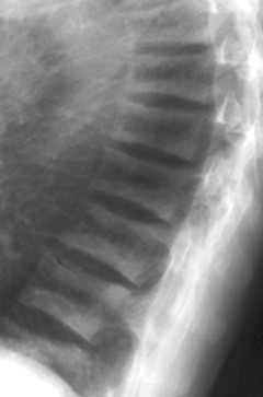
Lateral radiograph of the thoracic spine demonstrating increased sclerosis along the end plates and increased lucency of the central part, giving a “rugger jersey” spine appearance. Also note deformity of the vertebral bodies, with loss of height and increased diameter, in addition to wedge-shaped deformities at the upper thoracic spine related to decreased stability and weakness of the abnormal bone.

From Adam A et al: Grainger and Allison’s diagnostic radiology, ed 6, London, 2015, Elsevier. In Grant LA: Grainger & Allison’s diagnostic radiology essentials, ed 2, London, 2019, Elsevier.

Treatment

Modality of treatment depends on disease progression and which patients are more likely to suffer end-organ effects of hyperparathyroidism or benefit the most from surgery. Fig. 9 illustrates an algorithm for managing persistent or recurrent hyperparathyroidism.

BOX 2 Established Indications for Parathyroidectomy in Patients With Primary Hyperparathyroidism

  • All symptomatic patients (renal, bone, neurocognitive, or neuropsychiatric symptoms)
  • Serum calcium >1 mg/dl above the upper limit of normal
  • Age <50 yr
  • BMD T score <-2.5 (osteoporosis) or significant reduction in BMD
  • Vertebral compression fracture on spine imaging
  • Impaired renal function with GFR <60 ml/min
  • Nephrolithiasis or nephrocalcinosis
  • Hypercalciuria with increased stone risk (urine calcium >400 mg/24 h)
  • When active surveillance and routine long-term follow-up is not a good option

From Townsend CM et al: Sabiston textbook of surgery, ed 21, St Louis, 2022, Elsevier.

BOX 3 Indications for Parathyroidectomy in Patients With Secondary and Tertiary Hyperparathyroidism

Indications for Parathyroidectomy in Patients With Secondary Hyperparathyroidism (SHPT)

SHPT refractory to medical therapy:

  • Parathyroid hormone >1000 pg/ml
  • Calcium × phosphorus product >55

Renal osteodystrophy

Calciphylaxis

Other retractable symptoms, including uremic pruritus, persistent anemia, bone pain, muscle pain, abdominal pain, fatigue, and weakness

Indications for Parathyroidectomy in Patients With Tertiary Hyperparathyroidism

Severe hypercalcemia (calcium >12.5 mg/dl)

Persistent hypercalcemia 2 yr after renal transplantation, associated with:

  • Decline in renal function, without graft rejection
  • Nephrolithiasis
  • Progressive bone disease
  • Pancreatitis

From Cameron JL, Cameron AM: Current surgical therapy, ed 12, Philadelphia, 2017, Elsevier.

Figure 9 Algorithm for managing persistent or recurrent hyperparathyroidism (HPT).

!!flowchart!!

4D CT, Four-dimensional computed tomography; FNA, fine-needle aspiration; IONM, intraoperative neurophysiologic monitoring; IOPTH, intraoperative parathyroid hormone; PTH, parathyroid hormone; sestamibi, technetium (99mTc) sestamibi imaging; US, ultrasonography.

From Cameron JL, Cameron AM: Current surgical therapy, ed 12, Philadelphia, 2017, Elsevier.

Acute General Rx

Severe and/or symptomatic hypercalcemia may require hospitalization, especially if serum calcium >12 mg/dl. Acute management of hypercalcemia includes:

  • Vigorous hydration with IV normal saline (2 to 4 L/day). Fluid status must be monitored in patients with cardiac dysfunction or renal insufficiency to avoid fluid overload. Loop diuretics are often administered to increase calcium excretion or in patients who have risk of volume overload.12
  • Bisphosphonates can effectively decrease calcium levels. Zoledronate (4 mg IV over 15 min) or pamidronate (60 to 90 mg IV over 4 h) are both effective. Onset of action is 24 to 48 h.
  • Calcitonin (4 units/kg IM/SC every 12 h) may be used with bisphosphonates to achieve a more rapid reduction of calcium levels. Onset of action is within hours; however, efficacy is limited to 48 h.
  • Denosumab, a human monoclonal antibody that binds RANK ligand, also has been used in patients with severe hypercalcemia and parathyroid carcinoma because it lowers serum calcium and bone turnover. Unlike bisphosphonates, no dose adjustment is needed for patients with renal dysfunction.12

Pearls & Considerations

Comments

  • Parathyroidectomy should be considered for all patients with symptomatic hyperparathyroidism. Surgery also should be considered in older adults with reasonable life expectancy because fracture risk is lower with parathyroidectomy than with nonoperative management.13 If surgery is contraindicated or not desired, cinacalcet and bisphosphonates can be used.
  • Asymptomatic patients can be monitored with serial calcium, creatinine, estimated glomerular filtration rate, and BMD measurements. Disease progression may result in surgery.
  • Most patients can be managed medically by limiting factors that result in hypercalcemia (e.g., dehydration, immobilization, thiazide diuretics) and maintaining normal calcium and vitamin D intake.
  • Patients with osteopenia and high fracture risk may require antiresorptive therapy such as bisphosphonates.
  • Patients with nephrolithiasis and elevated serum calcium should be evaluated for primary hyperparathyroidism. A study of U.S. veterans with kidney stones revealed hypoparathyroidism in 38% of hypercalcemic patients, and 26% of these patients underwent parathyroidectomy within 2 yr after their kidney stone episode.14
Related Content

Hyperparathyroidism (Patient Information)

Related Content

    1. Insogna K.L. : Primary hyperparathyroidismN Engl J Med. ;379(11):1050-1059, 2018.
    2. Pallan S. : Diagnosis and management of primary hyperparathyroidismBMJ. ;344, 2012.
    3. Bilezikian J.P. : Primary hyperparathyroidismJ Clin Endocrinol Metab. ;103(11):3993-4004, 2018.
    4. Werners R., Clarke B. : Epidemiology of primary hyperparathyroidism Bilezikian J., editors : The parathyroids. ed 3:297-308, 2015.
    5. Bilezikian J.P. : Primary hyperparathyroidismEndocr Pract. ;18(5):781-790, 2012.
    6. Silva B.C. : Primary hyperparathyroidismBest Pract Res Clin Endocrinol Metab. ;32:593-607, 2018.
    7. Bilezikian J.P. : Primary hyperparathyroidismNat Rev Dis Primers. ;2, 2016.
    8. Udelsman R. : The surgical management of asymptomatic primary hyperparathyroidism: proceedings of the Fourth International WorkshopJ Clin Endocrinol Metab. ;99(10):3595-3606, 2014.
    9. Peacock M. : Cinacalcet HCl reduces hypercalcemia in primary hyperparathyroidism across a wide spectrum of disease severityJ Clin Endocrinol Metab. ;96(1):E9-E18, 2011.
    10. Rubin M.R. : The natural history of primary hyperparathyroidism with or without parathyroid surgery after 15 yearsJ Clin Endocrinol Metab. ;93:3462-3470, 2008.
    11. Bilezikian J.P. : Guidelines for the management of asymptomatic primary hyperparathyroidism: summary statement from the Fourth International WorkshopJ Clin Endocrinol Metab. ;99(10):3561-3569, 2014.
    12. Betea D. : Parathyroid carcinoma: challenges in diagnosis and treatmentAnn Endocrinol. ;76:169-177, 2015.
    13. Morris A.M. : Sigmoid diverticulitis: a systematic reviewJAMA. ;311(3):287-297, 2014.
    14. Ganesan C. : Analysis of primary hyperparathyroidism screening among US veterans with kidney stonesJAMA Surg. ;155:861-868, 2020.