section name header

Basic Information

AUTHOR: Relindis Azenwi Fru, MD

Definition

Porphyrias are rare inherited (mostly autosomal dominant) disorders due to deficiencies of heme synthesis enzymes. Table E1 classifies the various types. They are named after the Greek word porphurus (“purple”), after the red fluorescence of porphyrins exposed to ultraviolet light.

Table E2 summarizes the various types of porphyrias.

Synonyms

Acute intermittent porphyria (AIP)

Porphyria cutanea tarda (PCT)

Variegate porphyria (VP)

Hereditary coproporphyria (HCP)

Congenital erythropoietic porphyria (CEP)

Hepatoerythropoietic porphyria

Erythropoietic protoporphyria

ALA-dehydrase porphyria

ICD-10CM CODE
E80.20Unspecified porphyria
Epidemiology & Demographics
Incidence

0.13 cases/million/yr (Europe) with a peak incidence in Sweden (0.51 cases/million/yr)

Prevalence

1:75,000 population in Europe (1:1000 population in Sweden)

Predominant Sex & Age

Equal female-to-male ratio, except for AIP (female predominance). Pubertal onset is common.

Risk Factors

No risk factors are identified.

Triggers for acute crisis involve:

  • Alcohol
  • Barbiturates
  • Sulfonamides
  • Progesterone and other steroid hormones
  • Emotional/physical stress
  • Rapid weight loss
  • Hepatitis C infection can precipitate PCT
Genetics

Acute intermittent porphyria is more prevalent in northern Sweden (1:1000); variegate porphyria is more common in descendants of Dutch settlers from South Africa (both due to founder effect).

Classification

There are two broad categories of acute porphyrias. These include:

  1. Acute hepatic porphyrias (Neurovisceral porphyrias): Comprising four subtypes, AIP, VP, HCP, and ALA-D
  2. Cutaneous porphyrias: There are two main types, sporadic and familial forms. PCT is the most common subtype of porphyria. PCT is associated with hepatitis C. PCT is an acquired inhibition in uroporphyrinogen decarboxylase (UROD), the fifth enzyme in the pathway. A heterozygous disease variant in the UROD gene predisposes up to 20% of individuals (low penetrance).
Physical Findings & Clinical Presentation

  • Porphyrias can present as skin stigmata (porphyria cutanea), neurologic symptoms (acute intermittent porphyria), or both (variegata). Skin stigmata (Figs. E1, E2,E3) are the result of absorption of solar radiation by porphyrin and consequent blistering skin findings. Porphyrins do not cross the blood-brain barrier and only affect the autonomic and peripheral nervous systems. However, vasospasm can cause CNS findings. Neurologic symptoms in acute attacks include abdominal pain, vomiting, constipation, and urinary retention. Pain in the back or extremities is common. There is no convincing evidence that delta-aminolevulinic acid is the cause of pain in acute porphyrias. Motor neuropathy affects proximal muscles of upper extremities and the sensory component affects the trunk. Seizures associated with severe hyponatremia may occur. Hallucination, insomnia, restlessness, and psychosis are described. Porphyrias can be acute or chronic. Precipitating factors in acute porphyria are summarized in Table E3.
AHP (Neurovisceral porphyrias)

AIP

AIP is the most common form of the four neurovisceral porphyrias. It is inherited in an autosomal dominant fashion. The disorder occurs via deficiency of the enzyme porphobilinogen deaminase, which is the third enzyme in the heme biosynthetic pathway. Patients typically present with severe abdominal pain, nausea, vomiting, dark urine, +/–anemia, and otherwise a negative workup. They can also present with urinary retention, neurologic symptoms (sensory, motor, autonomic dysfunction).

Oral contraceptive pills or sex hormones, tobacco abuse, malnutrition, and stress can exacerbate or precipitate symptoms of AIP.

The diagnosis of AIP is confirmed by decreased erythrocyte PBG deaminase activity and/or mutation in the gene encoding PBGD (hydroxymethylbilane synthase).

These disorders are in the differential of acute abdomen; however, characteristically, rebound tenderness is not present.

TABLE E1 Porphyrias: Clinical Involvement, Enzymatic Etiology, and Chromosomal Location

Porphyria (Synonym)Acute Attack, Skin and Organ InvolvementEnzyme of Heme Biosynthesis AffectedChromosome Location
X-linked sideroblastic anemiaBone marrow5-Aminolevulinate synthase, erythroid-specific, mitochondrial (ALAS2)Xp11.21
X-linked dominant protoporphyriaSkin, red cells, liver5-Aminolevulinate synthase, erythroid-specific, mitochondrial (ALAS2)Xp11.21
ALA dehydratase deficiency porphyria (plumboporphyria)Acute liverALA dehydratase (porphobilinogen synthase)9q33.1
Acute intermittent porphyria (intermittent acute porphyria)Acute liverPorphobilinogen deaminase (hydroxymethylbilane synthase)11q23.3
Congenital erythropoietic porphyria (Günther disease)Skin, red cells, bone marrowUroporphyrinogen III synthase10q25.2-q26.3
Porphyria cutanea tarda (symptomatic porphyria, cutaneous hepatic porphyria)Skin, liverUroporphyrinogen decarboxylase1p34
Hereditary coproporphyriaAcute skin, liverCoproporphyrinogen oxidase3q12
Variegate porphyria (porphyria variegata)Acute skin, liverProtoporphyrinogen oxidase1q22
Erythropoietic protoporphyria (erythrohepatic protoporphyria)Skin, red cells, liverFerrochelatase (heme synthase)18q21.3

ALA, 5-Aminolevulinate.From Hoffman R et al: Hematology: basic principles and practice, ed 7, Philadelphia, 2018, Elsevier.

Etiology

  • Porphyrin binds to iron to form heme, the core of the hemoglobin molecule. Porphyrins are synthesized in the cytosol and mitochondria, and mutations in any of the eight enzymes involved in heme synthesis can result in porphyrias. When the defect is physiologically significant, it results in overproduction of pathway precursors preceding the defective step that enter the circulation and are excreted into urine or bile.
  • This metabolic pathway is limited only by iron in the red blood cell and not by its end product, heme, which therefore is used in the treatment of hepatic porphyrias, where it inhibits the ALAS enzyme. Table E4 summarizes the distribution of porphyrins in red cells, urine, and feces in different forms of porphyria.

TABLE E2 Classification of Porphyrias

ClassificationDiseaseBiochemistryClinical Features
Acute porphyriaAcute intermittent porphyriaIncreased ALA and PBGAcute attack
Variegate porphyriaIncreased ALA and PBG; increased porphyrinAcute attack; photosensitivity
Hereditary coproporphyriaIncreased ALA and PBG; increased porphyrinAcute attack; photosensitivity
ALA dehydratase deficiency porphyriaIncreased ALA; increased porphyrinAcute and chronic neuropathy
Nonacute porphyriaPorphyria cutanea tardaIncreased porphyrinPhotosensitivity
Erythropoietic protoporphyriaIncreased porphyrinPhotosensitivity
Congenital erythropoietic porphyriaIncreased porphyrinPhotosensitivity
X-linked dominant protoporphyriaIncreased porphyrinPhotosensitivity
PorphyrinuriasLead, alcohol, iron deficiency anemia, liver diseaseVarious biochemical manifestationsVarious clinical presentations

ALA, 5-Aminolevulinate; PBG, porphobilinogen.

From Hoffman R et al: Hematology: basic principles and practice, ed 7, Philadelphia, 2018, Elsevier.

Figure E1 Porphyria cutanea tarda.
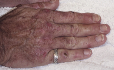
Eroded blisters and crusts.

Courtesy the Yale Residents’ Collection. From Skorecki K et al: Brenner & Rector’s the kidney, ed 10, Philadelphia, 2016, Elsevier.
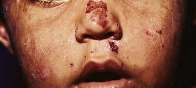
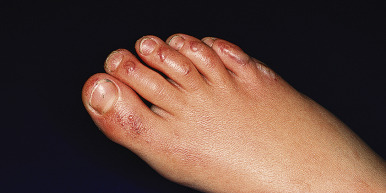
Figure E2 Congenital erythropoietic porphyria.

Vesicles, bullae, and crusts on sun-exposed areas.

From Paller AS, Mancini AJ: Hurwitz clinical pediatric dermatology: a textbook of skin disorders of childhood and adolescence, ed 5, Philadelphia, 2016, Elsevier.

Figure E3 Erythropoietic protoporphyria (EPP).

Vesicular lesions are only occasionally seen in patients with EPP.

From Paller AS, Mancini AJ: Hurwitz clinical pediatric dermatology: a textbook of skin disorders of childhood and adolescence, ed 5, Philadelphia, 2016, Elsevier.

Diagnosis

Differential Diagnosis

  • Lead toxicity
  • Paroxysmal nocturnal hemoglobinuria
  • Acute abdomen
  • Factitious disorder
Workup

Laboratory tests:

  • Screening: Urine porphobilinogen (PBG) is ordered first; if these results are normal, no further testing is indicated to exclude AIP, HCP, and VP.
  • 24-hr urine for delta-aminolevulinic acid (markedly elevated in AIP attacks) and porphobilinogen.
  • If urinary PBG and/or porphyrins are elevated, plasma and fecal porphyrins are measured.

Of note, if PBG is substantially elevated, plasma and stool samples can be obtained and sent for porphyrin measurements while treatment with hemin is started.

Confirmatory tests:

  • Plasma porphobilinogen (increased in AIP, VP, and HCP)
  • Erythrocyte zinc protoporphyrin, uroporphyrin, and coproporphyrin
  • Stool (coproporphyrins)
Imaging Studies

  • Usually performed to rule out causes of acute abdomen if the porphyria is to be diagnosed
  • Imaging if localized pain or rebound tenderness is present, when a primary surgical cause may be present

Treatment

Nonpharmacologic Therapy

  • Curative: Liver transplantation
  • High carbohydrate intake: 2000 kcal/24 hr daily oral or 300 kcal/day

TABLE E3 Changes in Porphyrins and Their Precursors in the Porphyrias, Porphyrinurias, and Hereditary Sideroblastic Anemia

Porphyrias and Other ConditionsALAPBGUrine UroporphyrinUrine CoproporphyrinFeces CoproporphyrinFeces ProtoporphyrinErythrocyte Protoporphyrin
Acute Porphyrias
Acute intermittent porphyriaRaised, very high in attackRaised, very high in attackUsually raisedaSometimes raisedSometimes raisedSometimes raisedNormal
Variegate porphyriaRaised in attackRaised in attackUsually raised in attackUsually raised in attackRaisedRaisedNormal
Hereditary coproporphyriaRaised in attackRaised in attackSometimes raised in attackUsually raised, always in attackRaisedUsually normalNormal
ALA dehydratase-deficiency porphyriaRaised in attackNormalNormalUsually raised in attackNormalNormalOccasionally raised
Nonacute Porphyrias
Porphyria cutanea tardaNormalNormalRaised (7-/8- carboxylate porphyrin levels very high in attack)Slightly raisedIsocoproporphyrin raised in remissionRaised in remissionNormal
Erythropoietic protoporphyriaNormalNormalNormalNormalNormalUsually raisedRaised, usually very high
Congenital erythropoietic porphyriaUsually normalUsually normalRaised, isomer IRaised, isomer INormalUsually raisedUsually raised
X-linked dominant protoporphyriaNormalNormalNormalNormalNormalUsually raisedRaised, usually very high
Other Conditions
Hereditary sideroblastic anemiaNormalNormalNormalNormalNormalNormalOccasionally raised
Lead poisoningRaisedNormalNormalSometimes raisedNormalNormalRaised when blood lead level >2 μM
Hereditary tyrosinemiaRaisedNormalNormalNormalNormalNormalNormal
Iron deficiency anemiaNormalNormalNormalNormalNormalNormalRaised

ALA, 5-Aminolevulinate; PBG, porphobilinogen.

From Hoffman R et al: Hematology: basic principles and practice, ed 7, Philadelphia, 2018, Elsevier.

TABLE E4 Precipitating Factors in Acute Porphyria

DrugsOther Stimuli
AlcoholFasting or dieting
BarbituratesHormones, stress
Angiotensin-converting enzyme (ACE) inhibitorsSmoking
Anticonvulsants
Antidepressants
Calcium channel blockers
Cephalosporins
Ergot derivatives
Erythromycin
Steroids or anabolic steroids
Contraceptives, hormone replacement therapy
Sulfonamides
Sulfonylureas

From Hoffman R et al: Hematology: basic principles and practice, ed 7, Philadelphia, 2018, Elsevier.

Acute General Rx

  • Opioids: Pain.
  • Phenothiazines: Nausea and psychiatric manifestations.
  • Propranolol: Tachycardia and hypertension.
  • Chloral hydrate or benzodiazepines: Insomnia.
  • Levetiracetam: Seizures.
  • Patients presenting with mild acute porphyria crisis can be treated with intravenous 10% dextrose infusion.
  • For patients with moderate to severe attacks, intravenous Hemin is needed. Hematin: Given IV 1 to 4 mg/kg once daily for 3 to 14 days. May repeat the dose every 12 hr for severe attacks, maximum dose 6 milligram/kilogram per day.
  • Hemin works by repression of ALAS 1 synthesis, thus decreasing urinary and plasma ALA and PBG.
  • PCT is effectively and readily treatable with the use of either repeated phlebotomy or use hydroxychloroquine, and both the treatments are equally effective and safe.
  • Givosiran, an RNA interference therapy, inhibits hepatic delta-aminolevulinic acid synthase 1 (ALAS1) expression. In a Phase III, placebo-controlled trial (ENVISION), patients with acute hepatic porphyria (AHP) who had recurrent attacks were randomized 1-1 to receive Givosiran at a dose of 2.5 mg/kg vs. placebo. Givosiran resulted in approximately 70% fewer attacks in the intention to treat population. Reductions in induced ALAS mRNA levels and nearly normalized levels of the neurotoxic intermediates delta ALA and porphobilinogen were seen with Givosiran treatment. Common adverse effects included injection site reactions, nausea, and vomiting. Givosiran also led to fewer days of hemin use and better daily scores for pain than placebo and is now FDA approved for use as a monthly subcutaneous injection in adults with AHP.
  • The FDA has granted marketing approval to use afamelanotide, a chemical analogue of alpha-melanocyte stimulating hormone, to increase pain-free light exposure in adult patients with a history of phototoxic reactions from erythropoietic protoporphyria (EPP).
Chronic Rx

  • It is advisable to carry a medical bracelet with the name of the porphyria and for specific avoidance of substances that may trigger porphyria in medical emergency or hospital admissions for other reasons.
  • Consume sufficient carbohydrates.
  • Avoid alcohol and other triggers, including rapid weight loss or emotional/physical stress.
Disposition

Admit all patients in acute attacks for hematin infusion.

Complementary & Alternative Medicine

Avoid herbs and toxins (alcohol) that may potentially interact with phenothiazines or cause hepatic interactions.

Referral

  • Centers that carry hematin
  • Outpatient follow-up with hematologist for education on triggers, screening of hepatocellular carcinoma, genetic counseling

Pearls & Considerations

Comments

  • The efficacy of hemin infusion is due to its inhibition of hepatic delta-aminolevulinic acid synthase-1, the enzyme that catalyzes delta-aminolevulic acid formation.
  • The inability to diagnose porphyrias puts patients at risk of unnecessary surgeries.
  • Attacks are triggered by prescribed drugs, alcohol, hormonal changes, fasting, or stress and may be prevented by avoiding triggers.
  • Cancer (hepatocellular carcinoma) may be diagnosed in an early stage. Screening for HCC in patients over the age of 50 with AIP is recommended. This is especially true for patients with continued elevations of ALA and PBG. Patient should have annual hepatic imaging (ultrasounds) for early detection of HCC.
  • Hyponatremia can be seen in AIP.
  • Iron overload from hemin therapy can be seen in patients requiring frequent hemin for acute crisis management.
  • Patients with AIP are increased risk for developing CKD.
  • Skin biopsies are not diagnostic and may cause harm.
Prevention

  • Use zinc oxide sunscreens.
  • Attacks can be prevented by avoiding triggers (see earlier).
  • Screen for hemachromatosis in PCT, as increasing liver iron triggers this disease.
  • Screen for hepatocellular carcinoma with liver ultrasound (alpha-fetoprotein is not usually increased).
Patient & Family Education

Genetic counseling may be offered. Most porphyrias are autosomal dominant with incomplete penetrance (half of carriers manifest symptoms).

Suggested Readings

  1. Balwani M. : Phase 3 trial of RNAi therapeutic givosiran for acute intermittent porphyriaN Engl J Med. ;382(24):2289-2301, 2020.
  2. Bissell D.M. : Role of delta-aminolevulinic acid in the symptoms of acute porphyriaAm J Med. ;128:313-317, 2015.
  3. Bissell D.M. : PorphyriaN Engl J Med. ;377:862-872, 2017.
  4. Singal A.K. : Liver transplantation in the management of porphyriaHepatology. ;60(3):1082-1089, 2014.
  5. Stein P.E. : Update review of the acute porphyriasBr J Haematol. ;176(4):527-538, 2017.
  6. Szlendak U. : Clinical, biochemical and molecular characteristics of the main types of porphyriaAdv Clin Exp Med. ;25(2):361-368, 2016.
  7. Wensink D. : Association of afamelanotide with improved outcomes in patients with erythropoietic protoporphyria in clinical practiceJAMA Dermatol. ;156(5):570-575, 2020.