AUTHOR: Fred F. Ferri, MD
Appendicitis is the acute inflammation of the vermiform appendix.
|
TABLE 1 Differential Diagnosis of Appendicitis
Diagnosis | Findings That Help Differentiate Entity From Appendicitis | ||
---|---|---|---|
Bacterial or viral enteritis | Nausea, vomiting, and diarrhea are severe; pain usually develops after vomiting. | ||
Epiploic appendagitis | Focal abdominal pain and tenderness without migration or progression of the pain; patients have a paucity of other GI symptoms such as anorexia or nausea. Laboratory findings are usually normal. | ||
Mesenteric adenitis | Duration of symptoms is longer; fever is uncommon; RLQ physical findings are less marked; WBC count is usually normal. | ||
Pyelonephritis | Pain is more likely to be felt in the right flank; high fever and rigors are common; marked pyuria or bacteriuria and urinary symptoms are present; abdominal rigidity is less marked. | ||
Renal colic | Pain radiates to the right groin; significant hematuria; character of the pain is clearly colicky. | ||
Acute pancreatitis | Pain and vomiting are more severe; tenderness is less well localized; serum amylase and lipase levels are elevated. | ||
Crohn disease | History of recurrent similar attacks; diarrhea is more common; palpable mass is more common; extraintestinal manifestations may have occurred or be present. | ||
Cholecystitis | History of prior attacks is common; pain and tenderness are greater; radiation of pain is to the right shoulder; nausea is more marked; liver biochemical tests are more likely to be abnormal. | ||
Meckel diverticulitis | Nearly impossible to distinguish preoperatively from appendicitis. | ||
Cecal diverticulitis | Difficult to distinguish preoperatively from appendicitis; symptoms are milder and of longer duration; CT is helpful; patients are usually older. | ||
Sigmoid diverticulitis | Usually occurs in older patients; changes in bowel habits are more common; radiation of the pain is to the suprapubic area, not RLQ; fever and WBC count are higher. | ||
Small bowel obstruction | History of abdominal surgery; pain is colicky; vomiting and distention are more marked; RLQ localization is uncommon. | ||
Ectopic pregnancy | History of menstrual irregularities; characteristic progression of symptoms is absent; syncope; positive pregnancy test. | ||
Ruptured ovarian cyst | Occurs in the middle of the menstrual cycle; pain is of sudden onset; nausea and vomiting are less common; WBC count is normal. | ||
Ovarian torsion | Vomiting is more marked and occurs at the same time as the pain; progression of symptoms is absent; abdominal or pelvic mass often is palpable. | ||
Acute salpingitis or tuboovarian abscess | Longer duration of symptoms; pain begins in the lower abdomen; often there is a history of STDs; vaginal discharge and marked cervical tenderness often are present. |
CT, Computed tomography; GI, gastrointestinal; IV, intravenous; RLQ, right lower quadrant; STD, sexually transmitted disease; WBC, white blood cell.
From Feldman M et al: Sleisenger and Fordtrans gastrointestinal and liver disease, ed 10, Philadelphia, 2016, Elsevier.
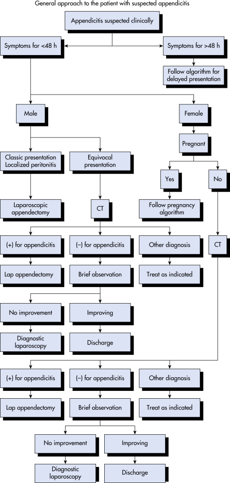
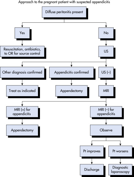
Patients with RLQ pain, nausea, vomiting, anorexia, and RLQ rebound tenderness should undergo prompt clinical and laboratory evaluation. Imaging studies are generally not necessary in typical appendicitis and generally reserved for patients with an equivocal likelihood of appendicitis. They are useful when the diagnosis is uncertain. Several clinical decision tools such as the Alvarado score (also known as the MANTRELS criteria) are available to assist in the diagnosis of appendicitis (Table 2). Laparoscopy may be useful as both a diagnostic and a therapeutic modality.
TABLE 2 Alvarado Score (MANTRELS Criteria)
Criterion | Point(s) | |
Symptoms | ||
M | Migration of pain to RLQ | 1 |
A | Anorexia | 1 |
N | Nausea and vomiting | 1 |
Signs | ||
T | Tenderness in RLQ | 2 |
R | Rebound pain | 1 |
E | Elevated temperature | 1 |
Laboratory Findings | ||
L | Leukocytosis | 2 |
S | Shift of WBCs to left | 1 |
Total Score | 10 | |
Interpretation | ||
1-4 | Appendicitis unlikely | |
5-6 | Appendicitis possible | |
7-8 | Probable appendicitis | |
9-10 | Surgery indicated |
RLQ, Right lower quadrant; WBCs, white blood cells.From Cameron P et al: Textbook of adult emergency medicine, ed 5, Philadelphia, 2019, Elsevier.
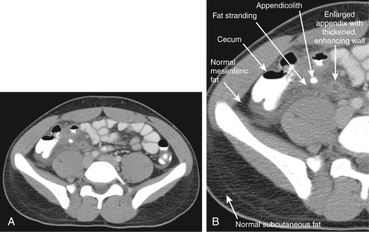
This CT Demonstrates Classic Findings of Appendicitis in an 18-Yr-Old Male with Right Lower Quadrant Pain, as Seen with CT with IV and Oral Contrast. Studies Suggest that CT Without Contrast Has Similar Sensitivity and Specificity. An Enlarged Appendix is Seen Near the Cecum as a Right Lower Quadrant Tubular Structure in Short-Axis Cross Section, Giving It a Circular Appearance. The Surrounding Fat Shows Stranding, a Smoky Appearance Indicating Inflammation (Compare with Normal Mesenteric and Subcutaneous Fat, Which is Nearly Black). The Appendiceal Wall Shows Enhancement, a Brightening after Administration of IV Contrast. This Slice Also Shows an Appendicolith, an Occasional Finding of Appendicitis. It Does Not Appear to Be Within the Appendix in This Slice, Because the Appendix Bends in and Out of the Plane of This Slice. An Appendicolith Usually Appears as a Calcified (White) Rounded Structure, Visible Without any Contrast. A, Axial CT Image. B, Close-Up.
From Broder JS: Diagnostic imaging for the emergency physician, Philadelphia, 2011, Saunders.
TABLE 3 Computed Tomography Findings of Appendicitis: SCALPEL Mnemonic
Term | Description | ||
---|---|---|---|
Stranding | Fat stranding suggests regional inflammation, possibly because of appendicitis. | ||
Cecum | The appendix originates from the cecum, which should be identified first to help localize the appendix. The cecum may show wall thickening, suggesting appendicitis. | ||
Air | Air outside of the lumen of the appendix is pathologic and suggests perforation. Air within the appendiceal wall is also abnormal. | ||
Large | The normal appendix is <6 mm; an enlarged appendix >6 mm suggests appendicitis. Wall thickening >1 mm also suggests appendicitis. | ||
Phlegmon | Inflammatory changes surrounding the appendix suggest a perforated appendix. A heterogeneous collection called a phlegmon may be seen. If the appendix has ruptured, a pericecal phlegmon may be the only remaining evidence, because the appendix itself may not be seen. | ||
Enhancement | The wall of an abnormal appendix enhances with IV contrast and appears brighter than the normal bowel or the normal psoas muscle. | ||
Lith | An appendicolith is a calcified stone sometimes found in the lumen of an inflamed appendix. |
IV, Intravenous.
From Broder JS: Diagnostic imaging for the emergency physician, Philadelphia, 2011, Saunders.
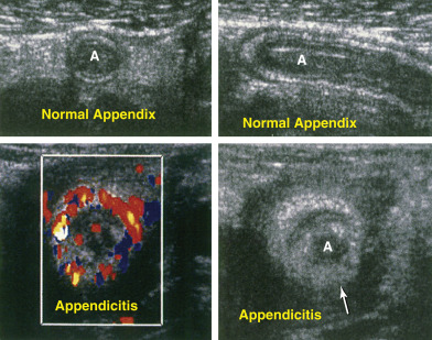
In Appendicitis, There is Distention and Wall Thickening (Bottom, Right Arrow), and Blood Flow is Increased, Leading to the So-Called Ring of Fire Appearance. A, Appendix.
From Townsend CM et al: Sabiston textbook of surgery, ed 21, St Louis, 2022, Elsevier.
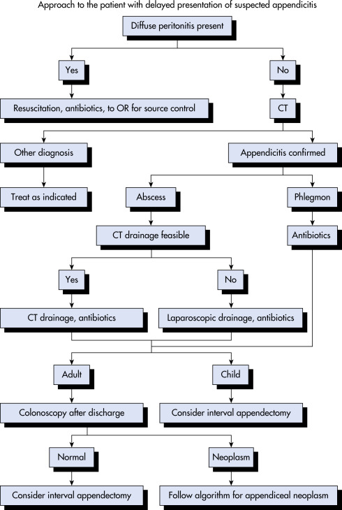
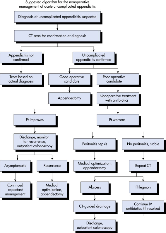
Management of Complicated Appendicitis:
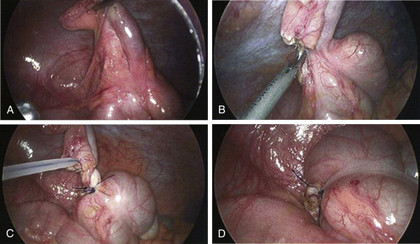
(A) Visualization and Upward Retraction of Appendix. (B) Division of the Mesoappendix Using Harmonic Scalpel. (C) Application of Endoloops to the Appendix. Two Loops are Used to Secure the Base; a Third Loop is Applied Distally to Avoid Spillage of the Luminal Contents. The Specimen is Then Divided Between the Endoloops. (D) View of Completed Appendectomy after Removal of the Specimen. (Note: Depending on the Surgeons Preference, an Endoscopic Stapling Device May Be Used to Divide the Mesoappendix and Appendix Instead of the Harmonic Scalpel and Endoloops.)
From Townsend CM et al: Sabiston textbook of surgery, ed 21, St Louis, 2022, Elsevier.
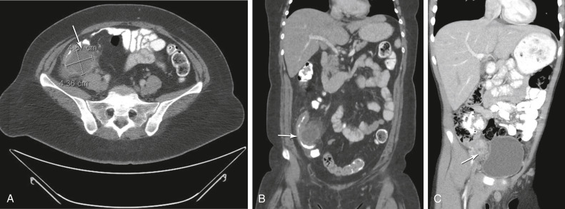
Sagittal (A) and Coronal (B) Computed Tomography Images Demonstrate an Appendiceal Abscess in a Patient Who Presented with a 2-wk History of Abdominal Pain and was Found to have a Palpable Mass on Examination. The Arrows Point to a Periappendiceal Abscess Cavity. She was Successfully Managed with Percutaneous Drainage and Antibiotic Therapy. Image C is a Similar Case in Which the Patient Presented with an Appendiceal Phlegmon and was Successfully Treated with Antibiotics Alone. The Arrow Points to the Phlegmon. (Note the Mass Effect on the Bladder.)
From Townsend CM et al: Sabiston textbook of surgery, ed 21, St Louis, 2022, Elsevier.
.