section name header

Basic Information

AUTHOR: Ali Ahmad, MD

Definition

  • Traumatic brain injury (TBI) is a broad term that encompasses multiple intracranial processes (including cerebral contusion, epidural hemorrhages, subdural hemorrhages, subarachnoid hemorrhages, skull fractures, diffuse axonal injury, and cerebral edema) that occur secondary to trauma to the head or if the head experiences a sudden deceleration injury without external trauma that results in injury to the brain.1 An overview of classification of TBI is summarized in Table 1. These injuries result in varying levels of cellular and macroscopic changes, detected with clinical examination and supplemented by neuroimaging.
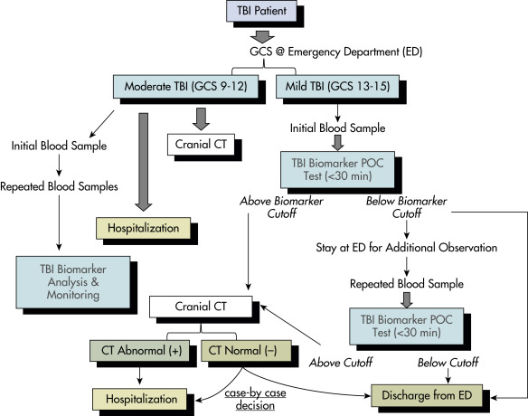
  • Mild TBI is defined as loss of consciousness <30 minutes, a Glasgow Coma Scale (GCS) of 13 to 15, and normal imaging.
  • Moderate TBI is defined as loss of consciousness for 30 minutes to 24 hr with even longer alteration in consciousness, a GCS of 9 to 12, and may or may not have abnormal imaging.
  • Severe TBI is defined as >24 hr of loss of consciousness with a GCS of 3 to 8 and prolonged posttraumatic amnesia with normal or abnormal imaging.1,2

TABLE 1 Overview of Classification of Traumatic Brain Injury


Mechanism
BluntHigh velocity (MVC)
Low velocity (fall, assault)
PenetratingGSW
Other (stab wounds, etc.)
BlastExplosive devices
SeverityMildGCS 14-15
ModerateGCS 9-13
SevereGCS 3-8
MorphologySkull fractureVaultLinear versus stellate
Depressed/nondepressed
Open/closed
BasilarWith/without CSF
With/without CN palsy
Intracranial lesionsFocalEpidural
Subdural
Intracerebral
DiffuseMild concussion
Classic concussion
Diffuse axonal injury

CN, Cranial nerve; CSF, cerebrospinal fluid; GCS, Glasgow Coma Scale; GSW, gunshot wound; MVC, motor vehicle collision.

From Jankovic J et al: Bradley and Daroff’s neurology in clinical practice, ed 8, Philadelphia, 2022, Elsevier.

Synonyms

TBI

Head injury

Concussion

Intracranial contusion

ICD-10CM CODES
S06.9 X0AIntracranial injury
S06.1X7ATraumatic cerebral edema with loss of consciousness of any duration with death due to brain injury prior to regaining consciousness, initial encounter
S06.2X9ADiffuse traumatic brain injury with loss of consciousness of unspecified duration, initial encounter
S06.300AUnspecified focal traumatic brain injury without loss of consciousness, initial encounter
S06.305AUnspecified focal traumatic brain injury with loss of consciousness greater than 24 hours with return to preexisting conscious level, initial encounter
S06.309AUnspecified focal traumatic brain injury with loss of consciousness of unspecified duration, initial encounter
S09.90Unspecified injury of the head
Epidemiology & Demographics

Traumatic brain injury (TBI) is a worldwide leading cause of mortality in the young and elderly. In the 2017 CDC surveillance report, unintentional falls have surpassed motor vehicle crashes as number one cause of TBI.3

Incidence

Globally, more than 69 million people suffer from TBI each year, with North America and Europe showing highest incidence and Southeast Asian and Western Pacific countries experiencing the greatest burden of disease. According to one estimate, up to 4.6 million individuals suffer from TBI in United States and Canada each year.4 In 2014, about 2.87 million emergency department visits, including deaths and hospitalizations, were associated with TBI. The financial burden of TBI has been estimated to be greater than $80 billion per yr in the United States alone and approaching $400 billion dollar worldwide in direct and indirect costs.5

Prevalence

In 2016, the global prevalence of TBI was estimated at 55.5 million. Between 1990 to 2016, the age-standardized prevalence of TBI increased by 8.4%.6 According to the CDC, there were 224,000 TBI-related hospitalization in 2017, decreased from 261,000 in 2016, while the number of deaths increased from 59,000 to 61,000.3

Peak Incidence

In the U.S. 792/100,000 individuals, including nonspecific head injury, removing which brings the number to 264/100,000.3

Predominant Sex & Age

TBI occurs more commonly in males and hospitalizations/deaths are the highest in older adults >75 yr of age.3

Risk Factors

  • Falls
  • Motor vehicle accidents
  • Physical violence
  • Sport injuries
  • Ballistic injuries (gunshot wounds, blast injuries)
Genetics

TBI and Apo E Ε4 synergistically are associated with a tenfold increased risk for Alzheimer disease. Apo E Ε4 is also associated with larger intracerebral hematomas and greater ischemia after TBI.7

Physical Findings & Clinical Presentation

TBI patients can present with a spectrum of clinical symptoms including nausea, vomiting, headache, seizures, altered mental status, and/or coma. Stigmata of trauma, including bruises, scalp lacerations, and periorbital or mastoid ecchymosis suggesting skull base fractures, are telltale signs of possible underlying traumatic brain injury. Box 1 describes risk stratification in patients with minor head trauma. The spectrum of TBI is most commonly assessed using the GCS, which ranges from 3 to 15 and utilizes eye, motor, and verbal exams (Table 2).

BOX 1 Risk Stratification in Patients With Minor Head Trauma

High Risk

  • Focal neurologic findings
  • Asymmetrical pupils
  • Skull fracture on clinical examination
  • Multiple trauma
  • Serious, painful, distracting injuries
  • External signs of trauma above the clavicles
  • Initial Glasgow Coma Scale score of 14 or 15
  • Loss of consciousness
  • Posttraumatic confusion or amnesia
  • Progressively worsening headache
  • Vomiting
  • Posttraumatic seizure
  • History of bleeding disorder or anticoagulation
  • Recent ingestion of intoxicants
  • Unreliable or unknown history of injury
  • Previous neurologic diagnosis
  • Previous epilepsy
  • Suspected child abuse
  • Age above 60 yr or below 2 yr
Medium Risk

  • Initial Glasgow Coma Scale score of 15
  • Brief loss of consciousness
  • Posttraumatic amnesia
  • Vomiting
  • Headache
  • Intoxication
Low Risk

  • Currently asymptomatic
  • No other injuries
  • No focality on examination
  • Normal pupils
  • No change in consciousness
  • Intact orientation and memory
  • Initial Glasgow Coma Scale score of 15
  • Accurate history
  • Trivial mechanism
  • Injury less than 24 hr ago
  • No or mild headache
  • No vomiting
  • No preexisting high-risk factors

TABLE 2 Useful Criteria to Assess the Severity of Head Injurya

!!flowchart!!
Quantifying the Degree of Head InjuryGlasgow Coma Scale (GCS)Score
  • Moderate TBI (GCS 9-13)
  • Severe TBI (GCS <8)
  • Significant head CT:
    • Cerebral edema
    • Midline shift
    • Subdural/epidural bleeding
    • Open head injury with intracranial air
Eye Opening
Spontaneous4
To speech3
To pain2
None1
Verbal Response
Oriented5
Confused conversation4
Inappropriate words3
Incomprehensible sounds2
None1
Best Motor Response
Obeys commands6
Localizes pain5
Flexion withdrawal to pain4
Abnormal flexion (decorticate)3
Extension (decerebrate)2
None (flaccid)1

TBI, Traumatic brain injury.

a Consultation with other specialists (i.e., neurosurgeon) may be valuable for the orthopedic surgeon unfamiliar with the process of “clearing” the patient’s head injury for ischemic monomelic neuropathy (IMN) fixation.

From Browner B et al: Skeletal trauma: basic science, management, and reconstruction, ed 6, Philadelphia, 2019, Elsevier.

Etiology

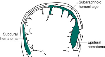
  • Mechanical falls (49.1%), motor vehicle accidents (24.5%), and assaults resulting in direct or indirect trauma to the head are the most common etiologies (Fig. E1, Fig. E2).

Figure E1 In This Drawing, a Hammer Blow to the Back of the Head Inflicts a Coup Injury to the Occipital Region and, as is Typical, a More Extensive Contrecoup Injury to the Inferior Surface of the Frontal and Anterior Tips of the Temporal Lobes

From Kaufman DM et al: Kaufman’s clinical neurology for psychiatrists, ed 9, Philadelphia, 2023, Elsevier.

Figure E2 Meningeal Arterial Bleeding, Which Usually Results from a Blow Forceful Enough to Fracture the Skull, Causes an Epidural Hematoma

In Contrast, Venous Bleeding, Usually Slower and Under Less Pressure, Causes Subdural Hematoma. Ruptured Aneurysms and Head Trauma Often Cause Subarachnoid Hemorrhage (SAH). In SAH, Blood Spreads Within the Subarachnoid Space over the Convexities, Between the Gyri, into the Interhemispheric Fissure, and Down into the Spinal Canal.

From Kaufman DM et al: Kaufman’s clinical neurology for psychiatrists, ed 9, Philadelphia, 2023, Elsevier.

  • Gunshot wounds are the most prevalent penetrating injuries (Fig. E3), accounting for 35% of deaths from TBI under the age of 45 yr in the United States. Self-inflicted injuries-for example, with nail guns-can also lead to penetrating injuries (Fig. E4). Gunshot wounds are the most lethal type of brain injury, 90% resulting in death.

Figure E3 Picture of a gunshot wound to the head-a penetrating injury.

The bullet leaves a path of destructions marked by blood, bone, and bullet fragments. Brain tissue around the bullet path is also damaged by the propagating pressure wave. However, this is not visible on computed tomography (axial computed tomography).

From Jankovic J et al: Bradley and Daroff’s neurology in clinical practice, ed 8, Philadelphia 2022, Elsevier.

Figure E4 Another Form of Penetrating Injury is Shown on This Anteroposterior x-Ray of the Skull, with Two Nails from a Nail Gun Stuck in the Skull and Penetrating the Right Frontal Lobe

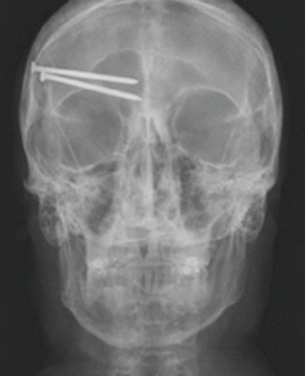
This patient was neurologically intact on presentation. The difficulty in such a case is that bleeding can occur deep in the brain when the nails are removed. This patient was also at high risk for developing pseudoaneurysm later on.

From Jankovic J et al: Bradley and Daroff’s neurology in clinical practice, ed 8, Philadelphia 2022, Elsevier.

  • The most common mechanisms of pediatric TBI vary according to age. Falls are the leading cause of TBI in children younger than 14 yr of age. Children younger than 4 yr of age are injured mainly by falls but are also affected by abusive injuries and motor vehicle accidents. Children 4 to 8 yr of age are injured in falls and motor vehicle accidents but also become more at risk for other transportation-related injuries such as bicycle-related incidents. Abusive head trauma (AHT) is particularly common in young infants aged less than 2 yr; approximately 30 of every 100,000 infants aged less than 1 yr were hospitalized for AHT.8
  • Sports-related TBIs account for roughly one third of all causes of TBI, with males, adolescents, and young adults with higher incidence. Some common sports associated with TBI are football, horse riding, cycling, skateboarding, hockey, water sports, and snow sports.9
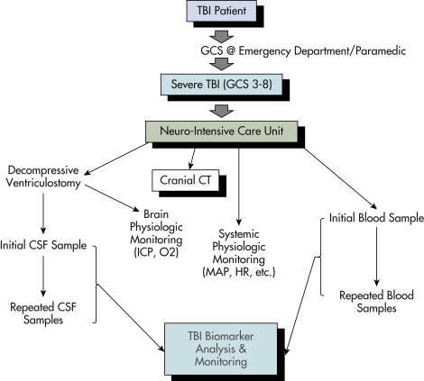
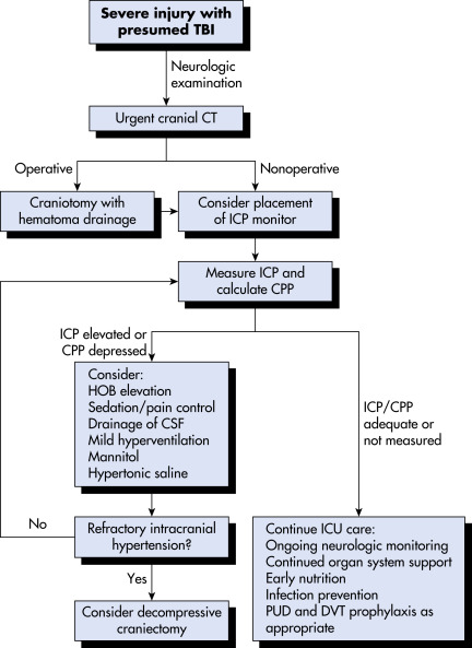
TREATMENT (FIG. 8)

Prevention of secondary injury is the primary goal of prehospital and early in-hospital management. Most common mechanisms of secondary injury are either intracranial (increased intracranial pressure [ICP], hematoma) or systemic (hypoxia, hypovolemia, hypotension). Early categorization of head trauma patients according to the severity (based on GCS) and transport to facilities equipped with personnel and technology to deal with issues pertaining to head trauma has improved the overall management of head injury patients and prevention of secondary injury.10 Assessment and treatment recommendations for mild TBI are summarized in Table 5. Airway, breathing, and circulation, however, still remain the most important parameters to be stabilized, and both directly and indirectly affect GCS and overall outcome. Trauma guidelines suggest intubation should be performed in any patient with a GCS of 8 or less to prevent hypoxemia and hypercapnia. Previously, patients were hyperventilated to decrease pCO2 in an effort to reduce ICP. Recent evidence suggests normo-ventilation for patients with severe TBI, with hyperventilation only being used as a temporary measure until other methods of reducing ICP are employed.11 Intravenous fluid resuscitation should also be started early to prevent hypovolemia resulting in hypotension, shown to double mortality. Transfer to and care in a Level 1 trauma center are associated with better outcomes. Monitoring and treatment recommendations for severe TBI are summarized in Table 6.

TABLE 6 Guidelines for the Management of Severe Traumatic Brain Injury

TopicLevel 1Level 2Level 3
Blood pressure and oxygenationInsufficient dataAvoid systolic blood pressure <90 mm HgAvoid hypoxia (Pao2 <60 mm Hg or O2 saturation <90%)
Hyperosmolar therapyInsufficient dataMannitol is effective for control of raised ICP at doses of 0.25 g/kg to 1 g/kg body weightRestrict mannitol use prior to ICP monitoring in patients with signs of transtentorial herniation
Prophylactic hypothermiaInsufficient dataInsufficient dataPooled data indicate that prophylactic hypothermia is not significantly associated with decreased mortality as compared with normothermic controls
Infection prophylaxisInsufficient dataPeriprocedural antibiotics for intubation should be administered to reduce the incidence of pneumonia
Early tracheostomy should be performed to reduce days on mechanical ventilation with pneumonia
To reduce infection, routine ventricular catheter exchange or prophylactic antibiotic use for ventricular catheter placement is not recommended
Deep venous thrombosis prophylaxisInsufficient dataInsufficient dataIntermittent pneumatic compression stockings are recommended
Low-molecular-weight heparin or low-dose unfractionated heparin should be used in combination with mechanical prophylaxis
Indications for ICP monitoringInsufficient dataICP should be monitored in all salvageable patients with a GCS score of 3-8 after resuscitation and an abnormal CT scanICP monitoring is indicated in patients with severe TBI with a normal CT scan if >40 yr of age with blood pressure <90 mm Hg
ICP pressure-monitoring technologyN/AN/AN/A
ICP thresholdsInsufficient dataTreatment should be initiated with ICP >20 mm HgA combination of ICP values and clinical and brain CT findings should be used to determine the need for treatment
Cerebral perfusion thresholdsInsufficient dataAggressive attempts to maintain CPP above 70 mm Hg with fluids and pressors should be avoided because of the risk of adult respiratory distress syndromeCPP of <50 mm Hg should be avoided
The CPP value to target lies within the range of 50-70 mm Hg. Patients with intact pressure autoregulation tolerate higher CPP values. Ancillary monitoring of cerebral parameters that include blood flow, oxygenation, or metabolism facilitates
CPP management
Brain oxygen monitoring and thresholdsInsufficient dataInsufficient dataJugular venous saturation (<50%) or brain tissue oxygen tension (<15 mm Hg) are treatment thresholds
Anesthetics, analgesics, sedativesInsufficient dataProphylactic administration of barbiturates to induce burst suppression electroencephalogram is not recommended. High-dose barbiturate administration is recommended to control elevated ICP refractory to maximum standard medical and surgical treatment. Hemodynamic stability is essential before and during barbiturate therapy. Propofol is recommended for the control of ICP but not for improvement in mortality or 6-mo outcomeN/A
NutritionInsufficient dataPatients should be fed to attain full caloric replacement by day 7 postinjuryN/A
Antiseizure prophylaxisInsufficient dataAnticonvulsants are indicated to decrease the incidence of early PTS (within 7 days of injury)N/A
HyperventilationInsufficient dataProphylactic hyperventilation (Paco2 of 25 mm Hg or less) is not recommendedHyperventilation is recommended as a temporizing measure for the reduction of ICP. Hyperventilation should be avoided during the first 24 hr after injury, when cerebral blood flow is often critically reduced. If hyperventilation is used, jugular venous oxygen saturation (SjO2) or brain-tissue oxygen tension (PbtO2) measurements are recommended to monitor oxygen delivery
SteroidsThe use of high-dose methylprednisolone is associated with increased mortality and is contraindicatedN/AN/A

CT, Computed tomography; CPP, cerebral perfusion pressure; ICP, intracranial pressure; GCS, Glasgow Coma Scale; PTS, posttraumatic seizures; TBI, traumatic brain injury.

From Jankovic J et al: Bradley and Daroff’s neurology in clinical practice, ed 8, Philadelphia, 2022, Elsevier.

TABLE 5 Assessment and Treatment Recommendations for Mild Traumatic Brain Injury

FunctionAssessmentTreatment
Overall recoveryStandardized symptom checklistPhysical rest 1-2 days1 followed by subsymptomatic aerobic exercise2
HeadacheDetermine the typeHA persisting more than 3-4 days may require abortive treatment tailored to phenotype (migraine, tension-type, occipital neuralgia, etc.)
VertigoRomberg test, dynamic standing, tandem gaitIf Hallpike Dix is normal, or if Epley maneuver does not relieve symptoms, consider physical therapy for vestibular rehabilitation
Eye movementsExamine cranial nerves 3, 4, 6 for tracking, saccades, diplopia, nystagmusPhysical therapy evaluation for vestibular rehabilitation
Near visionNear-point accommodation and binocular convergenceOphthalmologic evaluation for vision therapy
Cognitive functionSymptoms, cognitive testing, neuropsychologist evaluationSleep hygiene, neuropsychology evaluation for cognitive rehabilitation3

HA, Headache.

1 Thomas DG et al: Benefits of strict rest after acute concussion: a randomized controlled trial, Pediatrics 135:213-223, 2015.

2 Kurowski BG et al: Aerobic exercise for adolescents with prolonged symptoms after mild traumatic brain injury: an exploratory randomized clinical trial, J Head Trauma Rehabil 32:79-89, 2017.

3 Cooper DB et al: Cognitive rehabilitation for military service members with mild traumatic brain injury: a randomized clinical trial, J Head Trauma Rehabil 32:E1-E15, 2017.

From Goldman L, Schafer AI: Goldman-Cecil medicine, ed 26, Philadelphia, 2019, Elsevier.

Figure 8 Algorithm for the Management of Traumatic Brain Injury (TBI)

Cpp, Cerebral Perfusion Pressure; CSF, Cerebrospinal Fluid; CT, Computed Tomography; DVT, Deep Venous Thrombosis; Hob, Head of Bed; ICP, Intracranial Pressure; ICU, Intensive Care Unit; Pud, Peptic Ulcer Disease.

From Townsend CM et al: Sabiston textbook of surgery, ed 21, St Louis, 2022, Elsevier.

Details of in-hospital management including critical care and surgical intervention is beyond the scope of this text. Some important points are summarized below.

  • Advanced trauma life support (ATLS) protocol (airway, breathing, circulation, disability, exposure).
  • Ventilatory support.
  • Optimization of oxygenation, ventilation, and fluid status.
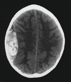
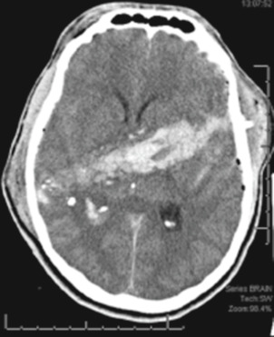
  • CT head (Fig. E9) to evaluate for mass lesion (hematoma) or cerebral edema. These findings may necessitate either surgical intervention or ICP monitor placement. ATLS guidelines recommend maximum 30 minutes between initial assessment and CT head.

Figure E9 Indications for acute seizure prophylaxis in severe head trauma.

From Marx JA et al: Rosen’s emergency medicine: concepts and clinical practice, ed 7, Philadelphia, 2010, Elsevier.

  • In case of either a severe TBI (GCS 8 or less) or a moderate TBI (GCS 8 to 13) with an unreliable neurologic exam, patients should be admitted to the intensive care unit for frequent neurologic checks. TBI guidelines suggest ICP monitor placement for GCS 8 or less to monitor intracranial pressure closely.11 ICP monitors are of various kinds, and the most commonly used include external ventricular drain, intraparenchymal pressure monitor, and a bolt device with brain tissue oxygen pressure monitoring with fiberoptic pressure monitor. Recent research has also supported the use of brain tissue oxygen monitoring for severe TBI patients.11
  • Surgical decompression may involve evacuation of hematoma (epidural, subdural, intraparenchymal, contusion) through craniotomy alone (replacement of bone after completion of operation) versus decompressive craniectomy (complete removal of bone without replacement). Skull fractures are treated depending on the morphology of the fracture. Open, depressed fractures require surgical debridement and elevation in most cases, in addition to broad-spectrum antibiotics.
  • Avoid electrolyte imbalance, especially hyponatremia and hyperglycemia, which may contribute to cerebral edema and increase intracranial pressure. A recent randomized controlled trial studied use of a continuous 20% hypertonic saline infusion vs standard therapy in patients with moderate to severe TBI (COBI trial) and did not improve neurologic status at 6 mo.12
  • Elevation of head of bed to allow better venous drainage to reduce intracranial pressure.
  • ICP management (Table 7), which may include the following: Drainage of cerebrospinal fluid via external ventricular drain, surgical hematoma evacuation, administration of hyperosmotic fluids to reduce edema, pharmacologic sedation and paralysis, pentobarbital-induced coma, and surgical decompression of the brain.11
  • Decompressive craniectomy in patients with TBI and refractory intracranial hypertension may reduce ICU stay but does not clearly improve outcomes. DECRA13 and RESCUEicp14 are two randomized controlled trials that assessed decompressive craniectomy (DC) for reduction in ICP vs. medical management. Both trials were unable to provide definite evidence for or against DC, indicating that the decision to proceed with DC should be made on case-by-case basis, after assessment of individual risks and benefits.11
  • Normothermia: In patients with TBI, hypothermia can reduce intracranial hypertension, but recent trials in patients with an intracranial pressure of more than 20 mm Hg after TBI, therapeutic hypothermia, plus standard care to reduce intracranial pressure did not result in outcomes better than those with standard care alone. The most recent TBI guidelines do not recommend hypothermia.11
  • Prevention of seizures in the acute setting. Seizure prophylaxis can be considered for the first 7 days. Indications for acute seizure prophylaxis in severe head trauma are described in Box 3. Usually phenytoin or levetiracetam is used.15 Seizure prophylaxis, however, has not shown to prevent long-term development of traumatic epilepsy.
  • Deep vein thrombosis (DVT) prophylaxis is recommended in almost all patients on hospital day 1 in addition to sequential compression devices (SCDs) for immobile or bedbound patients to prevent DVTs.11
  • Early initiation of parenteral nutrition.
  • Early tracheostomy for ventilator-dependent patients is recommended to reduce mechanical ventilation days.11

BOX 3 Indications for Acute Seizure Prophylaxis in Severe Head Trauma

  • Depressed skull fracture
  • Paralyzed and intubated patient
  • Seizure at the time of injury
  • Seizure at emergency department presentation
  • Penetrating brain injury
  • Severe head injury (Glasgow Coma Scale score 8)
  • Acute subdural hematoma
  • Acute epidural hematoma
  • Acute intracranial hemorrhage
  • Prior history of seizures

TABLE 7 Elevated ICP Management

  • Verify ICP
    1. Check if EVD is still patent
    2. Check to see if EVD waveform is present and adequate
    3. Check to see if EVD ICP correlates with intraparenchymal monitor if present
  • Check for 30-degree head elevation
  • Loosen cervical collar if in place
  • Open EVD for ICP >20 mm Hg for 10 min and then close and transduce ICP
    1. Repeat once
    2. If ICP >20 mm Hg, keep open at 15 mm Hg above midbrain and proceed with ICP module
  • Treat temperature >37.5°C with 650 mg of acetaminophen once
  • Sedation
    1. Titrate propofol to a Ramsay score of 4
      1. Do not exceed 5 mg/kg/h for more than 24 h
    2. Check potassium, triglycerides, creatine kinase, and urinalysis for myoglobinuria q 8 h for 24 h
    3. If maximal dose of propofol is reached and ICP >20 mm Hg
    4. Start fentanyl drip at 0.8 μg/kg/h
    5. Apply bispectral index (BIS) monitor
    6. Titrate fentanyl drip to a bispectral index of 30 or to a maximum of 5 μg/kg/h
    7. Start chlorhexidine gluconate (Peridex) with a loading infusion of 1 μg/kg over 10 min
      1. Continue maintenance infusion of 0.2 to 0.7 μg/kg/h
  • Hyperosmolar therapy
    1. 3% hypertonic saline bolus of 250 mL
    2. Before administering 3% hypertonic saline bolus, check if Na <130 mEq/L
    3. In emergency, administer mannitol 1-0.5 mg/kg bolus once
    4. Check sodium and serum osmolality q 4 h × 2 after every bolus
    5. Start 3% hypertonic saline drip at 0.5 mL/h if three 3% hypertonic saline boluses within 6 h
    6. Check sodium and serum osmolality q 2 h while on drip
    7. If sodium >160 mEq/L or serum osmolality >320 sOsm/L, call physician
    8. If serum sodium has increased to >10 mEq/L within the last 24 h, call physician
    9. If CBF >35 mL/min/100 g white matter or >80 mL/min/100 g gray matter refer to CBF module high flow
  • If core body temperature 37.5°C, start normothermia protocol
  • Hyperventilation
    1. Do not hyperventilate in the first 24 hr (goal of Paco2 of 35-40 mm Hg)
    2. If PbtO2 is <20 mmHg, go to hypoxia module
    3. If CBF <18 mL/min/100 g white matter or <67 mL/min/100 g gray matter, go to CBF module
    4. If PbtO2 and CBF are optimized, hyperventilate to 33-35 mm Hg
  • Radiology
    1. Refractory ICP >20 mm Hg despite intervention, obtain portable head CT without contrast immediately if no head CT since ICP is elevated despite maximal therapy
  • Consider surgery
  • Induce pentobarbital coma
    1. Only for diffuse nonoperative injuries
    2. Only with attending approval
    3. Order continuous EEG monitoring if not already in place
    4. Have norepinephrine drip ready at the bedside for MAP <80 mm Hg/CPP <60 mm Hg
    5. Pentobarbital bolus/loading: 10 mg/kg once over 60 min, then 5 mg/kg qh × 4 or until burst suppression
    6. Pentobarbital maintenance dose: 1 mg/kg/h titrated to burst suppression

CBF, Cerebral blood flow; CPP, cerebral perfusion pressure; EEG, electroencephalography; EVD, external ventricular drain; ICP, intracranial pressure; MAP, mean arterial pressure; PbtO2, Brain tissue oxygen.

From Jankovic J et al: Bradley and Daroff’s neurology in clinical practice, ed 8, Philadelphia, 2022, Elsevier.

Diagnosis

Differential Diagnosis

Differential diagnosis of TBI is quite limited; however, there are several considerations and diagnoses considered as a possibility within the realm of TBI. These are enumerated in the imaging section.

Workup

TBI workup is always a part of the advanced trauma life support (ATLS) protocol. Primary and secondary survey followed by imaging studies constitutes the standardized approach to TBI. Focused TBI workup includes:

  • History: Including timing of injury, duration of loss of consciousness if applicable, seizures (if any), comorbidities, use of anticoagulants and antiplatelet agents (requires reversal in the event of intracranial blood on imaging).
  • Neurologic examination: Glasgow Coma Scale, cranial nerves, motor/sensory exam. Assess for scalp lacerations, specifically overlying a skull fracture as well as cerebrospinal fluid (CSF) otorrhea or rhinorrhea.
  • CT imaging of the head if there is a significant history of impact to the head, polytrauma, positive loss of consciousness, or stigmata of trauma to the head. Factors to consider regarding the need for CT imaging in head-injured patients are described in Table 3. The American College of Emergency Physicians Clinical Policy Regarding Neuroimaging in adults with mild TBI is summarized in Box 2.

TABLE 3 Factors to Consider Regarding the Need for Computed Tomography in Head-Injured Patients

Indications for urgent CT include the following:
  • Evidence of skull fracture-basal, depressed, or open
  • Abnormal results of neurologic examination
  • Seizure
  • Vomiting more than once
  • High-risk mechanism (e.g., ejection from vehicle; injury to pedestrian or cyclist vs. car occupant)
  • Decreasing GCS score or persistently decreased GCS score below 15
  • Indications for lower threshold for CT scan include the following:
  • Age >60 yr
  • Persistent anterograde amnesia
  • Retrograde amnesia >30 min
  • Coagulopathy
  • Fall >5 stairs or >3 ft
  • Intoxication (examination unreliable)
  • LOC >30 min
  • Mechanism and location of injury
  • Social factors (e.g., abusive situation at home, language barriers precluding an accurate history)

CT, Computed tomography; GCS, Glasgow Coma Scale; LOC, loss of consciousness.

From Jankovic J et al: Bradley and Daroff’s neurology in clinical practice, ed 8, Philadelphia, 2022, Elsevier.

BOX 2 American College of Emergency Physicians Clinical Policy Regarding Neuroimaging in Adults With Mild Traumatic Brain Injury

A noncontrast head computed tomography (CT) is indicated (level-1 recommendation) in adults with level of consciousness (LOC) or posttraumatic amnesia only if one or more of the following is present:

  • Headache
  • Vomiting
  • Age >60 yr
  • Drug or alcohol intoxication
  • Deficits in short-term memory
  • Physical evidence of trauma above the clavicle
  • Posttraumatic seizure
  • Glasgow Coma Scale (GCS) score <15
  • Focal neurologic deficit
  • Coagulopathy

A noncontrast head CT should be considered (level-2 recommendation) in head trauma patients with no LOC or posttraumatic amnesia if there is:

  • Focal neurologic deficit
  • Vomiting
  • Severe headache
  • Age 65 yr
  • Physical signs of a basilar skull fracture
  • GCS score <15
  • Coagulopathy
  • A dangerous mechanism (e.g., ejection from motor vehicle, pedestrian struck, fall of more than 3 feet or 5 stairs)

From Marx JA et al: Rosen’s emergency medicine, ed 8, Philadelphia, 2014, Elsevier.

Laboratory Tests

  • Basic labs including CBC, basic metabolic panel, prothrombin time, activated partial thromboplastin time, urine drug screen, and ethanol blood level.
  • Consider tests for platelet function analysis for unknown antiplatelet use.
  • No blood biomarker currently exists (Figs. E5 and E6).

Figure E5 Envisioned Uses of Brain Biomarkers for Severe Traumatic Brain Injury (TBI) Patient Management

!!flowchart!!

CSF, Cerebrospinal fluid; CT, computed tomography; GCS, Glasgow Coma Scale; HR, heart rate; ICP, intracranial pressure; MAP, mean arterial blood pressure.

From Vincent JL et al: Textbook of critical care, ed 7, Philadelphia, 2017, Elsevier.

Figure E6 Envisioned Uses of Brain Biomarkers for Mild-to-Moderate Traumatic Brain Injury (TBI) Patient Management

CT, Computed tomography; ED, emergency department; GCS, Glasgow Coma Scale; POC, point-of-care.

From Vincent JL et al: Textbook of critical care, ed 7, Philadelphia, 2017, Elsevier.

Imaging Studies (TABLE 4

CT imaging forms the current cornerstone of imaging modalities for head trauma; however, it is not always necessary. Patients over the age of 16 with minimal head injury (i.e., no history of loss of consciousness, amnesia, and confusion), not on blood thinners, and without associated seizure generally do not need a CT scan. Canadian CT head rules for patients with GCS 13 to 15 who do have history of loss of consciousness, amnesia, and/or confusion are a useful guide in determining utility of obtaining a CT scan. If any of these risk factors are present, it is recommended that a CT scan of the head be considered.

TABLE 4 Comparison of Head Imaging Modalities

Computed Tomography ScansMagnetic Resonance ImagingAngiographySkull Radiography
AdvantagesFast
Patient accessible for monitoring
Defines acute hemorrhages, mass effects, bone injuries, hydrocephalus, intraventricular blood, edema
Defines contusions and pericontusion edema, posttraumatic ischemic infarction, brainstem injuriesHelps localize acute traumatic lesions
Defines vascular injuries, injuries to venous sinuses
Detects mass effects
Readily available
May help screen some patients for further imaging studies
DisadvantagesArtifacts arise from patient’s movement, foreign bodies
Streak artifacts may obscure brainstem or posterior fossa
Slow
Patients not easily accessible for monitoring
Does not define most acute hemorrhagic lesions
Not useful for bone injuries
Does not define nature of acute lesion
Does not detect infratentorial masses
Does not indicate presence or absence of intracranial injury
IndicationsAcute severe head trauma
Acute moderate head trauma
Suspected depressed skull fracture
High-risk minor head trauma
Suspected child abuse in minor head trauma
Deteriorating neurologic status
Persistent symptoms with postconcussive syndrome
Suspected posttraumatic ischemic infarction
Suspected contusions not seen on CT scan
Suspected vascular injury
CT scan not available
CT scan may not be done
Penetrating head trauma

CT, Computed tomography.

From Marx JA et al: Rosen’s emergency medicine: concepts and clinical practice, ed 7, 2010, Elsevier.

High risk

  • Failure to reach GCS of 15 within 2 hr
  • Suspected open or depressed skull fracture
  • Any signs of basal skull fracture (hemotympanum, “raccoon” eyes, CSF otorrhea/rhinorrhea, Battle’s sign)
  • Two or more episodes of vomiting
  • Age older than 65
Medium risk

  • Dangerous mechanism of injury or polytrauma
  • Retrograde amnesia to the event >30 minutes

Usually, in addition to a plain CT of the head (Fig. E7), computed tomography angiography (CTA) head/neck or CT of the spine is helpful if arterial or C-spine injury is suspected, respectively. Other imaging modalities such as MRI can be helpful in certain situations but are typically adjuncts in the acute setting to CT-guided management.

Figure E7 Non-Contrast-Enhanced Computed Tomography Scan of Acute Epidural Hematoma at the Level of Right Midconvexity

There is an associated mass effect and moderate midline shift.

From Marx JA et al: Rosen’s emergency medicine: concepts and clinical practice, ed 7, 2010, Elsevier.

Pathologies that can be identified with imaging are noted in the following:

  • Primary extraaxial: Epidural, subdural, subarachnoid hemorrhage
  • Primary intraaxial: Axonal injury, cortical contusion, intracerebral or intraventricular hemorrhage, encephalomalacia (from prior TBI or vascular insult)
  • Skull fracture: Linear, depressed, open, involving frontal sinus or skull base
  • Penetrating brain injury: Gunshot wounds, sharp objects resulting in parenchymal and vascular injury
  • Vascular injury: Dissection, traumatic carotid-cavernous fistula (CCF), dural arteriovenous fistula (dAVF), pseudoaneurysm formation
  • Secondary acute injury: Diffuse cerebral swelling/dysautoregulation (seen more commonly in children from posttraumatic hyperemia), infarction, infection from penetrating trauma, brain herniation from mass lesion or cerebral edema
  • Secondary chronic injury: Hydrocephalus (posttraumatic due to disruption of normal CSF absorption pathways), encephalomalacia, CSF leak (from skull base fractures, manifests as otorrhea or rhinorrhea, leptomeningeal cyst (seen most commonly in infants, skull fracture resulting in underlying dural injury)

Pearls & Considerations

TBI is major healthcare issue. Guidelines (Table 9) have been developed to address TBI in a timely and effective fashion.11 Clinical acumen and judgment, however, is irreplaceable and should be exercised for better patient care and outcomes. Early recognition of high-risk patients and early imaging and early evaluation at a Level 1 Trauma Center by a specialist are associated with improved outcomes. The goal of healthcare providers in the field or in the community is to identify patients who need this attention.

TABLE 9 Brain Trauma Foundation Recommendations for Traumatic Brain Injury

ParameterGuideline
Hyperosmolar therapyMannitol effective for control of raised ICP (0.25-1 g/kg)
Prophylactic hypothermiaEarly (within 2.5 hr), short term (48 hr postinjury) hypothermia not recommended to improve outcomes in patients with diffuse injury
Infection prophylaxisRoutine external ventricular catheter exchange not recommended; oral care is not recommended to reduce ventilator-associated pneumonia; antimicrobial ventricular EVD catheters decrease infection
ICP monitoringIndicated if GCS score = 3-8 on admission and abnormal CT. In severe traumatic brain injury and normal CT, indicated with two or more of the following: Age >40 yr, unilateral posturing, hypotension with SBP <90 mm Hg
CPP thresholdCPP <50 mm Hg should be avoided; aggressive interventions to maintain it above 70 mm Hg have a considerable risk of acute respiratory distress syndrome
Brain oxygen monitoring and thresholdsJugular venous saturation (50%) or above
Blood pressure and oxygenationMaintain SBP >100 mm Hg in patients 50-69 yr of age, >110 mm Hg in patients 15-49 and >70 yr of age; hypoxia (saturation <90% or PO2 <60 mm Hg) should be avoided
NutritionShould be initiated within at least by day 5 and at most day 7 postinjury
SedativesHigh-dose barbiturates recommended to control refractory ICP in the hemodynamically stable patient; propofol recommended for ICP control but does not improve mortality
Seizure prophylaxisDecreases early posttraumatic seizures (<7 days after injury); insufficient evidence to recommend levetiracetam over phenytoin
HyperventilationRecommended as temporizing measure; PCO2 below 25 mm Hg not recommended; avoid in first 24 hr after injury
SteroidsNot recommended, contraindicated

CPP, Cerebral perfusion pressure; CT, computed tomography; EVD, external ventricular drain; GCS, Glasgow Coma Scale; ICP, intracranial pressure; SBP, systolic blood pressure.

From Townsend CM et al: Sabiston textbook of surgery, ed 21, St Louis, 2022, Elsevier.

Patient & Family Education

Related Content

Concussion (Related Key Topic)

Postconcussion Syndrome (Related Key Topic)

CHRONIC Rx

DISPOSITION

TABLE 8 Common Neurocognitive Sequelae of Moderate to Severe Traumatic Brain Injury

Cognitive DomainClinical Manifestation of Impairment
AttentionDifficulty with sustained attention
Poor concentration
Psychomotor impersistence
MemoryProblems with acquiring and retaining new verbal or nonverbal information
Problems in retrieving verbal and nonverbal memories
Speed of information processingSlowed sensorimotor skills and information processing
Executive functioningProblems in convergent and divergent reasoning
Poor judgment
Difficulty planning
Problems in self-monitoring and self-correcting behavior
Awareness of symptomsDifficulty recognizing deficits
Unrealistic expectations concerning the recovery of functions
Problems related to poor treatment compliance
Language and communicationProblems in word comprehension
Impaired reading, spelling, and writing ability
Tendency to become fragmented in free speech
Integrative functionsProblems in adequate or time-efficient execution of various perceptual-motor-spatial-sequential tasks

From Jankovic J et al: Bradley and Daroff’s neurology in clinical practice, ed 8, Philadelphia, 2022, Elsevier.

REFERRAL

Early transfer to a Level 1 Trauma Center with neurosurgical personnel if high-risk findings are noted on clinical exam or CT head, and is associated with better outcomes.10

Related Content

    1. Robinson C.P. : Moderate and severe traumatic brain injuryContinuum (Minneap Minn). ;27(5):1278-1300, 2021.doi:10.1212/CON.0000000000001036
    2. Brasure M et al: Multidisciplinary postacute rehabilitation for moderate to severe traumatic brain injury in adults [Internet], Rockville, MD, 2012, Agency for Healthcare Research and Quality. (Comparative Effectiveness Reviews, No. 72.) Introduction. Retrieved August 22, 2022, from www.ncbi.nlm.nih.gov/books/NBK98986/.
    3. Surveillance report of traumatic brain injury-related hospitalizations and deaths by age group, sex, and mechanism of injury-United States, 2016 and 2017. Retrieved August 22, 2022, from www.cdc.gov/traumaticbraininjury/pdf/TBI-surveillance-report-2016-2017-508.pdf.
    4. Dewan M.C. : Estimating the global incidence of traumatic brain injuryJ Neurosurg. ;1-18, 2018.
    5. Maas A. : InTBIR Participants and Investigators: Traumatic brain injury: integrated approaches to improve prevention, clinical care, and researchLancet Neurol. ;16(12):987-1048, 2017.
    6. GBD 2016 Traumatic Brain Injury and Spinal Cord Injury Collaborators: Global, regional, and national burden of traumatic brain injury and spinal cord injury, 1990-2016: a systematic analysis for the Global Burden of Disease Study 2016Lancet Neurol. ;18(1):56-87, 2019.
    7. Nathoo N. : Genetic vulnerability following traumatic brain injury: the role of apolipoprotein EMol Pathol. ;56(3):132-136, 2003.doi:10.1136/mp.56.3.132
    8. Araki T. : Pediatric traumatic brain injury: characteristic features, diagnosis, and managementNeurol Med Chir (Tokyo). ;57(2):82-93, 2017.doi:10.2176/nmc.ra.2016-0191
    9. Theadom A. : Incidence of sports-related traumatic brain injury of all severities: a systematic reviewNeuroepidemiology. ;54(2):192-199, 2020.
    10. Stiver S.I., Manley G.T. : Prehospital management of traumatic brain injuryNeurosurgical Focus. ;25(4), 2008.doi:10.3171/FOC.2008.25.10.E5
    11. Carney N. : Guidelines for the management of severe traumatic brain injury, ed 4Neurosurgery. ;80(1):6-15, 2017.
    12. Roquilly A. : Atlanrea Study Group and the Société Française d’Anesthésie Réanimation (SFAR) Research Network: Effect of continuous infusion of hypertonic saline vs standard care on 6-month neurological outcomes in patients with traumatic brain injury: the COBI Randomized Clinical TrialJAMA. ;325(20):2056-2066, 2021.
    13. Cooper D.J. : Patient outcomes at twelve months after early decompressive craniectomy for diffuse traumatic brain injury in the randomized DECRA clinical trialJ Neurotrauma. ;37(5):810-816, 2020.
    14. Hutchinson P.J. : RESCUEicp Trial Collaborators: Trial of decompressive craniectomy for traumatic intracranial hypertensionN Engl J Med. ;375(12):1119-1130, 2016.
    15. Inaba K. : A prospective multicenter comparison of levetiracetam versus phenytoin for early posttraumatic seizure prophylaxisJ Trauma Acute Care. ;74:766-771, 2013.