AUTHORS: Jerry Yee, MD, MACP, FRCP, FASN, FNKF and Mark A. Perazella, MD, FASN, FNKF

An increase of serum creatinine of >0.5 mg/dl or >25% from baseline that occurs within 48 h after iodinated contrast media (CM) administration (Table E1).
TABLE E1 Classification of Iodinated Contrast Media
| Chemical Property | Relative Osmolality | Contrast Agent | Osmolality (mOsm/kg H2O) |
|---|---|---|---|
| Ionic | High osmolality | Diatrizoate | 1500-1860 |
| Ioxitalamate | |||
| Ioxitalamate | |||
| Ionic | Low osmolality | Ioxaglate | 600 |
| Nonionic | Low osmolality | Iobitridol | 521-695 |
| Iohexol | |||
| Iomeprol | |||
| Iopamidol | |||
| Iopromide | |||
| Ioversol | |||
| Nonionic | Iso-osmolal | Ioxaglate | 270-320 |
| Iotrolan | |||
| Iodixanol |
Adapted from Vincent JL et al: Textbook of critical care, ed 6, Philadelphia, 2011, Saunders.
Contrast-induced nephropathy (CIN)
Radiocontrast-induced nephropathy (RCIN)
Contrast-induced acute kidney injury (CI-AKI)
| ||||||||
- The term contrast-induced AKI (CI-AKI), formerly called contrast-induced nephropathy (CIN), implies that contrast medium is the sole etiologic factor of AKI during circumstances of contrast administration. However, frequently, multiple potential etiologic factors are present. The term CI-AKI should only be used when contrast medium administration is the sole putative risk factor for development of AKI. Thus the term contrast-associated AKI (CA-AKI) is more appropriate in the majority of cases of AKI that occur in the context of CM administration. Therefore CI-AKI is a subset of CA-AKI, which encompasses any AKI occurring with 48 h of CM administration.
- Low osmolar CM (LOCM) are hyperosmolar (∼600 mOsm/kg H2O) relative to isosmolar CM (IOCM; ∼290 mOsm/kg) and serum (∼285 mOsm/kg H2O to 295 mOsm/kg H2O). The dimeric structure of IOCM renders them more viscous than LOCM. The majority of contemporary iodinated CM are categorized as LOCM. High osmolar iodinated CM (HOCM) have greater osmolalities than LOCM and IOCM. HOCM have been replaced by LOCM and IOCM for intravenous (IV) administration in modern clinical practice.
The overall incidence of CA-AKI is reported as approximately 7%, and 11% of acquired AKI cases during hospitalization have been attributed to CA-AKI. However, recent retrospective studies using statistical techniques to minimize confounding factors suggest that CA-AKI incidence has been greatly overestimated. The following opinions and recommendations apply to IV, for example, contrast-enhanced computed tomography (CT), versus intraarterial (e.g., coronary artery angiography) CM administration. The latter has unique considerations that do not apply to IV CM administration (e.g., requirement for arterial access, atheroembolic complications, and population-specific AKI risk factors).
Current controversies regarding the true incidence of CA-AKI call into question the relationship of age to CA-AKI risk. The frequency of CA-AKI appears to increase with age, but age may in fact be simply a risk factor for other causes of AKI that occur in patients receiving CM, in particular, declining glomerular filtration rate (GFR). There is no sex predilection for CA-AKI.
Incidence rates of CA-AKI vary widely in the literature, the result of discrepancies in type and dose of CM used, rationale for ordering the imaging study, and varying definitions of CA-AKI. In one risk prediction model of CA-AKI, 24% of patients in the highest-risk decile undergoing percutaneous coronary intervention (PCI) developed CA-AKI. In another large, retrospective study using propensity-matched controls, no increased risk of AKI occurred in patients who received CT with IV contrast compared to patients who did not receive CT. The difference is likely explained by the risk factors present in those getting intraarterial contrast for a coronary intervention.
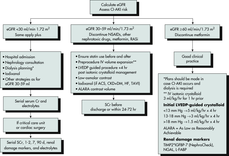
Patient-related factors include chronic kidney disease (CKD) with an estimated glomerular filtration rate (eGFR) <30 ml/min per 1.73 m2 (CKD stage 4 or greater), age >75 yr, diabetes mellitus with CKD, albuminuria, abnormal volemic states such as heart failure and volume depletion, and hemodynamic instability. Nonpatient-related factors include contrast osmolality, exposure to ionic contrast (rarely used), total contrast dose, sequential CM dosing, concomitant exposure to nephrotoxic agents, and requirement for an intraaortic balloon pump treatment. It is unclear if the intraarterial route of CM administration (e.g., cardiac catheterization) bears greater risk for AKI than IV CM administration (e.g., CT). Inpatients have a higher risk of developing CA-AKI than outpatients (Box E1).
BOX E1 Recommendations to Reduce the Risk of Contrast-Associated Acute Kidney Injury
|
Adapted from Vincent JL et al: Textbook of critical care, ed 6, Philadelphia, 2011, Saunders.
Elevation in serum creatinine generally occurs 24 to 48 h after administration of contrast. Typically, there are no accompanying symptoms or exam findings. AKI usually resolves within 10 days of radiocontrast exposure. Less than 1% of patients with CA-AKI will require dialysis, typically patients with a history of advanced kidney disease.
- Nephrotoxicity occurs from the vasoconstrictive effect of CM on large and small renal arteries and arterioles with consequent renal medullary ischemia.
- Contrast also exerts a direct cytotoxic effect on the vascular endothelium and renal tubular cells, leading to cell injury and death from reactive oxygen species.
- CM viscosity may play a role in pathogenesis. Low- and iso-osmolal radiocontrast is more viscous than high-osmolal CM.
- CM may also cause tubular injury through an effect known as “osmotic nephropathy.”