section name header

Basic Information

AUTHOR: Patricia Cristofaro, MD

Definition

Q fever is a zoonotic systemic febrile illness caused by Coxiella burnetii that may be acute or chronic. The concept of persistent localized disease may soon replace that of chronic Q fever.

Synonym

C. burnetii infection

ICD-10CM CODE
A78Q fever
Epidemiology & Demographics

  • C. burnetii is found worldwide.
  • Common animal reservoirs are cattle, sheep, and goats.
  • Pets such as cats, rabbits, and pigeons are also reservoirs.
  • A parturient cat caused an outbreak in Nova Scotia just by delivering in the same room as a card game.
  • Most cases are found in individuals who have direct contact with infected animals (e.g., farmers, veterinarians) or who are exposed to contaminated animal urine, feces, milk, or placental tissues.
  • Q fever is seen more often in men than in women (ratio of 3:1).
  • Its incidence in the U.S. is increasing, with more than 30 cases recently reported in U.S. due to military personnel recently deployed to Iraq and Afghanistan.
  • Travel to Middle Eastern and African countries poses the greatest risk.
  • C. burnetii is a CDC class B bioterrorism agent.
  • Q fever may be acquired from ingestion of unpasteurized milk or dairy products.
Physical Findings & Clinical Presentation

  • Acute Q fever presentation:
    • 1. Fever
    • 2. Pneumonia (usually mild and atypical)
    • 3. Hepatitis
    • 4. Meningoencephalitis (rare)
    • 5. Many cases are asymptomatic
  • Chronic/persistent localized Q fever presentation (duration of infection >6 mo):
    • 1. Chronic endocarditis
    • 2. Vascular infection (aneurysms and vascular prostheses)
    • 3.Bone and joint infections (culture-negative osteomyelitis or prosthetic joint infection)
  • Most common clinical symptoms:
    • 1. Chills
    • 2. Sweats
    • 3. Nausea
    • 4. Vomiting
    • 5. Cough, nonproductive
    • 6. Headache (usually severe and persistent)
    • 7. Fatigue
  • Most frequent physical findings:
    • 1. Fever
    • 2. Inspiratory rales
    • 3. Purpuric rash
    • 4. Hepatomegaly
    • 5. Splenomegaly
  • Infected pregnant women may be asymptomatic acutely, yet fetal wastage may occur
Etiology

  • Q fever is caused by the Proteobacteria C. burnetii.
  • C. burnetii is a gram-negative coccobacillus transmitted from arthropods to animals to human beings.
  • The disease is acquired most often by inhalation of aerosols. In the lungs, it proliferates in macrophages and then gains access to the bloodstream, producing a transient bacteremia. Thereafter it can invade many organs, most commonly the lungs and liver (Fig. E1).
  • There is an incubation period of 3 to 30 days before systemic symptoms manifest.
  • Persons with abnormal heart valves, prosthetic valves, and endovascular grafts are at high risk of progression to chronic Q fever. This includes individuals with bicuspid aortic valves.

Figure E1 Histology of the liver in a patient with acute Q fever.

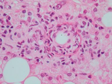
Hepatic ring granuloma of Q fever with peripheral epithelioid macrophages and lymphocytes admixed with neutrophils, characteristic central “doughnut” hole, and ring of fibrin. Note the “fatty liver” parenchyma (hematoxylin and eosin, original magnification ×400).

From Ryan ET: Hunter’s tropical medicine and emerging infectious diseases, ed 10, Philadelphia, 2020, Elsevier.

Diagnosis

Differential Diagnosis

Q fever can have various presentations and must be in the differential diagnosis of fever, hepatitis, pneumonia, endocarditis, and meningitis. Situations that should prompt serologic testing for Q fever are described in Table E1.

TABLE E1 Situations That Should Prompt Serologic Testing for Q Fever

Acute Q fever (phase II antigen and immunoglobulin [Ig]G 200 and IgM 50)
Fever in a patient in contact with ungulates
Unexplained prolonged fever (>7 days)
Granulomatous hepatitis
Fever and thrombocytopenia
Meningoencephalitis
Myocarditis
Erythema nodosum
Fever during pregnancy
Fever in a patient in contact with a parturient pet
Unexplained atypical pneumonia
Fever and an increase in transaminases (2-5 times the normal level)
Aseptic meningitis
Guillain-Barré syndrome
Pericarditis
Spontaneous abortion
Chronic Q fever (phase I antigen and IgG 800 and IgA 100)
Blood culture-negative endocarditis
Patient with a valvulopathy and unexplained fever
Weight loss
Fatigue
Increased erythrocyte sedimentation rate
Increased transaminases
Thrombocytopenia
Patient with unusually rapid degradation of a prosthetic valve
Fever in a patient with a vascular aneurysm or prosthesis
Aseptic osteomyelitis
Chronic pericarditis
Multiple spontaneous abortions

From Goldman L, Schafer AI: Goldman’s Cecil medicine, ed 24, Philadelphia, 2012, Saunders.

Workup

  • CBC, erythrocyte sedimentation rate (ESR), and liver function tests
  • Urinalysis
  • Serology
  • Chest radiography
Laboratory Tests

In acute Q fever:

  • CBC and white blood cell count are usually normal.
  • Thrombocytopenia can occur (25%).
  • Elevation of hepatic transaminases (two to three times the normal range) may be seen.
  • Antibody detection by immunofluorescence assay is the most commonly used method because of its high sensitivity and specificity. Complement fixation (CF) shows a fourfold rise in titer between acute and convalescent samples.
  • Acute Q fever (phase II antigen and IgG 200 and IgM 50).
  • Polymerase chain reaction is desirable in early disease.
  • Testing has been approved by the FDA and can be obtained from the CDC through the state health department.

In chronic Q fever (almost always endocarditis):

  • ESR is elevated.
  • Anemia is present.
  • Microscopic hematuria.
  • Blood cultures are almost always negative.
  • MIF (microimmunofluorescence) titer of >1:800 to phase I antigen is diagnostic.
Imaging Studies

  • Chest radiographs may be abnormal, showing segmental lobe consolidation.
  • Pleural effusions (35%).
  • Transthoracic echocardiography after the diagnosis of acute Q fever can be used to stratify risk of progression to endocarditis.
  • PET/CT imaging may be instrumental in determining the focus of persistent localized or chronic Q fever.

Treatment

Nonpharmacologic Therapy

Oxygen as needed in patients with pneumonia

Acute General Rx

  • Acute Q fever can be treated with doxycycline (100 mg bid) for 14 to 21 days or
  • Clarithromycin is the macrolide of choice for acute infection given intolerance of doxycycline
  • Ofloxacin 200 mg PO tid or ciprofloxacin 875 mg PO bid for 14 to 21 days
  • Hydroxychloroquine plus doxycycline for endocarditis associated with Q fever
  • Fluoroquinolones are recommended for suspected meningoencephalitis because of excellent penetration into the CNS
  • Pregnant women may be treated with trimethoprim/sulfamethoxazole until the seventh month of gestation
Chronic Rx

  • Chronic Q fever is treated with a combination of two antibiotics; doxycycline 100 mg bid and hydroxychloroquine 200 mg PO tid is the preferred therapy.
  • Duration of treatment: 2 to 3 yr, at minimum 18 mo.
  • Strongly consider prophylactic treatment for exposed individuals with preexistent high-risk cardiac lesions, pregnant women even if asymptomatic, and persons with persistently elevated anticardiolipin antibodies, lupus anticoagulant, antismooth muscle antibodies, or antimitochondrial antibodies.
Disposition

  • Patients with acute Q fever respond well to antibiotics, with deaths rarely reported.
  • Up to 20% of patients develop a post-Q fever fatigue syndrome.

Mortality rate in chronic Q fever endocarditis is high (24%). Many patients will need valve replacement surgery.

Referral

Referral to an infectious disease expert is recommended in any cases of suspected acute or chronic Q fever.

Drug levels must be monitored and serial serologies evaluated.

Reporting

Q fever has been a reportable disease in the U.S. since 1999. The CDC must be notified of cases.

Pearls & Considerations

Comments

  • An acellular (CMR) vaccine is available in the U.S. However, vaccines that are available in other countries such as Australia have been more thoroughly studied.
  • Infected patients do not require specific isolation precautions.
  • Q fever derived its name in 1935 from Derrick, who was suspicious of a new disease during a series of acute febrile illnesses in abattoir workers of Queensland, Australia, justifying the name of Q fever (for “query”).

Suggested Readings

  1. Bjork A. : First reported multi-state human Q fever outbreak in the United States, 2011Vector Borne Zoonotic Dis. ;14(2):111-117, 2014.
  2. Chieng D. : 18-FDG PET/CT scan in the diagnosis and follow-up of chronic Q-fever aortic valve endocarditisHeart Lung Circ. ;25, 2016.
  3. Eldin C. : Rainfall and sloth births in May, Q fever in July. Cayenne French GuianaAm J Trop Hyg. ;92, 2015.
  4. Eldin C. : From Q fever to Coxiella burnetii infection: a paradigm changeClin Microbiol Rev. ;30, 2017.
  5. Kampscheur L.M. : Chronic Q fever diagnosis-consensus guidelines versus expert opinionEmerg Infect Dis. ;21, 2015.
  6. Keijmel S.P. : Differentiation of acute Q fever from other infections in patients presenting to hospital, The NetherlandsEmerg Infect Dis. ;21, 2015.
  7. Keijmel S.P. : Effectiveness of long-term doxycycline treatment and cognitive behavioral therapy in fatigue severity (Q fever), a RCTCID. ;64, 2017.
  8. de Lange : Should acute Q fever patients be screened for valvulopathy to prevent endocarditis?CID. ;67, 2018.
  9. Million M. : Reevaluation of the risk of fetal death and malformation after Q feverCID. ;59, 2014.
  10. Million M. : Antiphospholipid antibody syndrome with valvular vegetations in acute Q feverCID. ;62, 2016.
  11. Million M., Raoult D. : Recent advances in the study of Q fever epidemiology, diagnosis and managementJ Infect. ;71(Suppl 1), 2015.
  12. Straily A. : Surveillance for Q fever endocarditis in the United States, 1999-2015CID. ;65, 2017.
  13. Van Roeden S.E. : Treatment of chronic Q fever: clinical efficacy and toxicity of antibiotic regimensCID. ;66, 2018.
  14. Wegdam-Blans M.C. : Chronic Q fever: review of the literature and a proposal for new diagnostic criteriaJ Infect. ;64:247-259, 2012.