section name header

Basic Information

AUTHOR: Fred F. Ferri, MD

Definition

Testicular torsion is a twisting of the spermatic cord leading to cessation of testicular blood flow, ischemia, and infarction if left untreated.

Synonym

Spermatic cord torsion

ICD-10CM CODE
N44.03Torsion of testis, unspecified
Epidemiology & Demographics
Incidence

Affects 1 in 4000 males <25 yr.

Predominant Age

Two thirds of all cases occur between the ages of 12 and 18 yr but may occur at any age, including antenatally.

Physical Findings & Clinical Presentation

  • Typical sequence is sudden onset of hemiscrotal pain, then swelling, nausea, and vomiting without fever or urinary symptoms.
  • Physical examination may reveal a tender firm testis, high-riding testis, horizontal lie of testis, absent cremasteric reflex, and no pain with elevation of testis. Absence of the cremasteric reflex (stroking or pinching the medial thigh causing contraction of cremaster muscle and elevation of testis) is the most sensitive physical finding.
  • Painless testicular swelling occurs in 10%.
  • One out of three patients reports previous episodes of spontaneously remitting scrotal pain.
  • In the neonate, testicular torsion should be presumed in patients with a painless, discolored hemiscrotal swelling.
  • In rare cases, torsion may involve an undescended testicle. In such situations an empty hemiscrotum is palpated with a tender lump in the inguinal area.
Etiology

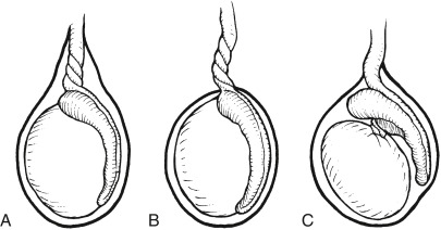
  • There are three types of testicular torsion: Extravaginal, caused by nonadherence of the tunica vaginalis to the dartos layer; intravaginal torsion, caused by malrotation of the spermatic cord with the tunica vaginalis; and torsion of the testis below the epididymis (Fig. E1). Intravaginal torsion accounts for 90% of cases.
  • Torsion usually occurs in the absence of any precipitating events. Trauma accounts for <10% of cases.

Figure E1 Types of testicular torsion.

A, Intravaginal torsion above the epididymis. B, Extravaginal torsion. C, Torsion of the testis below the epididymis.

From Rumack CM et al: Diagnostic ultrasound, ed 4, Philadelphia, 2011, Elsevier.

Diagnosis

Diagnosis made mainly by clinical suspicion (Table E1). Color Doppler ultrasound evaluation or a nuclear testicular scan (Figs. E2 and E3) may help with diagnosis. Ultrasonography shows absent or decreased blood flow; scintigraphy reveals decreased perfusion on symptomatic side.

TABLE E1 Differentiation of Testicular Torsion, Epididymitis, and Appendage Torsion

Testicular TorsionEpididymitisAppendage Torsion
Historical Features
AgePeak incidence in neonatal and adolescent groups but may occur at any agePrimarily adolescents and adults but may occur at any ageTypically prepubertal boys
Risk factorsUndescended testicle (neonate), rapid increase in testicular size (adolescent), failure of previous orchiopexySexual activity or promiscuity, GU anomalies, GU instrumentationPresence of appendages
Pain onsetSuddenGradualGradual or sudden
Previous episodes of similar painPossible (spontaneous detorsion)UnlikelyOccasional
History of traumaPossiblePossiblePossible
Nausea, vomitingMore likelyLess likelyLess likely
DysuriaLess likelyMore likelyLess likely
Physical Findings
FeverLess likelyMore likely, particularly with advanced disease (epididymo-orchitis)Less likely
Location of swelling and tendernessTesticle, progressing to diffuse hemiscrotal involvementEpididymis, progressing to diffuse hemiscrotal involvementLocalized to head of affected testicle or epididymis
Cremasteric reflexTesticular torsion less likely if presentMay be present or absentMay be present or absent
Testicle positionHigh-riding testicle, transverse alignmentNormal position, vertical alignmentNormal position, vertical alignment
PyuriaLess likelyMore likelyLess likely

GU, Genitourinary.

Including epididymo-orchitis

From Adams JG et al: Emergency medicine, clinical essentials, ed 2, Philadelphia, 2013, Elsevier.

Figure E2 Testicular torsion.

Evaluation of blood flow to the testicle has been done by giving an intravenous bolus of radioactive material. The right and left iliac vessels are clearly identified, and sequential images are obtained every 3 seconds. Here, increased flow is seen to the rim of the left testicle (arrows), and there is no blood flow centrally. This is the appearance of a testicular torsion in which the torsion has been present for more than approximately 24 hr. Ant, Anterior.

From Mettler FA [ed]: Primary care radiology, Philadelphia, 2000, Saunders.

Figure E3 Acute testicular torsion.

A to C, This 13-yr-old boy presented with acute, excruciating right-sided scrotal pain. A, Transverse gray-scale image of both testes demonstrates enlargement of the avascular right testis. B, Absence of both color and pulsed Doppler flow is noted on this sagittal image of the right testis. C, Normal pulsed and color Doppler flow confirmed in asymptomatic, normal-appearing left testis. D, Teenage boy with acute left-sided testicular torsion. Closed manual detorsion of the left testis was done at diagnosis in the ultrasound suite. Color Doppler sonogram of the left testis obtained immediately after detorsion shows dramatic hyperemia throughout the left testis. LT, Left; RT, right; SAG, sagittal; TRV, transverse.

From Rumack CM et al: Diagnostic ultrasound, ed 4, Philadelphia, 2011, Elsevier.

Differential Diagnosis

  • Torsion of the testicular appendages (appendix testis)
  • Testicular tumor
  • Epididymitis
  • Incarcerated inguinoscrotal hernia
  • Orchitis
  • Spermatocele
  • Hydrocele, varicocele
Workup

The diagnosis is usually based on history and physical examination.

Imaging Studies

  • Doppler ultrasonic stethoscope (Doppler flowmetry) (Fig. E3)
  • Radionuclide scrotal scanning (technetium-99m): Cold testicle (Fig. E2)

Treatment

Surgical derotation of the spermatic cord followed by bilateral testicular fixation with nonabsorbable sutures. If affected testis is nonviable, orchiectomy of affected testis and orchiopexy of contralateral side are performed. Attempts at manual detorsion should not delay surgical consultation.

Prognosis

  • The degree of ischemia depends on the duration of torsion and the degree of rotation of the spermatic cord.
  • There is an 80% testicular salvage rate if detorsion occurs within 12 hr of onset.
  • After 24 hr, irreversible testicular infarction is expected.
  • Because contralateral testes can be affected (immunologic process), if treatment delayed and blood flow does not return after detorsion, some recommend orchiectomy of the infarcted testicle.

Pearls & Considerations

Related Content

Testicular Torsion (Patient Information)