section name header

Basic Information

Author: Vishal I. Patel, MD and Pranav M. Patel, MD, FACC, FAHA, FSCAI

Definition

An abdominal aortic aneurysm (AAA) is a segmental full-thickness dilation of 1.5 times or greater than expected normal diameter of the abdominal aorta, defined as the fifth part of the aorta between the diaphragm and the aortic bifurcation. The average diameter of a human infrarenal aorta is approximately 2 cm, thus a threshold of 3 cm is commonly considered aneurysmal.1,2 Due to a lack of definition uniformity, the term "ectasia" has fallen out of favor and is not commonly used to describe the imaging interpretation of aortic enlargement.

ICD 10-CM CODES
I71.4Abdominal aortic aneurysm, without rupture
I71.3Abdominal aortic aneurysm, ruptured
Epidemiology & Demographics

  • Lifetime risk for AAA is 8.2% in men and 10.5% in current smokers.1
  • Approximately 13,000 deaths/yr in the U.S. are attributed to AAA.2
  • AAA is predominantly a disease of older adults and affects males four times more than females.3
  • The prevalence of AAA ranges 1.2% to 3.3% in males older than 60 yr, which is lower than had been previously reported, likely due to overall reductions in smoking. The prevalence in the U.S. specifically is unclear given that screening is rare in nonsmokers.4
  • More than half of males with subaneurysmal aorta (2.6 to 2.9 cm) develop an AAA >3.0 cm within 5 yr of their original ultrasound. 28% of male patients with a subaneurysmal aorta will develop a large AAA >5.5 cm within 15 yr.5
Natural History

  • AAAs tend to develop in the infrarenal aorta and expand at a faster rate the larger the diameter of the AAA, with each 0.5 cm increase in baseline AAA diameter increasing the rate of expansion by 0.59 mm/yr.6
  • Larger aneurysms are associated with increased rate of expansion, and thus current guidelines recommend more frequent surveillance for larger aneurysms. Frequency of surveillance for aneurysms is as follows: 3.0 to 3.9 cm every 3 yr, 4.0 to 4.9 cm every yr, 5.0 to 5.4 every 6 mo.6 Aneurysmal size is the greatest predictor of rupture.2
  • Out of hospital AAA rupture is often lethal with mortality ranging from 80% to 90%.2,4 Female sex, current smoking, and older age are associated with an increased risk of AAA rupture. Current smokers are two times more likely than former smokers to suffer from AAA rupture.4
  • Annual risk for rupture of the AAA depends on the size of the AAA2:
    1. <1% for AAA 5.4 cm or under
    2. 9.4% for AAA between 5.5 cm and 5.9 cm
    3. 10.2% for AAA between 6.0 cm to 6.9 cm
    4. 32.5% for AAA 7.0 cm or greater
  • Heavily calcified AAAs expand at a slower rate than less calcified AAAs.7 The presumed mechanism may be stabilization of the aortic wall by calcification.
Screening & Monitoring

Major societal guidelines share similar screening recommendations. These include the U.S. Preventive Services Task Force (USPSTF), the Society for Vascular Surgery (SVS), the American Heart Association (AHA), and the American College of Cardiology (ACC).

  • The USPSTF recommends one-time screening for AAA by ultrasonography in males ages 65 to 75 who have a history of smoking, and selectively offer screening to males 65 yr of age or older who have never smoked but have certain risk factors, including family history of AAA in a parent or sibling. This latter population has been shown to have a higher prevalence of AAA, and selectively screening this group has been shown to decrease AAA-specific mortality.4
  • The USPSTF and AHA/ACC have found little benefit in repeat screening in males with a negative ultrasound and has determined that males over the age of 75 are unlikely to benefit from screening. It was also concluded that the current evidence is insufficient to assess the balance of the harms and benefits of screening for AAA in females ages 65 to 75 who have ever smoked or with family history of AAA, thus screening may be reasonable in certain cases.1,4
  • The SVS recommends one-time ultrasonography screening for AAAs in males and females ages 65 to 75 yr with a history of tobacco use; in males and females 65 to 75 yr with a first-degree relative with an AAA; and in patients 75 yr or older in good health who have either a history of smoking or a first-degree relative with an AAA.8
  • The SVS also recommends monitoring by ultrasound or CT scan should be performed every 6 mo for patients with AAAs measuring 5.0 to 5.4 cm in diameter, every 12 mo for AAAs measuring 4.0 to 4.9 cm in diameter, and every 3 yr for AAAs 3.0 to 3.9 cm in diameter.8
Physical Findings & Clinical Presentation

  • Most aneurysms are asymptomatic and incidentally discovered on imaging studies.9,10
  • Physical examination has moderate sensitivity for detection of AAA. Abdominal palpitation may reveal a pulsatile mass and is less than 50% sensitive in detection of AAA. Detection is further limited in individuals with abdominal girth >100 cm. Palpation of the aneurysm does not increase the risk of rupture.10
  • Symptomatic patients may present with pain of the abdomen, back, flank, or groin or sequelae secondary to compression or mass effect of adjacent organs. Early satiety, nausea, and vomiting may be caused by compression of adjacent bowel. Venous thrombosis or insufficiency may occur from iliocaval venous compression. Thromboembolization can cause lower extremity pain and discoloration. Abdominal bruits can be present in case of renal or visceral arterial stenosis. Ureteral obstruction and hydronephrosis can cause flank and groin pain and lead to obstructive renal failure.10 Additionally, aneurysmal formation can lead to development of arteriovenous fistulas and aortoenteric fistulas, which may present as high-output heart failure and gastrointestinal blood loss, respectively.10
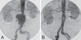
  • Approximately half of patients with AAA rupture will classically present as a triad of abdominal or back pain, hypotension, and a pulsatile abdominal mass. Ruptured aneurysms (Fig. E1) lead to rapid progression to hemorrhagic shock and require emergent surgery within hours to increase chances of survival.2,9

Figure E1 A 68-yr-old man with a ruptured abdominal aortic aneurysm.

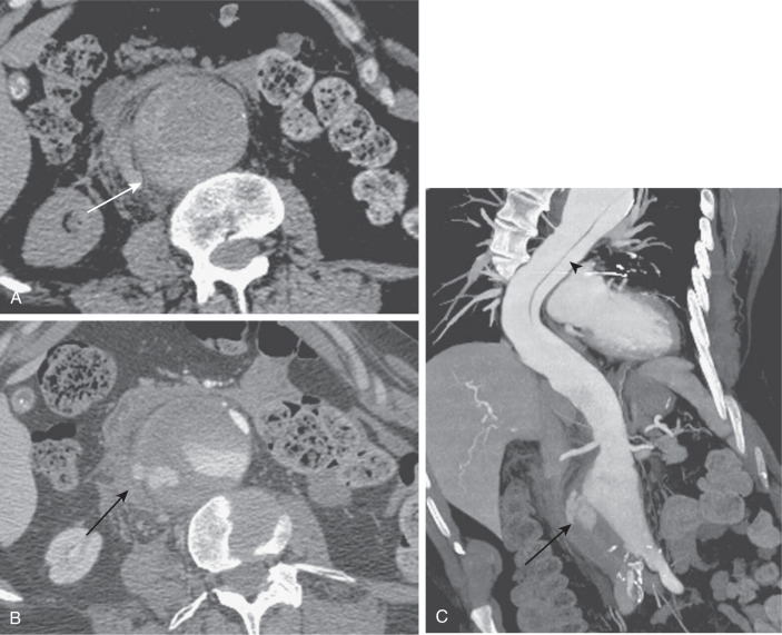
An axial unenhanced computed tomography (CT) image (A) reveals hyperattenuation within the wall of the aorta (arrow), the so-called "crescent" sign. An axial arterial phase CT image (B) reveals extravasation of contrast material beyond the confines of the aortic wall (arrow). A coronal maximum intensity projection image (C) reveals extraluminal extravasation of contrast material (arrow), as well as an incidentally noted thoracic aortic dissection (arrowhead).

(From Soto JA, Lucey BC: Emergency radiology, the requisites, ed 2, Philadelphia, 2017, Elsevier.)

Etiology

  • AAA development is characterized by medial degeneration of the aortic wall via various pathophysiologic mechanisms including smooth muscle cell apoptosis and media layer thinning, vascular inflammation from lymphocyte and macrophage infiltration, and extracellular matrix degradation.2,11 These degenerative processes deplete the normal lamellar elastin matrix leading to vascular remodeling and aneurysmal formation, expansion, and eventual rupture.11 Matrix metalloproteinases have been implicated in the development of aneurysms as well and have been tested with variable success as a therapeutic target for prevention of AAA.12
  • Most AAAs develop an intraluminal thrombus that contributes to wall degradation through oxidative stress, cell apoptosis, and inflammation.1
  • Advanced age is significantly associated with AAA. Individuals over the age of 65, compared to under age 55, are 9.4 times as likely to develop AAA.3
  • Smoking is considered one of the strongest modifiable risk factors for AAA development. A history of smoking was associated with an odds ratio of 5.07 for formation of AAA over 4 cm. Smoking was considered to be responsible for 75% of the excess prevalence of AAAs greater than 4 cm.13
  • The association of family history of AAA suggests a role of inherited connective tissue diseases such as Marfans and Ehlers-Danlos in the pathogenesis of AAA formation.3
  • Additional risk factors include hypertension, hyperlipidemia, obesity, prexisting peripheral arterial disease, cerebrovascular disease, coronary artery disease, and other aneurysmal vessels.
  • Regular consumption of fruits, vegetables, and nuts along with regular moderate intensity exercise were associated with reduced risk of AAA formation.3
  • Ethnically, Caucasian race is associated with increased risk of AAA development compared to African American, Hispanic, and Asian origins.3

Diagnosis

Differential Diagnosis

Most patients with AAA are asymptomatic, and the condition is discovered on routine examination or incidentally diagnosed when ordering studies for other symptoms.9 Diagnosis of AAA should be considered in the differential with the following symptoms: Abdominal, back, or flank pain and/or a pulsatile abdominal mass. The differential diagnosis of these symptoms can include aortic dissection, ulcerated aortic plaque, renal colic, mesenteric ischemia, pancreatitis, diverticulitis, peptic ulcer disease, biliary tract disease, and others.

Laboratory Tests

In general, laboratory studies are not routinely indicated. For suspected infected or inflammatory aneurysms, white blood cell (WBC), erythrocyte sedimentation rate (ESR), C-reactive protein (CRP), and blood cultures can be considered. An elevated d-dimer may indicate a thrombus within the aneurysm. Fig. 2 describes an algorithm for the diagnosis and treatment of abdominal aortic aneurysms.

Figure 2 Algorithm for the diagnosis and treatment of abdominal aortic aneurysms (AAAs).

!!flowchart!!

BP, Blood pressure; CT, computed tomography; MRI, magnetic resonance imaging; NS, normal saline; PRBCs, packed red blood cells; SBP, systolic blood pressure; US, ultrasonography.

(From Adams JG et al: Emergency medicine, clinical essentials, ed 2, Philadelphia, 2013, Elsevier.)

Imaging Studies

  • Abdominal ultrasound (Fig. 3) is 94% to 100% sensitive and 98% to 100% specific in identifying an aneurysm. Ultrasound is readily available, noninvasive, and accurate. Increasing application in emergency settings as point-of-care imaging has led to a significant reduction in time to diagnosis and treatment of AAA.4,8
  • Computed tomography (CT) (Fig. 4) scan is recommended for preoperative aneurysm imaging due to reliability of size estimation of AAA within 0.2 mm.3 CT scan can identify extension to renal vessels with more precision than ultrasound. It is the imaging modality of choice for symptomatic AAA and can also detect the integrity of the wall (Fig. 5) and exclude rupture.10
  • Magnetic resonance angiography (MRA) may also be used and is more accurate than CT; however, it has a longer study acquisition time and may not be readily available.3
  • Plain radiographs may show the outline of an aneurysm in calcified aortas. This is an insensitive test for diagnosing AAA and is not routinely a first-line test.
  • Diagnostic aortography has essentially been replaced by other noninvasive imaging modalities such as CT or MRA. Intraoperative angiography is still used for determining treatment options and postprocedure efficacy (Fig. 6).
  • Endovascular aneurysm repair (EVAR) needs close and lifelong imaging surveillance of the aneurysm site for the timely detection of possible complications, including endoleaks, graft migration, fractures, graft infection, and enlargement of aneurysmal sac size with eventual rupture.8 Contrast-enhanced computed tomography (CTA) is considered the gold standard in EVAR follow-up (Fig. 7); however, routine use is limited given cumulative radiation exposure and risk of contrast-induced renal injury. Guidelines suggest serial ultrasound surveillance if neither enoleak nor AAA enlargement is seen on CTA at 1 yr after EVAR.8

Figure 7 Digital subtraction angiogram following endovascular aneurysm repair.

(From Fillit HM: Brocklehurst’s textbook of geriatric medicine and gerontology, ed 8, Philadelphia, 2017, Elsevier.)

Figure 6 A, Conventional Catheter Angiography with Bilateral Marked Catheters in Place Demonstrates a Large, Lobulated, Infrarenal Aortic Aneurysm (Arrowhead) with a 4-cm Proximal Neck Suitable for Endovascular Repair

B, An Image after Endovascular Repair Demonstrates Complete Exclusion of the Aneurysm (Arrowhead) with No Endoleak and Preservation of the Renal and Hypogastric Arteries.

(From Soto JA, Lucey BC: Emergency radiology, the requisites, ed 2, Philadelphia, 2017, Elsevier.)

Figure 4 Three-Dimensional Computed Tomography Image Illustrates the Presence of an Infrarenal Abdominal Aortic Aneurysm

An, Aneurysm; CIA, common iliac artery; EIA, external iliac artery; IIA, internal iliac artery; IN, infrarenal neck; LK, left kidney; RA, renal artery; RK, right kidney.

(From Townsend CM et al [eds]: Sabiston textbook of surgery, ed 17, Philadelphia, 2004, Saunders.)

Figure 5 Aneurysm of the Abdominal Aorta

A Large Aortic Aneurysm is Evident. The Aorta Exceeds 5 cm in Diameter. A Large Amount of Thrombus (T) Partially Surrounds the Contrast-Enhanced Patent Lumen (L).

Note the atherosclerotic calcification (arrowhead) in the wall of the aneurysm.

Figure 3 Transverse image of an abdominal aortic aneurysm.

Note the measurements of 3.33 × 3.85 cm. The inferior vena cava is seen to the patient’s right of the aorta, and the vertebral body is seen below the two vessels. Note also that there appears to be an echogenic flap within the aorta, possibly representing an aortic dissection.

(From Adams JG et al: Emergency medicine, clinical essentials, ed 2, Philadelphia, 2013, Elsevier.)

Treatment

Medical Management

  • The primary goal of medical therapy is to reduce growth rates, subsequently reducing the need for aortic repair and aortic-related mortality. The secondary goal is to decrease risk of nonaortic cardiovascular events given shared risk factors between aortopathy and atherosclerotic disease.1
  • Smoking cessation is critical for treatment of patients with AAAs and can decrease rate of expansion. Patients with known AAA or a family history of aneurysms should be advised to stop smoking and be offered smoking cessation interventions.13
  • Maintaining healthy blood pressure with beta-blockers or angiotensin-receptor blockers mitigates proteolysis pathways and reduces shear stress on the aortic wall.1
  • Use of lipid-lowering therapies such as HMG-CoA reductase inhibitors (statins) helps to target inflammatory pathways, reducing medial degeneration.1
  • Healthy diet and exercise have been observed to be associated with reduced risk of AAA via shared multifactorial mechanisms. Of note, moderate exercise does not increase the rate of aneurysm expansion or the risk of rupture.3
  • Definitive treatment depends on the size of the aneurysm (see "Chronic Therapy").
Acute Therapy

  • AAA can be treated with open surgical repair (OSR) or EVAR (Fig. E8). EVAR is recommended in patients with suitable anatomy given reduced short-term morbidity compared to OSR.8,14,15 Elective, symptomatic, or ruptured AAA repair can be performed via endovascular or open techniques.8,10 Ruptured AAA requires emergent repair, and symptomatic AAA requires urgent repair.8
  • Historically, emergent open repair had been the standard of care; however, trials now show no significant difference in 30-day mortality compared to EVAR.14 There was a higher incidence of reintervention for patients undergoing EVAR, although interventions to deal with procedural complications were generally less invasive and involved catheter-based approaches.
  • The major limitations for EVAR include anatomic issues such as tortuosity or small caliber iliac arteries prohibiting graft deployment, end-organ ischemia from endograft limb occlusion, endoleak from inadequate fixation or sealing of the graft to the vessel wall, and inability to follow up patients to exclude late failure of stent-grafts and development of endoleaks.

Figure E8 Endovascular Repair (Ipsilateral and Contralateral) of Abdominal Aortic Aneurysm with a Modular Endograft

(From Hornacky A, Phillips N: Vascular surgery. In Hornacky A, Phillips N [eds]: Berry & Kohn’s operating room technique, ed 14, Philadelphia, 2021, Elsevier.)

Chronic Therapy

  • Optimizing blood pressure and lipid profile is recommended for patients with hypertensive and atherosclerotic disease. AAA formation has often been recognized as a distinct degenerative process from atherosclerosis, but emerging data suggest some shared pathophysiology. Outcomes data from clinical trials of medical therapy are limited, thus conflicting guidelines regarding medical therapy still persist.1,2,8
  • Data are also sparse as to the protective effects of antibiotics such as doxycycline and roxithromycin to limit the expansion of small AAAs.8,18
  • AAA repair to eliminate the risk for rupture should be performed for patients with infrarenal or juxtarenal AAA of approximately 5.5 cm or larger in diameter. Repair in females can be considered at diameters larger than 5 cm per Society of Vascular Surgery guidelines. Additionally, AAAs with a rate of enlargement greater than 0.5 cm over 6 mo should be considered for repair. All patients who are symptomatic should undergo repair, regardless of size.8
  • Smoking cessation is recommended for at least 2 wk before surgery.8 It is reasonable to delay elective AAA repair in an effort to optimize comorbid medical conditions. Preoperative nutritional status should be optimized prior to undergoing repair. Intravenous antibiotics with a first-generation cephalosporin should be administered 30 min to 1 h prior to either OSR or EVAR.8
  • There is no clear advantage to early repair (open or endovascular) for small asymptomatic AAAs. Meta-analysis has demonstrated no significant difference in all-cause mortality or AAA-related mortality with early treatment (EVAR vs. OSR) compared with surveillance for small AAAs.19
  • Historically, EVAR was thought to be associated with better short-term outcomes than OSR (lower 30-day mortality and myocardial infarction [MI] rates, shorter hospital stays, and better health-related quality of life up to 12 mo postoperatively) but increased rates of graft-related complications and long-term all-cause mortality. However, there is conflicting evidence about the long-term relative benefits of OSR, with some recent trials demonstrating no significant difference in long-term mortality.8,10,
  • In patients who have undergone EVAR, long-term surveillance is required to assess for an endoleak, stent migration, change in aneurysm size, and need for reintervention.8 Surveillance for endovascular AAA repair has typically involved use of periodic CT scans, but abdominal ultrasound is gaining widespread adoption for postprocedure monitoring. Surveillance is recommended to occur at least 1 mo and 12 mo postoperatively, followed by annually thereafter.8
  • EVAR with proximally fenestrated grafts (FEVAR) is an alternative to open repair in the management of juxtarenal aortic aneurysms and short-neck abdominal aortic aneurysms, with the "neck" defined as the distance from the lowest main renal artery to the beginning of the aneurysm. Contemporary literature shows it is a safe and efficacious treatment, particularly for those deemed surgically high risk.15
  • For patients with limited life expectancy, elective AAA repair is not recommended.6
Referral

  • Vascular surgical referral is recommended at the time of diagnosis of AAA.8
  • It is important to optimize any comorbid conditions along with surgical referral, including cardiology and endocrinology referrals as indicated.8

Pearls & Considerations

Comments

  • Most AAAs are infrarenal. This is thought to be due in part to decreased lamellar structural proteins in the vascular wall below the renal arteries leading to decreased vascular wall strength.2,11
  • Surgical risk is increased in patients with coexisting coronary artery disease, pulmonary disease, or chronic renal failure. Evaluation for ischemia and aggressive perioperative hemodynamic monitoring can help identify high-risk patients to help decrease postoperative complications.8
  • AAAs expand at a faster rate the larger the diameter of the AAA, with each 0.5 cm increase in baseline AAA diameter increasing the rate of expansion by 0.59 mm/yr.6
Related Content

  • Abdominal Aortic Aneurysm (Patient Information)

Related Content

    1. Isselbacher E : 2022 ACC/AHA guideline for the diagnosis and management of aortic diseasehttps://doi.org/10.1016/j.jacc.2022.08.004J Am Coll Cardiol. 80(24):e223-393, 2022.
    2. Kent KC : Clinical practice. Abdominal aortic aneurysmshttp://dx.doi.org/10.1056/NEJMcp1401430N Engl J Med. 371(22):2101-2108, 2014.
    3. Kent KC : Analysis of risk factors for abdominal aortic aneurysm in a cohort of more than 3 million individualshttp://dx.doi.org/10.1016/j.jvs.2010.05.090J Vasc Surg. 52(3):539-548, 2010.
    4. Guirguis-Blake JM : Primary care screening for abdominal aortic aneurysm: a systematic review for the U.S. Preventive Services Task Force. Agency for Healthcare Research and Quality (US), January 2014.
    5. Oliver-Williams C : Lessons learned about prevalence and growth rates of abdominal aortic aneurysms from a 25-year ultrasound population screening programmehttp://dx.doi.org/10.1002/bjs.10715Br J Surg. 105(1):68-74, 2018.
    6. Bown MJ : Surveillance intervals for small abdominal aortic aneurysms: a meta-analysishttp://dx.doi.org/10.1001/jama.2013.950J Am Med Assoc. 309(8):806-813, 2013.
    7. Klopf J : The prognostic impact of vascular calcification on abdominal aortic aneurysm progressionhttp://dx.doi.org/10.1016/j.jvs.2021.11.062J Vasc Surg. 75(6):1926, 2022.
    8. Chaikof EL : The Society for Vascular Surgery practice guidelines on the care of patients with an abdominal aortic aneurysmhttps://doi.org/10.1016/j.jvs.2017.10.044J Vasc Surg. 67(1):2-77.e2, 2018.
    9. Sakalihasan N : Abdominal aortic aneurysmshttps://doi.org/10.1038/s41572-018-0030-7Nat Rev Dis Primers. 4(1):34, 2018.
    10. Wanhainen A : Editor’s choice - European Society for Vascular Surgery (ESVS) 2019 clinical practice guidelines on the management of abdominal aorto-iliac artery aneurysmshttp://dx.doi.org/10.1016/j.ejvs.2018.09.020Eur J Vasc Endovasc Surg. 57(1):8-93, 2019.
    11. Raffort J : Monocytes and macrophages in abdominal aortic aneurysmhttp://dx.doi.org/10.1038/nrcardio.2017.52Nat Rev Cardiol. 14:457-471, 2017.
    12. Baxter BT : Effect of doxycycline on aneurysm growth among patients with small infrarenal abdominal aortic aneurysms: a randomized clinical trialhttp://dx.doi.org/10.1001/jama.2020.5230J Am Med Assoc. 323(20):2029-2038, 2020.
    13. Norman PE, Curci JA : Understanding the effects of tobacco smoke on the pathogenesis of aortic aneurysmhttp://dx.doi.org/10.1161/ATVBAHA.112.300158Arterioscler Thromb Vasc Biol. 33(7):1473-1477, 2013.
    14. Badger S : Endovascular treatment for ruptured abdominal aortic aneurysmhttp://dx.doi.org/10.1002/14651858.CD005261.pub4Cochrane Database Syst Rev. 5:CD005261, 2017.
    15. Jones AD : Meta-analysis of fenestrated endovascular aneurysm repair versus open surgical repair of juxtarenal abdominal aortic aneurysms over the last 10 yearshttp://dx.doi.org/10.1002/bjs5.50178BJS Open. 3(5):572-584, 2019.
    16. Chen ZG : The long-term outcomes of open and endovascular repair for abdominal aortic aneurysm: a meta-analysishttp://dx.doi.org/10.1016/j.asjsur.2019.01.014Asian J Surg. 42(10):899-906, 2019.
    17. Lederle FA : Open versus endovascular repair of abdominal aortic aneurysmhttp://dx.doi.org/10.1056/NEJMoa1715955N Engl J Med. 380(22):2126-2135, 2019.
    18. Rughani G : Medical treatment for small abdominal aortic aneurysmshttp://dx.doi.org/10.1002/14651858.CD009536.pub2Cochrane Database Syst Rev. 9:CD009536, 2012.
    19. Ulug P : Surgery for small asymptomatic abdominal aortic aneurysmshttp://dx.doi.org/10.1002/14651858.CD001835.pub5Cochrane Database Syst Rev. 7:CD001835, 2020.