section name header

Basic Information

Author: Tanvi Rana, MD and Nima R. Patel, MD, MS

Definition

Abnormal uterine bleeding (AUB) is a broad term for variations in normal menses. Normal duration of menstrual flow is 5 days, with normal menstrual cycles lasting 21 to 35 days. Heavy menstrual bleeding with normal cycles, historically called menorrhagia, is a large volume of menstrual blood loss quantified as >80 ml per cycle. Bleeding also may be prolonged, intermenstrual, frequent, and irregular. This language replaces prior terms such as metrorrhagia (bleeding between cycles); polymenorrhea (menses <21 days apart); and oligomenorrhea (menses >35 days apart).

The PALM-COEIN classification of AUB was adopted in 2011 to standardize terminology and reflect etiology: Polyp, Adenomyosis, Leiomyoma, Malignancy and hyperplasia, Coagulopathy, Ovulatory dysfunction, Endometrial, Iatrogenic, and Not yet classified. The PALM-COEIN terminology is used to classify the etiology of the bleeding; for instance, AUB-P would refer to AUB due to polyps.

Synonyms

  • Menorrhagia
  • Menometrorrhagia
  • Dysfunctional uterine bleeding
  • Irregular menstrual cycle
ICD 10-CM CODES
N92.0Excessive and frequent menstruation with regular cycle
N92.1Excessive and frequent menstruation with irregular cycle
N92.2Excessive menstruation at puberty
N92.4Excessive bleeding in the premenopausal period
N92.6Irregular menstruation, unspecified
N93.9Abnormal uterine and vaginal bleeding, unspecified
Epidemiology & Demographics
Incidence:

AUB occurs in 10% to 15% of reproductive-age patients, 5% of emergency room visits in nonpregnant patients, 30% of office visits, and 70% of gynecologic consults.2,3

Prevalence:

9% to 14%1

Predominant Sex & Age:

Females ages 13 to 50, with peak in adolescence and perimenopausal periods.1

Risk Factors:

Genetic predisposition, anticoagulation treatment, obesity, endocrinopathies, autoimmune disease, liver disease, renal disease, and sex hormone secreting tumors.

Genetics:

Hereditary coagulopathy (most commonly von Willebrand disease, a platelet dysfunction disorder) can be seen in 20% of women with heavy menstrual bleeding and can present in adolescents with prior undiagnosed coagulopathy. This can lead to hospitalization in up to 19% of affected women.1,3

Physical Findings & Clinical Presentation
Pertinent History:

Age, age at menarche or menopause, menstrual bleeding patterns, severity of bleeding, pain, hirsutism, acne, symptoms of thyroid dysfunction or other endocrinopathies, underlying medical conditions, surgical history, medications, and family history.

  • If heavy bleeding since menarche, screen for signs and symptoms of an underlying coagulopathy, such as postpartum hemorrhage, surgery-related bleeding, bleeding from dental work, easy bruising, epistaxis, and frequent gum bleeding1,2
Physical Examination:

Weight, hirsutism, acne, thyroid nodules, signs of insulin resistance (acanthosis nigricans), signs of coagulopathy (petechiae, ecchymoses, pallor, swollen joints), pelvic examination (including external, speculum, and bimanual examinations)1,2

Etiology

  • PALM-COEIN
  • Additional etiologies: Pregnancy/miscarriage; atrioventricular malformation (AVM); cervical/endometrial infections; foreign body, such as intrauterine device (IUD) malposition; use of hormones, including oral contraceptive pills (OCPs) and long-acting reversible contraceptives (LARCs); anticoagulants; severe kidney or liver disease; thyroid disorders
Diagnosis

Differential Diagnosis

In addition to the PALM-COEIN classification, it is also important to consider etiologies listed earlier as well as urinary or GI tract sources.

Differential diagnosis of AUB can be further narrowed by age.

  • Ages 13 to 18: Most commonly caused by anovulation due to immaturity or dysregulation of the hypothalamic-pituitary-ovarian axis; this also can be when coagulopathies are first identified
  • Ages 19 to 39: Most commonly caused by pregnancy or structural issues such as polyps and fibroids; cancer is relatively rare in this age range
  • Ages 40+: Most commonly caused by ovulatory changes with perimenopause, although cancer has an increased incidence in this age range
Workup

  • History and physical examination
  • Laboratory, pathology, and imaging studies
Laboratory Tests

  • Pregnancy test
  • Complete blood count (CBC) ± iron studies if anemia suspected
  • Thyroid-stimulating hormone (TSH)
  • Prolactin level
  • Targeted screening for coagulopathies if suspected
  • Pap smear if indicated
  • Endometrial sampling by endometrial biopsy or hysteroscopic sampling if >45 yr OR <45 yr with history of unopposed estrogen (polycystic ovary syndrome, obesity), failed medical management, or persistent abnormal bleeding
Imaging Studies

  • Pelvic ultrasound. Transvaginal provides better assessment than transabdominal and is generally preferred.
  • Sonohysterography or hysteroscopy if ultrasound is not adequate or if further evaluation of the cavity is required, especially to assess for intracavitary lesions; superior to ultrasonography in detection of intracavitary lesions.
  • MRI if needed for surgical planning or further evaluation of structural abnormality.2

Treatment

Acute General Rx

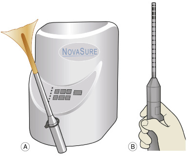
First-line therapy for acute AUB without known coagulopathy is hormonal management, such as oral progestins, high-dose combined OCPs, intravenous (IV) estrogen, and tranexamic acid. Blood transfusion as indicated. Surgical management includes dilation and curettage, endometrial ablation (Fig. E1), uterine artery embolization, or hysterectomy (Table E1). A comparison of effective reduction of blood loss with each treatment modality is summarized in Table E2.

TABLE E2 Reduction of Menstrual Blood Loss with Treatment

Agent UsedDecrease in Menstrual Blood Loss (%)
EACA47
AMCA44-54
NSAID21-50
Oral contraceptives52
Levonorgestrel IUD at 3, 6, and 12 mo82, 88, 96
Endometrial ablationUp to 100; 68%-78% of patients achieve normal menses
Hysterectomy100

AMCA, Tranexamic acid; EACA,Ε-aminocaproic acid; IUD, intrauterine device; NSAID, nonsteroidal antiinflammatory drug.

From Gershenson DM et al: Comprehensive gynecology, ed 8, Philadelphia, 2022, Elsevier.

TABLE E1 Pros and Cons of Different Hysterectomy Methods

ProsCons
Total abdominal hysterectomy (TAH)
  • Cervix is removed; therefore no further need for smears and no further risk of cervical malignancy (thus particularly suitable for those with a history of abnormal cytology)
  • Good access to ovaries
Increased surgical morbidity
Subtotal abdominal hysterectomy
  • Fewer complications than TAH ( bleeding, infection, bladder injury, ureteric damage)
  • Good access to ovaries
Risk of cervical cancer remains as before
Vaginal hysterectomy
  • May be lower incidence of bladder and bowel injury in straightforward cases (compared with abdominal hysterectomy)
  • No painful abdominal wound
  • Limited ovarian access
  • Contraindicated with:
  • Large uterus
  • Restricted uterine mobility
  • Limited vaginal space
  • Adnexal pathology
  • Cervix flush with vagina

From Magowan BA: Clinical obstetrics and gynecology, ed 4, Philadelphia, 2019, Elsevier.

Figure E1 Conservative surgical treatments for menorrhagia.

A, impedance-controlled ablation; B, microwave endometrial ablation.

(From Magowan BA: Clinical obstetrics and gynecology, ed 4, Philadelphia, 2019, Elsevier.)

Chronic Rx

Several long-term medical management options exist, some of which can be used together as part of a multimodal regimen.

  • Nonsteroidal antiinflammatory drugs
  • Combined hormonal contraceptives (pill, transdermal patch, vaginal ring) in a cyclic or continuous regimen
  • Progesterone IUD
  • Oral progesterone
  • Tranexamic acid
  • Gonadotropin-releasing hormone (GnRH) analogue

Elagolix (GnRH agonist) was FDA approved in 2020 for heavy premenstrual bleeding associated with uterine fibroids; currently not recommended for use >24 mo due to concern for bone density loss.5

Surgical Management

Need for surgical intervention depends on stability of patient, bleeding severity, contraindication to medical management, or lack of response to medical management.

  • Dilation and curettage
  • Hysteroscopic resection of uterine pathology including endometrial polyps and submucosal leiomyomas
  • Endometrial ablation
  • Uterine artery embolization
  • Myomectomy
  • Hysterectomy
Referral

  • Refer to gynecologist if:
    1. Unresponsive to initial hormonal management.
    2. Concern for a structural etiology or malignancy.
    3. Endometrial sampling reveals endometrial hyperplasia or malignancy. Resampling is necessary with progestin therapy for hyperplasia. If endometrial hyperplasia associated with complex glands or atypia, consultation with gynecologist is warranted due to high degree of progression to malignancy.
Related Content

Related Content

  1. Bradley LD, Gueye NA : The medical management of abnormal uterine bleeding in reproductive aged womenAm J Obstet Gynecol. 214(1):31-44, 2016.
  2. Practice bulletin no. 128: diagnosis of abnormal uterine bleeding in reproductive-aged womenReaffirmed 2021. https//doi.10.1097/AOG.0b013e318262e320Obstet Gynecol. 120(1):197-206, 2012.
  3. Practice bulletin no. 136: management of abnormal uterine bleeding associated with ovulatory dysfunctionReaffirmed 2022. https://doi.10.1097/01.AOG.0000431815.52679.bbObstet Gynecol. 122:176-185, 2013.
  4. ACOG committee opinion no: 557: management of acute abnormal uterine bleeding in nonpregnant reproductive-aged womenReaffirmed 2020. https://doi.10.1097/01.AOG.0000428646.67925.9aObstet Gynecol. 121(4):891-896, 2013.
  5. Carr B : Elagolix alone or with add-back therapy in women with heavy menstrual bleeding and uterine leiomyomashttps://doi:10.1097/AOG.0000000000002933.Obstet Gynecol. 132(5):1252-1264, 2018.