section name header

Basic Information

Author: Ajay S. Koti, MD and Emily C.B. Brown, MD, MS

Definition

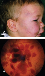
Abusive head trauma (AHT) refers to a distinct form of traumatic brain injury that occurs as a consequence of inflicted, or nonaccidental, injury to a child, typically under the age of 4 yr. Formerly known as "shaken baby syndrome," AHT arises from shaking, blunt impact, or a combination thereof.1 The injury pattern may not clearly differentiate between these mechanisms. AHT is a life-threatening condition, and survivors commonly suffer long-term sequelae. The constellation of findings most often includes subdural hemorrhage, and extensive retinal hemorrhages and/or encephalopathy frequently accompany the intracranial findings. Children with AHT may present with or without evidence of additional cutaneous, musculoskeletal, or visceral injuries (Fig. E1). AHT is often challenging to diagnose because the clinical manifestations may be vague or nonspecific, caregivers may not report a history of trauma, and there may be no external sign of injury.

Figure E1 Abusive head trauma.

A, Facial bruising. B, Fundus hemorrhages involving different levels.

(Courtesy R. Bates. From Kanski JJ, Bowling B: Clinical ophthalmology, a systematic approach, ed 7, Philadelphia, 2010, Saunders.)

Synonyms

  • Shaken baby syndrome
  • Infant whiplash syndrome
  • Inflicted pediatric neurotrauma
  • Nonaccidental head injury
  • AHT
ICD-10CM CODES
T74.4Shaken infant syndrome
T74.12Child physical abuse, confirmed
T76.12Child physical abuse, suspected
Epidemiology & Demographics
Incidence:

Incidence is estimated at 32 to 38 cases per 100,000 children under 1 yr of age.1

Prevalence:

AHT is the most common cause of fatal head injuries in children under 2 yr old, accounting for 53% of serious or fatal traumatic injuries. It is the leading cause of abusive fatalities overall.2 Morbidity and mortality are higher for victims of AHT than for children injured accidentally. Approximately one in four cases are fatal.1 Long-term neurologic sequelae are common, occurring in 70% of survivors. These include visual deficits, neurocognitive problems, seizure disorders, cerebral palsy, and intellectual disability.1

Predominant Sex:

Males are found to be at higher risk for AHT than females, by as much as a 2-to-1 margin.

Peak Incidence:

Victims of AHT are typically younger than 1 yr of age, with a median age of 4 mo. The peak incidence of fatal AHT occurs even earlier, at 1 to 2 mo, which overlaps with the peak of infant crying; this has often been reported as a trigger of abuse.2 Infants are particularly susceptible to injuries from shaking and/or impact due to their unique body proportions, neural immaturity, and neuroanatomy.3

Risk Factors:

Risk factors for AHT include prematurity, perinatal illness, multiple gestations, male sex, and excessive crying. Other psychosocial stressors, such as parental substance use or mental illness, lack of access to child care, and poverty, have been associated with AHT on a population level. Those who inflict AHT are more often males.4,5 It is important to note, however, that AHT is a medical diagnosis that affects infants of every socioeconomic circumstance and caregiving environment, and a diagnosis of abuse should not be influenced by psychosocial risk factors.

Physical Findings & Clinical Presentation

Infants with AHT often present with nonspecific symptoms (e.g., drowsiness, lethargy, irritability, vomiting, seizures, irregular respirations, or apnea) and either no history of trauma or an inconsistent report of a low-height fall.3 Symptoms can range in severity, and it is not unusual for infants to present with an unexplained coma or arrest. Examination findings include bruising, intraoral injuries such as a torn frenulum, macrocephaly, and a bulging fontanelle, but many infants have no external signs of trauma. Subdural hemorrhage is the most common radiologic finding in AHT, and indeed, AHT is one of the most common causes of subdural hemorrhage in infants. Other neuroimaging findings include cerebral edema, loss of gray-white differentiation, parenchymal injury, torn/thrombosed bridging veins, cervical spine ligamentous injuries, and spinal subdural hematoma.2 Fractures, such as posterior rib fractures and classic metaphyseal lesions, may be detected on skeletal survey, and dilated retinal examination may reveal retinal hemorrhages. Often these are bilateral, too numerous to count, in multiple layers, and extending to the ora serrata (retinal periphery), though a fraction of patients will have no retinal hemorrhages at all. Some will have retinoschisis. Delayed diagnoses are common, and AHT is missed by medical providers approximately 30% of the time on initial presentation.6 This often leads to recurrent and escalating injuries to the child until the abuse is recognized.7 Physicians often fail to detect AHT among children of intact, white families, likely reflecting bias in the suspicion of abuse.6 It should be noted that the diagnosis of abuse is based on a recognizable constellation of clinical findings and not the family’s psychosocial circumstances.

Etiology

AHT is thought to occur from rotational acceleration-deceleration forces, such as those generated in shaking; however, blunt impact trauma can cause overlapping findings. These forces are typically not concordant with typical infant caregiving or minor accidental trauma. Subdural hemorrhages arise from bridging vein trauma. Children with more severe presentations may have traumatic axonal injury. Vitreoretinal traction accounts for retinal hemorrhages, which may occur with retinoschisis. Victims of AHT will frequently experience multiple episodes of trauma, highlighting the importance of diagnosis to prevent further harm.

Diagnosis

Differential Diagnosis

Accidental head trauma, severe infection, bleeding disorder, metabolic disorder, brief resolved unexplained event (BRUE), birth trauma

Workup

Detailed history and physical; occipital-frontal circumference; noncontrast head CT; delayed brain and spine MRI; dilated eye examination by an ophthalmologist; laboratory testing and imaging to evaluate for additional injuries and alternative medical diagnoses; social work assessment. Although cranial ultrasound can identify large chronic subdural collections in infants with macrocrania, it is poorly sensitive for most subdural hemorrhages and it does not replace the need for more advanced imaging with CT or MRI.

Laboratory Tests

  • CBC, prothrombin time, partial thromboplastin time, factors VIII and IX (and consider factor XIII), d-dimer, fibrinogen, electrolytes, blood urea nitrogen, creatinine, aspartate aminotransferase, alanine transaminase, lipase
  • For patients with fractures, evaluate bone health: Calcium, phosphorus, alkaline phosphatase, parathyroid hormone, and 25-OH vitamin D
  • If normal newborn screen cannot be confirmed or there is concern for frontal atrophy on imaging: Consider testing for glutaric aciduria
Imaging Studies

Head computed tomography (CT) for patients with acute symptoms, skeletal survey, and MRI of the brain and spine (ideally obtained on day 3 to 5 after presentation). If concern for intraabdominal injury, consider chest/abdomen/pelvis CT. Repeat skeletal survey 2 to 3 wk later to evaluate for healing fractures that may not have been evident on the initial study.

Treatment

Treatment is centered on the care of the patient’s specific injuries and on the prevention of future injuries.

Nonpharmacologic Therapy

Admission to a hospital for multidisciplinary evaluation and determination of safe disposition

Acute General Rx

Supportive care and interventions as dictated by the clinical, laboratory, and imaging findings. Many infants require intensive care unit admission for neurologic monitoring.

Chronic Rx

Consistent primary care with developmental surveillance, social work involvement, parenting services, and additional referrals as clinically indicated.

Referral

Child abuse pediatrics, ophthalmology, and (as indicated) the following: Neurology, neurosurgery, physical medicine and rehabilitation, gastroenterology, nutrition, and developmental services (i.e., early intervention services).

Health care providers in all states are mandatory reporters of suspected abuse. All cases of suspected AHT require reporting to child protective services for investigation, and most will also involve law enforcement. Other children in the home should be referred for medical evaluation. Abuse is frequently a repetitive phenomenon, and involvement of child protective services (CPS) is important to ensure the child’s safety.7

Pearls & Considerations

Comments

Legal proceedings have given rise to several alternate theories that question the diagnosis and biomechanical mechanisms of AHT, but these theories are not supported by the medical literature.2 It is important to note that the role of the medical team is to objectively establish a diagnosis, not to investigate or assign responsibility for the child’s injuries.

Prevention

Several prevention programs focus on educating new parents about the risk of shaking a baby. Some are focused on younger men prior to the peak ages of child-rearing. Advocacy for public policy to mitigate family stress may also prevent AHT and other forms of maltreatment. These include food assistance, expanded access to health services, paid family leave, and affordable child care.1

Patient & Family Education

The Period of Purple Crying, which seeks to help parents and other caregivers understand normal crying in young infants and the risks of AHT, is an education program that has been implemented in hospitals across the U.S. and internationally. Similar programs include Take 5, All Babies Cry, and Calm Baby Gently. Public health nurses may be engaged for more longitudinal education in the community setting. Historically, the efficacy of these interventions has yielded mixed results.8,9

Related Content

  1. Narang SK : Council on Child Abuse and Neglect: abusive head trauma in infants and childrenPediatrics. 145(4):e20200203, 2020.
  2. Choudhary AK : Consensus statement on abusive head trauma in infants and young childrenPediatr Radiol. 48(8):1048-1065, 2018.
  3. Boos SC, Dias MS : Abusive head trauma Laskey A, Sirotnak A, editors : Child abuse: medical diagnosis and management. ed 4American Academy of Pediatrics-Itasca:199-284, 2020.
  4. Keenan HT, Jenny C : Epidemiology of abusive head traumaChild abuse and neglect: diagnosis, treatment, and evidence. Elsevier-St Louis:35-38, 2011.
  5. Rorke-Adams L : Head trauma Reece RM, Christian CW, editors : Child abuse: medical diagnosis and management. American Academy of Pediatrics-Itasca:53-119, 2009.
  6. Jenny C : Analysis of missed cases of abusive head traumaJ Am Med Assoc. 281(7):621-626, 1999.
  7. Hymel KP, Deye KP : Abusive head trauma Jenny C, editors : Child abuse and neglect: diagnosis, treatment, and evidence. Elsevier-St Louis:349-358, 2011.
  8. Spector L : Evidence-based child abuse and neglect prevention programs Laskey A, Sirotnak A, editors : Child abuse: medical diagnosis and management. ed 4American Academy of Pediatrics-Itasca:1111-1128, 2020.
  9. Wood JN : Challenges in prevention of abusive head traumaJAMA Pediatr. 169(12):1093-1094, 2015.