section name header

Basic Information

Author: Bethany K. Sederdahl, MPH, MD and Philip A. Shlossman, MD

Definition

Hypertensive disorders of pregnancy represent a spectrum of disease ranging from gestational hypertension to severe preeclampsia. Preeclampsia is defined as new-onset hypertension (systolic blood pressure 140 or diastolic 90) after 20 wk gestation or up to 6 wk postpartum in the presence of proteinuria or evidence of end-organ injury.1 In the absence of proteinuria, other criteria, including thrombocytopenia, acute kidney injury, impaired liver function, pulmonary edema, and new-onset neurologic symptoms, can be used for diagnosis. In 2013, the American College of Obstetricians and Gynecologists (ACOG) Task Force on Hypertension in Pregnancy revised the criteria, making the presence of proteinuria unnecessary to make the diagnosis.2 The task force reinforced the importance of hypertension as a necessary condition and deemphasized proteinuria.

Hypertensive disorders of pregnancy can be categorized as follows:1

  • Gestational hypertension: Hypertension in pregnancy after 20 wk without proteinuria or evidence of end-organ injury
  • Preeclampsia: Hypertension in pregnancy after 20 wk, with proteinuria but without evidence of end-organ injury
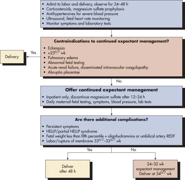
  • Preeclampsia with severe features (Table 1)
  • Chronic hypertension: Hypertension pre-dating pregnancy or present at <20 wk gestation
  • Chronic hypertension with superimposed preeclampsia: The presence of chronic hypertension with elevation in blood pressure from baseline often requiring increasing antihypertensives or with development of new onset of signs or symptoms meeting criteria for severe preeclampsia (Table 1)
  • Hemolysis, elevated liver enzymes, and low platelet count (HELLP) syndrome: A severe form of preeclampsia defined by lactate dehydrogenase (LDH) greater than 600 IU/L, liver enzymes (aspartate aminotransferase [AST], alanine transaminase [ALT]) greater than twice the upper limit of normal, and a platelet count less than 100 × 109/L

TABLE 1 Criteria for the Diagnosis of Preeclampsia with Severe Features

  • In patients with preeclampsia, severe features can be diagnosed if any one of the following criteria is present:
  • Blood pressure 160 mm Hg systolic or 110 mm Hg diastolic on two separate occasions at least 4 h apart (unless antihypertensive therapy is initiated before this time)
  • Serum creatinine >1.1 mg/dl or a doubling of the serum creatinine
  • New onset of visual disturbances
  • New-onset headache unresponsive to medication and not accounted for by alternative diagnoses
  • Pulmonary edema
  • Hepatocellular injury (serum transaminases at least twice the upper limit of normal) or severe persistent right upper quadrant or epigastric pain unresponsive to medications
  • Thrombocytopenia (platelet count <100 × 109/L)
Synonyms

  • Pregnancy-induced hypertension (PIH)
  • Hypertensive disorders of pregnancy (HDP)
  • Severe gestational hypertension
ICD-10CM CODES
O11.1Pre-existing hypertension with pre-eclampsia, first trimester
O11.2Pre-existing hypertension with pre-eclampsia, second trimester
O11.3Pre-existing hypertension with pre-eclampsia, third trimester
O11.9Pre-existing hypertension with pre-eclampsia, unspecified trimester
O14.00Mild to moderate pre-eclampsia, unspecified trimester
O14.02Mild to moderate pre-eclampsia, second trimester
O14.03Mild to moderate pre-eclampsia, third trimester
O14.10Severe pre-eclampsia, unspecified trimester
O14.12Severe pre-eclampsia, second trimester
O14.13Severe pre-eclampsia, third trimester
O14.90Unspecified pre-eclampsia, unspecified trimester
O14.92Unspecified pre-eclampsia, second trimester
O14.93Unspecified pre-eclampsia, third trimester
Epidemiology & Demographics
Incidence:

Hypertensive disorders of pregnancy are recognized as the second leading cause of maternal mortality (after hemorrhage) and are attributed to 18% of maternal mortality globally. The global incidence of preeclampsia is estimated to be 2% to 4% with significant regional variation.3,4 The disease burden is borne disproportionately by women in low- and middle-income countries or who are otherwise disadvantaged.5

Risk Factors:

See Table 2. Recently, SARS-COV-2 infection has been associated with a twofold increased risk of preeclampsia. Vaccination has been shown to be safe in pregnancy and reduce the likelihood of infection.6,7

TABLE 2 Risk Factors for Preeclampsia

  • Nulliparity
  • Multifetal gestations
  • Preeclampsia in a previous pregnancy
  • Chronic hypertension
  • Pregestational diabetes
  • Gestational diabetes
  • Thrombophilia
  • Systemic lupus erythematosus
  • Prepregnancy body mass index greater than 30
  • Antiphospholipid antibody syndrome
  • Maternal age 35 yr or older
  • Kidney disease
  • Assisted reproductive technology
  • Obstructive sleep apnea

From American College of Obstetricians and Gynecologists: Gestational hypertension and preeclampsia: ACOG Practice Bulletin No. 222, Obstet Gynecol 135:e237-e260, 2020.

Genetics:

Preeclampsia is a complex genetic disorder that is not yet well understood.8 However, large studies describing the epigenetics of maternal hypertension have implicated alterations of the fetal FLT1 locus in the development of preeclampsia.9 There are both fetal and maternal genetic factors influencing the risk of developing preeclampsia. The maternal genetic contribution has been estimated at 30% to 35% and the fetal contribution at 20%.10

Pathogenesis

The underlying causes of preeclampsia have been studied extensively for over a century; however, many uncertainties remain.11

Physical Findings & Clinical Presentation

Headache that persists despite treatment with acetaminophen is one of the classic neurologic symptoms of preeclampsia. Visual changes associated with preeclampsia, including scotoma, blurred vision, or aura.1,12 Classic neurologic exam findings of preeclampsia include hyperreflexia, clonus, or cranial nerve VI palsy.13

Endothelial dysfunction and elevated perfusion pressure accompanied by cerebral edema are considered the underlying cause of cerebral injury, including posterior reversible encephalopathy syndrome (PRES), in patients with preeclampsia. PRES is characterized by cerebral edema classically affecting the occipital and parietal lobes. Patients with PRES may manifest headache, visual loss or disturbance, altered sensorium, or altered consciousness.13 Severe cases of PRES may result in seizure or coma. PRES is also associated with increased risk of hemorrhagic and ischemic cerebrovascular accident. Importantly, patients with hypertensive disorders of pregnancy have a fivefold increased risk for occurrence of hemorrhagic or ischemic stroke.13

Patients with persistent neurologic symptoms are at highest risk for developing PRES. Management of PRES includes controlling hypertension, possible antiepileptic therapy, and indicated follow-up care.

Shortness of breath is often an indicator of underlying pulmonary edema from elevated pulmonary perfusion pressure accompanied by endothelial dysfunction. Rales may be auscultated, and a chest x-ray examination may be useful in confirming the presence of edema. Management includes fluid restriction and use of diuretics as clinically indicated.1

Dependent edema, which may affect the lower extremities, vulva, or back, is also frequently seen. It occurs due to increased vascular permeability and sodium retention related to renal injury and abnormal RAAS activation. It can be managed with compression and diuretics. Physiologic edema of pregnancy can sometimes be distinguished from pathologic edema by timing of onset. Rapidly progressing edema >4 to 5 lbs of weight gain per wk and facial edema are not typical of healthy pregnancy.1

The right upper quadrant or epigastric pain classically associated with preeclampsia is thought to be the result of periportal and parenchymal necrosis, with hepatocellular edema sometimes resulting in distension of the Glisson capsule. However, no strong correlation between symptoms and laboratory abnormalities exists.14

Oliguria in preeclampsia is due to decreased intravascular volume as a result of vascular endothelial dysfunction and RAAS activation.1,12

1% of patients with preeclampsia and 3% of patients with severe preeclampsia will develop placental abruption. Vaginal bleeding in a preeclamptic patient should prompt a workup for abruption.1

It is important to remember that patients can be asymptomatic with elevated blood pressures and high risk. Rarely, HELLP or eclampsia may present in a normotensive patient. It is important to maintain a high level of suspicion for preeclampsia in patients at increased risk.

Diagnosis

Differential Diagnosis

  • Acute fatty liver of pregnancy
  • Appendicitis
  • Autoimmune hepatitis
  • Chronic hypertension
  • Chronic kidney disease
  • Diabetic ketoacidosis
  • Gallbladder disease
  • Gastroenteritis
  • Glomerulonephritis
  • Hemolytic-uremic syndrome
  • Idiopathic thrombocytopenia
  • Thrombotic thrombocytopenic purpura
  • Pancreatitis
  • Peptic ulcer disease
  • Secondary hypertension
  • Systemic lupus erythematosus
  • Viral hepatitis
  • Medications or medication withdrawal
  • Migraine headache
Workup

Hypertension:

  • Two blood pressure measurements at least 4 h apart, with a systolic BP of 140 mm Hg or more or a diastolic blood pressure of 90 mm Hg or more.
  • Severe hypertension with systolic BP of 160 mm Hg or more or a diastolic blood pressure of 110 mm Hg or more.
  • Severe hypertension can be confirmed in a brief period to allow for immediate treatment of severe range blood pressure.

Proteinuria:

  • 300 mg per 24-h urine collection
  • Protein/creatinine ratio 0.3
  • 2+ on dipstick (only if other methods not available)

Other diagnostic labs:

Laboratory Tests

  • Generally, the recommended laboratory testing is consistent with labs that can be used to identify end-organ dysfunction.
  • CBC to assess for anemia or thrombocytopenia.
  • Liver function tests (aspartate aminotransferase, alanine aminotransferase) are useful in determining the presence of hepatic injury and are also useful in excluding HELLP syndrome.
  • Serum creatinine (and uric acid).
  • Prothrombin time, partial thromboplastin time, and fibrinogen can be checked to rule out disseminated intravascular coagulation, if clinically appropriate.
Imaging Studies

  • Computed tomography or MRI scan of head if persistent headache or neurologic symptoms raising concern for PRES or stroke
  • Chest x-ray examination if there is concern for pulmonary edema
  • Sonogram of fetus to evaluate for intrauterine growth restriction, amniotic fluid level, placental location
  • Sonogram of maternal liver if subcapsular hematoma suspected

Treatment

Acute General Rx

Delivery is the treatment of choice and the only cure for the disease. This must be taken in the context of the gestational age of the fetus and severity of the preeclampsia.

  • For preeclampsia with severe features, administer magnesium sulfate 4 to 6 g intravenous (IV) loading dose, with 2 g/h maintenance (adjust dosage for renal insufficiency). If there is a contraindication to magnesium sulfate, such as myasthenia gravis, then use phenytoin at 10 to 15 mg/kg loading dose, then 200 mg IV q8h starting 12 h after loading dose.
  • Treat blood pressure (BP) >160 mm Hg systolic or >110 mm Hg diastolic either with hydralazine 5 to 10 mg IV, then 10 mg, every 20 min to a max dose of 20 mg; or with labetalol hydrochloride 20, 40, 60, 80 mg IV, escalating dosage every 10 min to a max dose of 300 mg IV; or with nifedipine 10 to 20 mg orally every 20 min to a max dose of 180 mg/day for acute blood pressure control. If maximum dose of one medication is reached, add an additional medication to reach goal of BP 140 to 150/90 to 100 mm Hg.
  • Continuous fetal monitoring.
  • Epidural is anesthesia of choice for pain management in labor or cesarean delivery.
  • All patients undergoing induction of labor or cesarean delivery should receive antiseizure medications (magnesium sulfate) if disease with severe features, for delivery and 12 to 24 h postpartum.
Chronic Rx

Preeclampsia without severe features: If <37 wk, close observation for worsening maternal or fetal condition with delivery at 37 wk. If 24 to 36 wk, consider antenatal corticosteroids. If severe features develop or there is evidence of fetal compromise, earlier delivery is appropriate.

Preeclampsia with severe features: Delivery in the presence of maternal or fetal compromise, labor, or 34 wk. From 24 to 34 wk consider steroids with close monitoring, and at <24 wk offer termination of pregnancy (Fig. 1). Risks of expectant management include severe worsening of disease, eclampsia, abruptio placenta, stillbirth, HELLP syndrome, and intensive care unit (ICU) admission. Contraindications to expectant management include eclampsia, HELLP syndrome, pulmonary edema, disseminated intravascular coagulation, abruptio placenta, uncontrollable severe hypertension, fetal demise, and nonreassuring fetal status. Labetalol, hydralazine, and nifedipine are the drugs of choice for long-term blood-pressure control during pregnancy.

Figure 1 Management Plan for Patients with Preeclampsia with Severe Features Before 34 wk Gestation

!!flowchart!!

HELLP, Hemolysis, elevated liver enzymes, and low platelets; REDF, reversed end-diastolic flow.

(From Gabbe SG et al: Obstetrics: normal and problem pregnancies, ed 7, Philadelphia, 2017, Elsevier.)

Disposition

  • Preeclampsia is a progressive and unpredictable disease process; a course of expectancy should be managed with caution. Up to 20% of patients who have seizures are normotensive.
  • The reoccurrence rate for preeclampsia in a subsequent pregnancy is approximately 20%, and higher in cases with a second trimester presentation or complications. This risk may be decreased with the use of daily low-dose aspirin.
  • For high-risk patients, baseline assessment of renal function (24-h urine collection for total protein and creatinine clearance or protein:creatinine ratio), CBC, serum creatinine, liver function tests (LFTs), and uric acid should be obtained at the first prenatal visit.
Referral

Obstetric management with input from maternal-fetal subspecialists in severe cases. Transfer of care to facilities with appropriate level of care for all cases <34 wk.

Pearls & Considerations

Comments

  • Low-dose aspirin exposure during the early stages of placentation decreases the risk of preeclampsia, preterm birth, and intrauterine growth restriction in women who are at high risk of preeclampsia. The ACOG and the U.S. Preventive Services Task Force (USPSTF) recommend the use of low-dose aspirin as preventive medication starting after 12 wk of gestation (ideally before 16 wk) in women who are at high risk for preeclampsia.16,17
  • The development of preeclampsia may be one of the earliest identifiable risk markers for potential future cardiovascular disease in women. Women with a history of preeclampsia are at increased risk for cardiovascular disease, and appropriate follow-up should be encouraged.
Related Content

Related Content

    1. Gestational hypertension and preeclampsia: ACOG Practice Bulletin No. 222Obstet Gynecol. 135:e237-e260, 2020.
    2. Hypertension in pregnancy: executive summaryObstet Gynecol. 122(5):1122-1131, 2013.
    3. Khan KS : WHO analysis of causes of maternal death: a systematic reviewLancet. 367(9516):1066-1074, 2006.
    4. Abalos E : Global and regional estimates of preeclampsia and eclampsia: a systematic reviewEur J Obstet Gynecol Reprod Biol. 170(1):1-7, 2013.
    5. Magee LA : PreeclampsiaN Engl J Med. 386(19):1817-1832, 2022.
    6. Conde-Agudelo A, Romero R : SARS-CoV-2 infection during pregnancy and risk of preeclampsia: a systematic review and meta-analysisAm J Obstet Gynecol. 226(1):68-89.e3, 2022.
    7. Male V : SARS-CoV-2 infection and COVID-19 vaccination in pregnancyNat Rev Immunol. 22:277-282, 2022.
    8. Williams PJ, Broughton Pipkin F : The genetics of pre-eclampsia and other hypertensive disorders of pregnancyBest Pract Res Clin Obstet Gynaecol. 25(4):405-417, 2011.
    9. McGinnis R : Variants in the fetal genome near FLT1 are associated with risk of preeclampsiaNat Genet. 49:1255-1260, 2017.
    10. Gray KJ : Genetic predisposition to preeclampsia is conferred by fetal DNA variants near FLT1, a gene involved in the regulation of angiogenesisAm J Obstet Gynecol. 218:211-218, 2018.
    11. Rana S : Preeclampsia: pathophysiology, challenges, and perspectivesCirc Res. 124(7):1094-1112, 2019.
    12. Burton GJ : Pre-eclampsia: pathophysiology and clinical implicationshttp://dx.doi.org/10.1136/bmj.l2381. BMJ366:l2381.BMJ. 366:l2381, 2019.
    13. McDermott M : Association with posterior reversible encephalopathy syndrome and strokeStroke. 49(3):524-530, 2018.
    14. Barton JR : Hepatic histopathologic condition does not correlate with laboratory abnormalities in HELLP syndrome (hemolysis, elevated liver enzymes, and low platelet count)Am J Obstet Gynecol. 167:1538-1543, 1992.
    15. Committee Opinion No 692: emergent therapy for acute-onset, severe hypertension during pregnancy and the postpartum periodObstet Gynecol. 129(4):e90-e95, 2017.
    16. LeFevre ML : Low-dose aspirin use for the prevention of morbidity and mortality from preeclampsia: U.S. Preventive Services Task Force recommendation statementAnn Intern Med. 161:819-826, 2014.
    17. Committee Opinion No 692: low-dose aspirin use in pregnancyObstet Gynecol. 132:e44-e52, 2018.