Author: Vishnu Kadiyala, MD and Philip Stockwell, MD

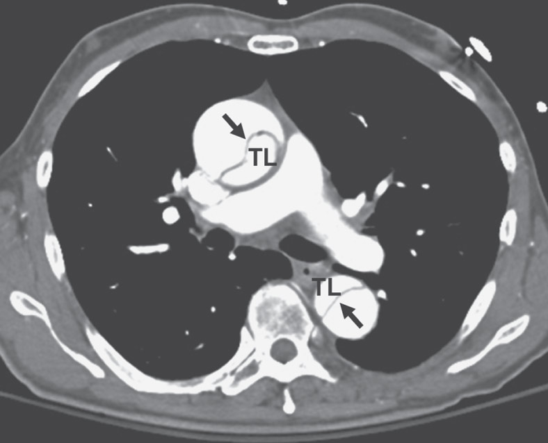
Acute aortic syndromes are a spectrum of aortic pathologies that include aortic dissection, intramural hematomas (IMHs), and penetrating atherosclerotic ulcers.1 Aortic dissections are the most common and occur when blood passes through an intimal tear, separating the intima from the medial layers and creating a false lumen.1 IMH and penetrating aortic ulcers are variants of the classically described aortic dissection. Fig. E1 illustrates acute aortic syndromes.
Figure E1 Acute aortic syndromes.
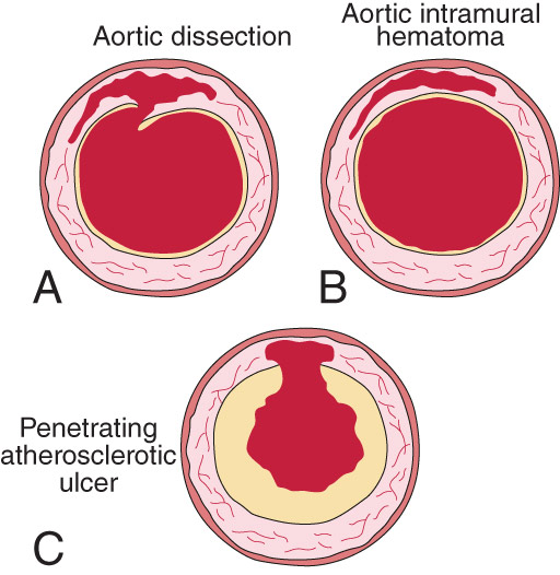
A, Classic aortic dissection. There is a tear in the intima with blood entering the media and a dissecting cleavage plane propagating for variable distances anterograde (and occasionally retrograde) throughout the aortic wall. B, Aortic intramural hematoma (IMH). A spontaneous hemorrhage of the vasa vasorum leads to bleeding within the media in the absence of an intimal tear or intimal flap. C, Penetrating atherosclerotic aortic ulcer (PAU). An ulcerated aortic plaque ruptures into the media, leading to an outpouching or ulceration in the aortic wall. This may be associated with IMH formation; pseudoaneurysm; or a focal, thick-walled aortic dissection.
(From Libby P et al: Braunwald’s heart disease, a textbook of cardiovascular medicine, ed 12, Philadelphia, 2022, Elsevier.)
Aortic dissection is generally classified according to anatomic location (Fig. E2). Table E1 summarizes classification schemes of acute aortic dissection.
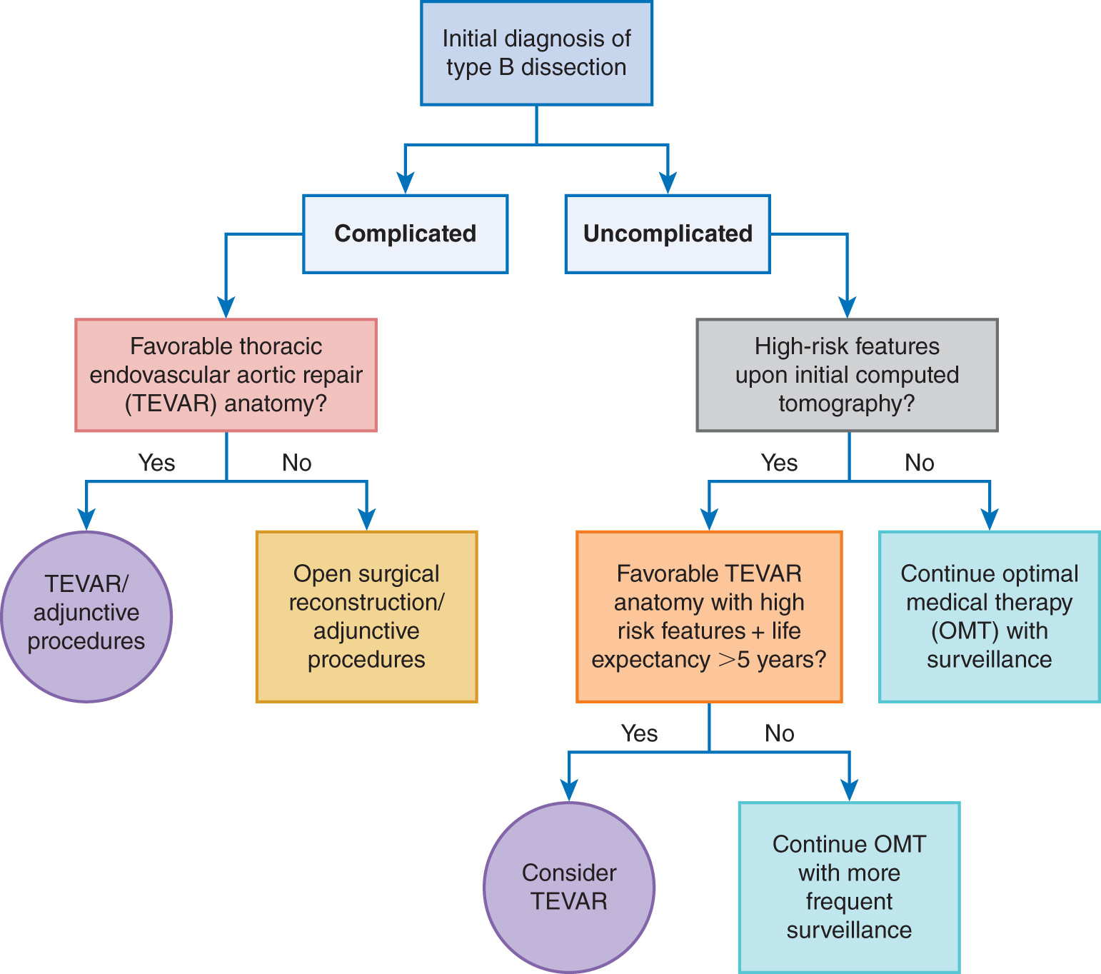
- •Stanford (more commonly used classification system): Type A ascending aorta (proximal), type B descending aorta (distal).
- •DeBakey: Type I ascending and descending aorta, type II ascending aorta, type III descending aorta.
- •The Society of Thoracic Surgery and Society of Vascular Surgery has recently proposed a classification system of dissection subtypes to more precisely define the location of disease as extending between two anatomic zones.2
- •Aortic dissection can also be classified by acuity of presentation: Hyperacute (<24 h), acute (2 to 7 days), subacute (8 to 30 days), and chronic (>30 days).
TABLE E1 Classification Schemes of Acute Aortic Dissection
From Libby P et al: Braunwald’s heart disease, a textbook of cardiovascular medicine, ed 12, Philadelphia, 2022, Elsevier.
Figure E2 Acute aortic dissection classifications.
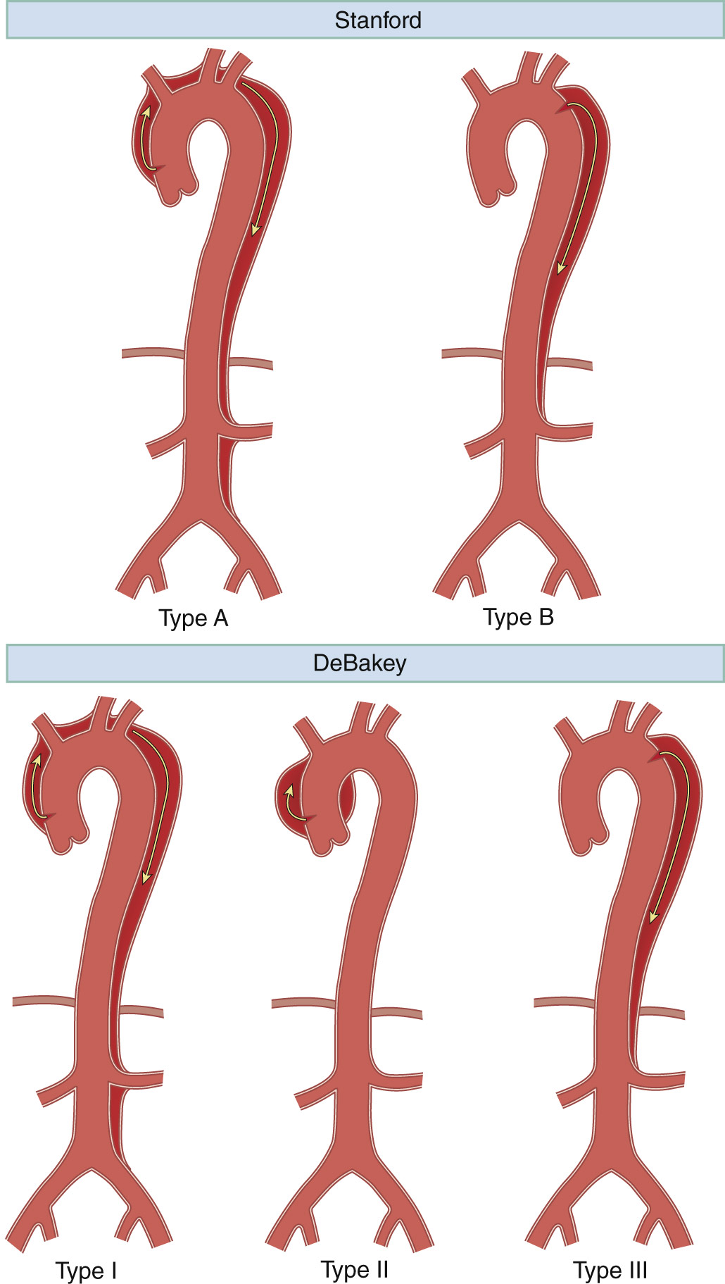
The Stanford classification divides dissections into those involving the ascending aorta from dissection originating there or proximal dissection of a more distal origin (Type A) or those limited to the descending aorta (Type B). The DeBakey classification divides dissections into those arising in the ascending aorta and extending distally beyond the innominate artery (Type I), arising in the ascending aorta with extension limited to the level of the innominate artery takeoff (Type II), and arising at or distal to the left subclavian artery takeoff with extension distally (Type III).
(From Walls RM et al: Rosen’s emergency medicine, concepts and clinical practice, ed 10, Philadelphia, 2023, Elsevier.)
2.6 to 3.5 per 100,000 person-yr; more frequent in winter as compared to other seasons, although no clear reason has been identified.3,4
- •Hypertension (found in up to 77% of patients with aortic dissection)3,5
- •Atherosclerosis (found in up to 27% of patients with aortic dissection)3,5
- •Preexisting aortic aneurysm (found in up to 16% to 20% of patients with aortic dissection)3,5
- •Age (60 to 80 yr)
- •Family history of aortic aneurysms/dissection
- •History of cardiac surgery, aortic valve replacement, intraaortic catheterization1,3,5
- •Disorders of collagen (Marfan syndrome, Loeys-Dietz syndrome, Ehlers-Danlos syndrome)3,5
- •Vascular inflammation (giant cell arteritis, Takayasu arteritis, rheumatoid arthritis, syphilitic aortitis)
- •Aortic coarctation, bicuspid aortic valve
- •Turner syndrome
- •Cocaine abuse (usually within 12 h of last use of cocaine)1,3,5
- •Trauma (∼20% road accident fatalities found to have ruptured aorta on autopsy)1
- •Pregnancy and delivery
- •Fluoroquinolone use1
- •Table E3 summarizes genetically triggered conditions associated with aortic dissection
TABLE E2 Risk Factors for Aortic Dissection
TAVR, Transcatheter aortic valve replacement.
From Libby P et al: Braunwald’s heart disease, a textbook of cardiovascular medicine, ed 12, Philadelphia, 2022, Elsevier.
TABLE E3 Thoracic Aortic Aneurysm Syndromes and Conditions Due to a Heritable or Genetic Cause
| Condition | Gene | Clinical Features |
| Syndromic HTAD* | ||
| Marfan syndrome | FBN1 | Aortic root aneurysm, AD, TAA, MVP, long bone overgrowth, scoliosis, pectus deformities, ectopia lentis, myopia, tall stature, PTX, dural ectasia |
| Loeys-Dietz syndrome | TGFBR1, TGFBR2,*SMAD3, TGFB2, TGFB3, SMAD2 | TAA, branch vessel aneurysms, AD, arterial tortuosity, MVP, craniosynostosis, hypertelorism, bluish sclera, bifid/broad uvula, translucent skin, visible veins, club feet, dural ectasia,* premature osteoarthritis |
| Vascular Ehlers-Danlos syndrome | COL3A1 | TAA, AAA, arterial rupture, AD, MVP, bowel and uterine rupture, PTX, translucent skin, atrophic scars, small joint hypermobility, easy bruising, carotid-cavernous fistula |
| Arterial tortuosity syndrome | SLC2A10 | Tortuous large- and medium-sized arteries, aortic dilation, craniofacial, skin and skeletal features |
| Shprintzen-Goldberg syndrome | SKI | Craniosynostosis, skeletal features, aortic dilation |
| Congenital contractural arachnodactyly (Beals syndrome) | FBN2 | MVP, arachnodactyly, Marfanoid habitus, digital contractures, mild aortic dilation |
| Cutis laxa | EFEMP2 (Fibulin-4) | TAA, arterial tortuosity, arterial stenosis, hypertelorism, arachnodactyly |
| EDS with periventricular nodular heterotopia (PVNH) | FLNA (filamin A) | X-linked, PVNH, TAA, BAV, MV disease, PDA, VSD, seizures, joint hypermobility |
| Meester-Loeys syndrome | BGN | X-linked, TAA, AD, skeletal abnormalities |
| LOX-related TAA | LOX (lysyl oxidase) | TAA, BAV, AD, Marfanoid habitus in some |
| Nonsyndromic HTAD (Familial TAA) | ||
| FTAA | ACTA2 (α-smooth muscle actin) | TAA, AD, BAV, Moya-Moya, premature CAD and CVD, livedo reticularis, iris flocculi |
| FTAA | MYH11 (myosin heavy chain-11) | TAA, AD, PDA |
| FTAA | MYLK (myosin light chain kinase) | AD at relatively small aortic size |
| FTAA | PRKG1 (protein kinase cGMP-dependent) | Aortic root aneurysm and AD |
| FTAA | MAT2A (MAT IIα) | TAA, AD, BAV |
| FTAA | MFAP5 (microfibrillar-associated protein 5) | TAA, AD, skeletal features may be present |
| FTAA | FOXE3 (forkhead transcription factor) | TAA, AD |
| Bicuspid Aortic Valve/Associated Ascending Aortic Aneurysm | ||
| Familial BAV/AS and TAA | NOTCH1 (NOTCH1) | Aortic stenosis, TAA |
| TGFBR1, TGFBR2, TGFB2, TGFB3, ACTA2, MAT2A, GATA5, SMAD6, LOX, ROBO4, TBX20 | BAV with TAA | Syndromic and nonsyndromic FTAA with an increased frequency of BAV |
| Turner syndrome | XO, Xp | BAV, COA, TAA, AD, short stature, lymphedema, webbed neck, premature ovarian failure, affects 1 in 2500 live-born girls |
AAA, Abdominal aortic aneurysm; AAT, aortic aneurysm syndrome; AD, aortic dissection; BAV, bicuspid aortic valve; CAD, coronary artery disease; COA, coarctation of the aorta; CVD, cerebrovascular disease; EDS, Ehlers-Danlos syndrome; FTAA, familial thoracic aortic aneurysm (and dissection) syndrome; HTAD, heritable TAA; LDS, Loeys-Dietz syndrome; MFS, Marfan syndrome; MV, mitral valve; MVP, mitral valve prolapse; PDA, patent ductus arteriosus; PTX, pneumothorax; TAA, thoracic aortic aneurysm; TGF, transforming growth factor; VSD, ventricular septal defect.
* Some individuals with pathogenic variants in a gene which can lead to syndromic HTAD have very few or no syndromic features, and variants in some genes causing syndromic HTAD may also lead to nonsyndromic HTAD.
From Libby P et al: Braunwald’s heart disease, a textbook of cardiovascular medicine, ed 12, Philadelphia, 2022, Elsevier.
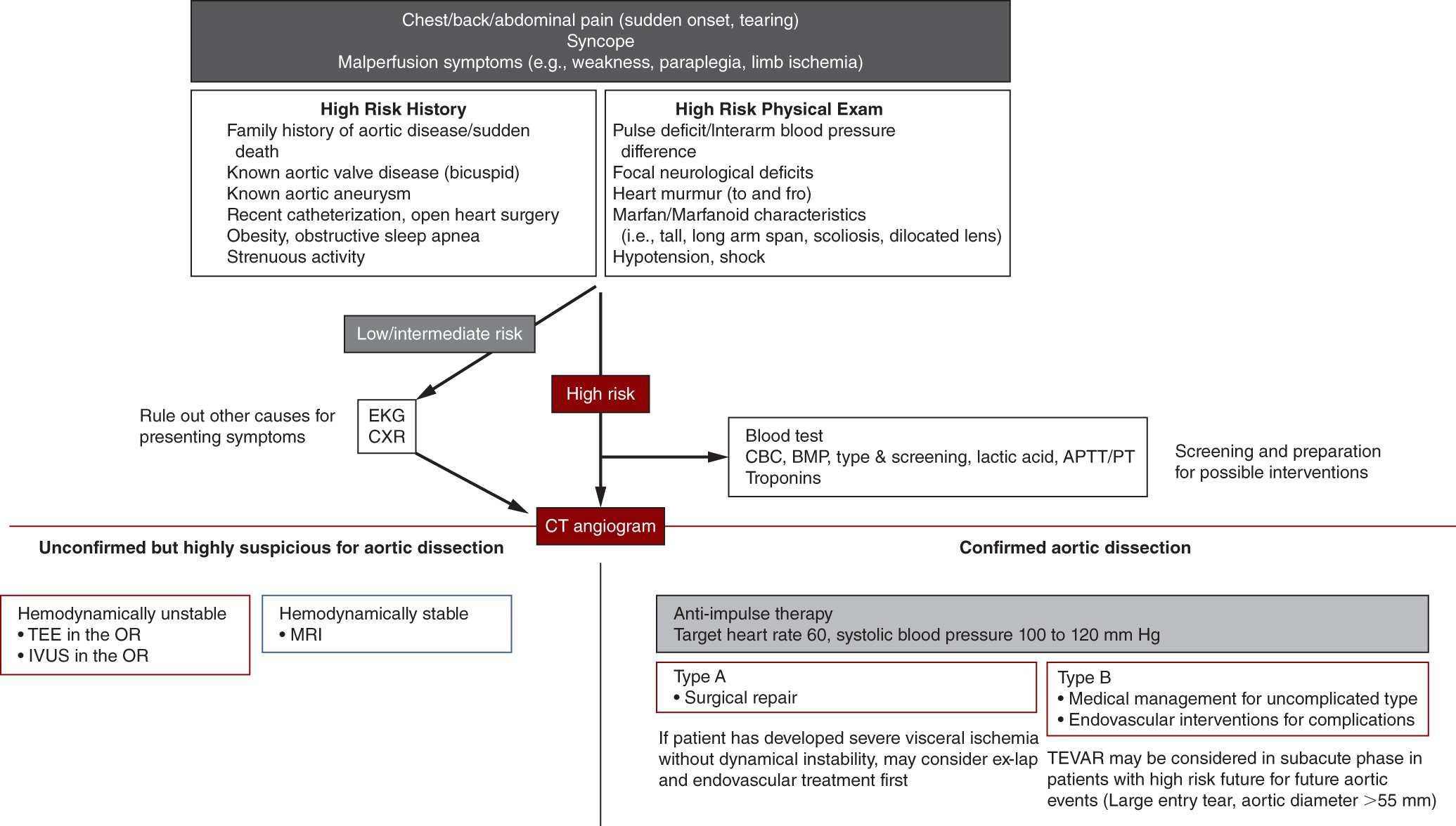
- •Sudden onset of severe sharp, tearing, or ripping chest, back, or abdominal pain.1,3,5 However, painless dissection occurs in ∼6.3% of cases2
- •Anterior chest pain (79% type A, 63% type B)3
- •Back pain, abdominal pain (43% type A, 64% type B)3
- •Syncope (19% type A, 3% type B), generally secondary to cardiac tamponade or stroke3
- •Congestive heart failure (CHF)1
- •May present with hypertension (28% for type A, 66% in type B dissection),5 although 25% present with hypotension (systolic blood pressure <100 mm Hg), which can indicate bleeding, cardiac tamponade, or severe aortic regurgitation.3 Patients presenting with very high systolic blood pressure (>180 mm Hg for type A, >200 mm Hg for type B dissection), as well as in those with systolic blood pressure ≤100 mm Hg, have significantly higher in-hospital mortality rates
- •Pulse and blood pressure differentials (>20 mm Hg between arms) in 19% to 31% of cases caused by partial compression of subclavian arteries6
- •Aortic regurgitation in 40% to 75% of cases of proximal dissection,1 often with diastolic decrescendo murmur
- •Myocardial ischemia caused by coronary artery occlusion, most commonly involving the right coronary artery
- •Stroke in 5% to 10% of patients (secondary to dissection into or decreased blood flow to the carotids)1
- •Mesenteric ischemia occurs in 3% to 5% of cases, with external compression, flap prolapse, or involvement of arterial ostia1
- •Horner syndrome (ptosis, miosis, anhidrosis)
- •Vocal cord paralysis or hoarse voice (caused by compression of the left recurrent laryngeal nerve)1
Genetics, in addition to other risk factors (Tables E4, E5, E6, and E7), contribute to the development of aortic aneurysms and dissection.
TABLE E4 Ascending Thoracic Aortic Aneurysms
| Degenerative | Associated With Atherosclerosis | ||
| Inheritable/connective tissue | Marfan syndrome | ||
| Ehlers-Danlos syndrome | |||
| Loeys-Dietz syndrome | |||
| Turner syndrome | |||
| Osteogenesis imperfecta | |||
| Rheumatoid arthritis | |||
| Bicuspid aortic valve | |||
| Aneurysm of sinus of Valsalva | |||
| Arteritis | Giant cell | ||
| Takayasu | |||
| Behçet disease | |||
| Relapsing polychondritis | |||
| Infectious | Syphilis | ||
| Mycotic aneurysms |
From Soto JA, Lucey BC: Emergency radiology: the requisites, ed 2, Philadelphia, 2017, Elsevier.
TABLE E5 Aortic Arch Aneurysms
| Degenerative | Resulting From Atherosclerosis | ||
| Arteritis | Giant cell | ||
| Takayasu | |||
| Behçet disease | |||
| Infectious | Syphilis |
From Soto JA, Lucey BC: Emergency radiology: the requisites, ed 2, Philadelphia, 2017, Elsevier.
TABLE E6 Descending Thoracic Aortic Aneurysm
| Degenerative | Resulting From Atherosclerosis | ||
| Focal pseudoaneurysm resulting from a penetrating aortic ulcer | |||
| Arteritis | Giant cell | ||
| Takayasu | |||
| Behçet disease | |||
| Inherited/connective tissue | Marfan syndrome | ||
| Ductus aneurysm | |||
| Loeys-Dietz syndrome | |||
| Infectious | Mycotic aneurysms | ||
| Traumatic | Posttraumatic pseudoaneurysms | ||
| Chronic aortic transection |
From Soto JA, Lucey BC: Emergency radiology: the requisites, ed 2, Philadelphia, 2017, Elsevier.
TABLE E7 Crawford Classification of Thoracoabdominal Aneurysms
| I | Descending thoracic aorta to suprarenal aorta | ||
| II | Proximal descending thoracic aorta to infrarenal aorta (below the diaphragm) | ||
| III | Mid descending thoracic aorta to infrarenal aorta | ||
| IV | Supravisceral aorta to infrarenal aorta |
From Soto JA, Lucey BC: Emergency radiology: the requisites, ed 2, Philadelphia, 2017, Elsevier.