Author: Fred F. Ferri, MD
Lichen planus (LP) refers to an idiopathic pruritic, papulosquamous disease with characteristic histopathologic and clinical features, manifesting with a papular skin eruption characteristically found over the flexor surfaces of the extremities, genitalia, and mucous membranes.
| ICD-10CM CODES | |||
| L43 | Lichen planus | ||
| L43.0 | Hypertrophic lichen planus | ||
| L43.1 | Bullous lichen planus | ||
| L43.9 | Lichen planus, unspecified | ||
| L43.8 | Other lichen planus | ||
LP often manifests with pruritic, flat-topped violaceous papules, but the clinical presentation varies depending on the area involved. Classically, lesions are described with the six "P"s: Purple, pruritic, planar, polygonal, papule, and plaque. In some patients, fine white lines are visible across the surface of the lesions, known as Wickham striae.
Figure E1 Lichen planus of the vulva with a white, lacelike pattern and erythema.
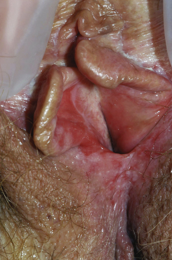
(From Crum CP et al: Diagnostic gynecologic and obstetric pathology, ed 3, Philadelphia, 2018, Elsevier.)
Figure E2 Lichen planus of the penis.
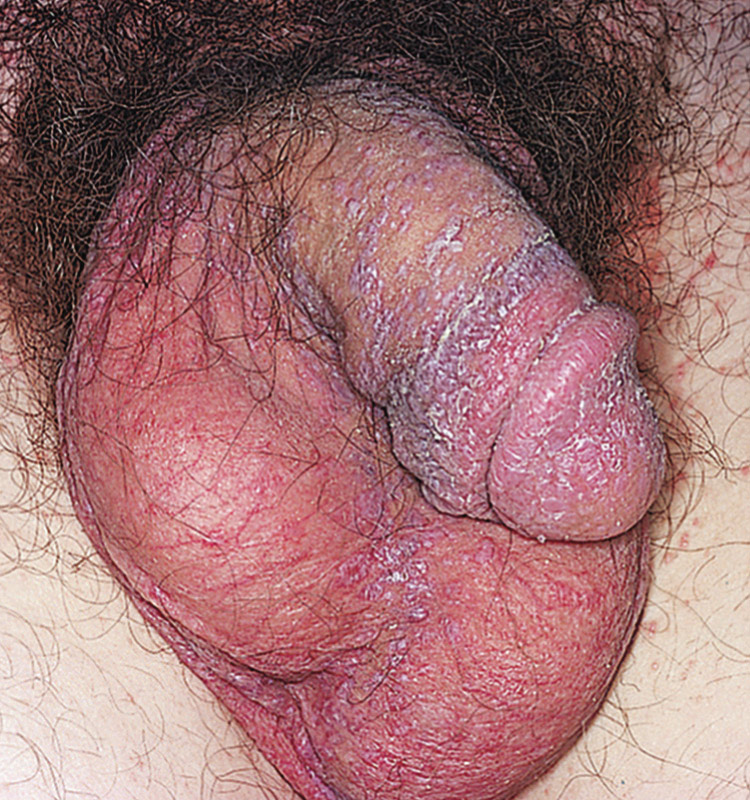
(From Swartz MH et al: Textbook of physical diagnosis: history and examination, ed 8, Philadelphia, 2020, Elsevier.)
Figure E3 Flat-topped, purple, polygonal papules of lichen planus.
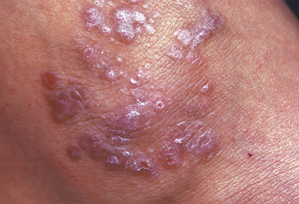
(From Kliegman RM et al: Nelson textbook of pediatrics, ed 22, Philadelphia, 2025, Elsevier.)
Figure E4 Shiny, flat-topped, polygonal, violaceous papules of lichen planus.
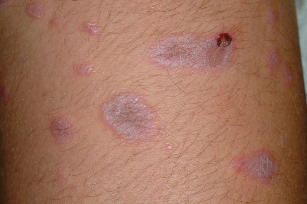
Note the Wickham striae (linear, whitish-gray streaks) on the surface.
(From Paller AS, Mancini AJ: Hurwitz clinical pediatric dermatology: a textbook of skin disorders of childhood and adolescence, ed 5, Philadelphia, 2016, Elsevier.)
Figure E5 A and B, Lichen planus.
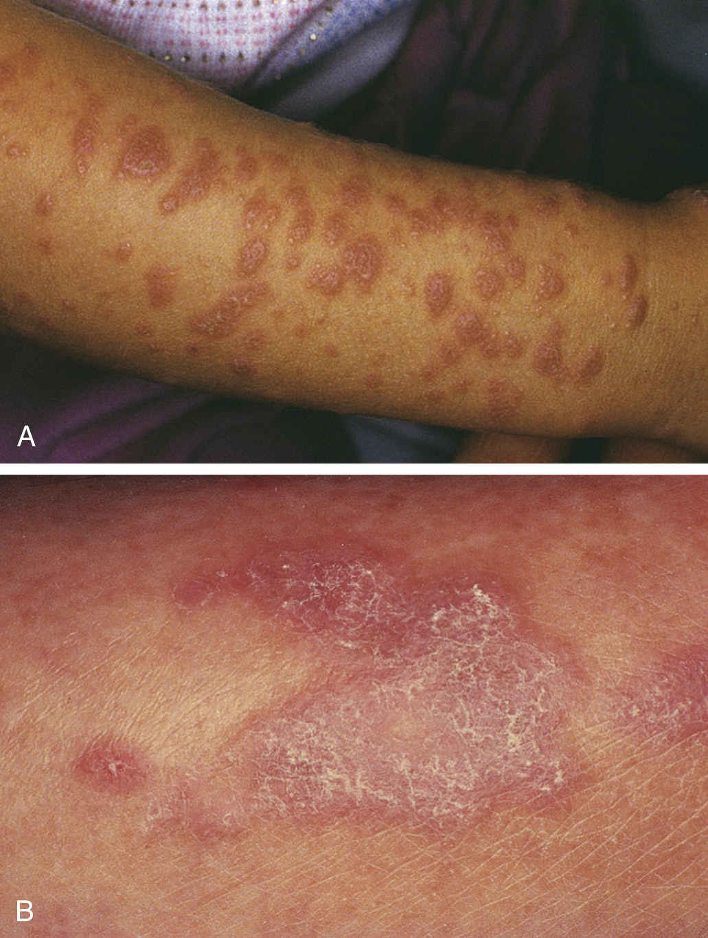
Note the fine, reticulated white scales.
(From Swartz MH et al: Textbook of physical diagnosis, history and examination, ed 8, Philadelphia, 2020, Elsevier.)
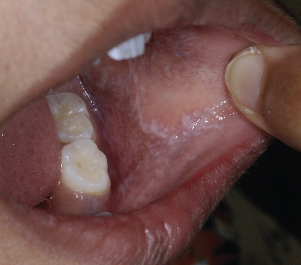
Wickham striae on the buccal mucosa.
(From Paller AS, Mancini AJ: Hurwitz clinical pediatric dermatology: a textbook of skin disorders of childhood and adolescence, ed 5, Philadelphia, 2016, Elsevier.)
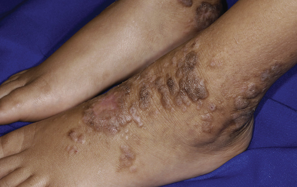
TABLE E1 Key Features of LP Variants
| Acute (exanthematous) LP | Rapid onset of disseminated lesions; heals with PIH; rapidly self-resolves (3-9 mo) | ||
| Actinic LP (LP subtropicus) | Most common in Middle Eastern and Indian patients (also Africans); young adults or children; onset in spring or summer on sun-exposed sites (face, forehead > dorsal UE, neck, intertriginous sites); comprised of discoid papules/plaques (hyperpigmented focus with hypopigmented rim) or melasmalike patches (less common) | ||
| Annular LP | Usually asymptomatic; annular plaques with raised violaceous-white edge with central clearing; resembles GA but is scaly; axilla is most common site, followed by penis | ||
| Atrophic LP | Enlarging small violaceous, annular plaques with centrally depressed/atrophic, hyperpigmented area; clinically resembles early morphea or LS&A legs most common site | ||
| Bullous LP | Blisters develop on longstanding LP lesions due to extensive epidermal damage (expanded Max-Joseph spaces) | ||
| Drug-induced LP (lichenoid drug eruption) |
| ||
| Genital LP | |||
| Hypertrophic LP (aka "LP verrucosus") | Extremely pruritic, thick, scaly plaques; most commonly on dorsal feet/shins, wrists; symmetric; lasts longer (avg. duration 6 yr); may → multiple keratoacanthomas or follicular-based SCCs; biopsy may show many eosinophils | ||
| Inverse LP | Axilla > inguinal and inframammary folds > antecubital and popliteal fossae; poorly defined hyperpigmentation usually present (thus may overlap with LP pigmentosus) | ||
| Linear LP | Refers to lesions that appear spontaneously (not due to koebnerization) in a Blaschkoid distribution; favors younger patients (20-30 yo); likely due to somatic mosaicism | ||
| Oral LP | Over half of patients with cutaneous LP have oral involvement Reticular LP: most common; lacy white raised linear lines; usually asymptomatic; most commonly on bilateral buccal mucosa > gingivae > tongue > lips Atrophic, erosive, and bullous oral LP: more painful, F > M; must check for esophageal and genital involvement; may progress to SCC (1%-2%) | ||
| Nail LP | Seen in 10% of LP patients; usually affects several nails; classic findings = longitudinal ridging, lateral thinning, fissuring, and dorsal pterygium; kids lack these other nail findings but may present as 20-nail dystrophy (rare in adults) | ||
| LP/LE overlap | Acral sites with bullae, ulceration, nail loss, and pain; overlapping features of lupus and LP seen clinically and on H&E/DIF | ||
| Palmoplantar LP | Commonly ulcerative (esp. on soles); occurs in 30-40 yo age group; extremely painful and recalcitrant to therapy; usually with typical LP elsewhere | ||
| LP pemphigoides | Vesicobullous lesions occur anywhere on skin (most commonly on uninvolved skin) due to circulating IgG antibodies against BPAG2 (180-kD antigen, type XVII collagen); occurs weeks to months after onset of LP; pathogenesis: LP damages epidermis → exposes hidden antigens that are recognized by T cells | ||
| LP pigmentosus | Skin types 3 and 4; brown or slate gray macules on sun-exposed face, neck, and flexures; lacks preceding erythema and minimally pruritic; evolves into reticulate hyperpigmented patches; classic LP lesions in only 20%; occurs later in life (30-40 yo) than ashy dermatosis (childhood to late 20s); discussed further in Section 3.26 | ||
| Lichen planopilaris (LPP; follicular LP) | Perifollicular hyperkeratosis with narrow violaceous rim on scalp (> other hair-bearing areas) → scarring hair loss; frontal fibrosing alopecia: variant in elderly women along the frontal hairline | ||
| Graham-Little-Piccardi-Lasseur syndrome | Variant of LPP; classic triad = nonscarring pubic and axillary hair loss w/ disseminated spiny follicular papules (KP-like), cutaneous or mucosal LP, and scarring alopecia on scalp |
ACE, Angiotensin-converting enzyme; DIF, direct immunofluorescence; GA, geographic atrophy; H&E, hematoxylin and eosin staining; HCTZ, hydrochlorothiazide; IgG, immunoglobulin G; KP, keratosis pilaris; LE, lupus erythematosus; LP, Lichen planus; LPP, lichen planopilaris; LS&A, lichen sclerosus et atrophicus; NSAIDs, nonsteroidal antiinflammatory drugs; PIH, pregnancy-induced hypertension; SCC, squamous cell carcinoma; TNF, tumor necrosis factor; UE, upper extremity; UVB, ultraviolet B radiation.
From Alikhan A, Hocker TLH: Review of dermatology, ed 2, Philadelphia, 2024, Elsevier.