Author: Julia Probert, MD and Felicia Smith, MD


The American Psychiatric Association’s Diagnostic and Statistical Manual, 5th edition, text revision (DSM-5-TR), defines delirium as:
- •Disturbance of consciousness with reduced ability to focus, sustain, or shift attention.
- •The disturbance develops over a short period of time (usually hours to days) and tends to fluctuate during the course of a day.
- •An additional disturbance in cognition (e.g., memory deficit, disorganization, language, visuospatial ability, or perception).
- •A change in cognition or development of a perceptual disturbance that is not better accounted for by a preexisting, established, or evolving dementia.
- •There is evidence from history, physical exam, or laboratory findings that the disturbance is caused by a medical condition, substance intoxication or withdrawal (i.e., due to a drug of abuse or to a medication), exposure to a toxin, or is due to multiple etiologies.
Hyperactive, hypoactive, and mixed subtype
| ICD-11 CODES | |||
| 6D70.0 | Delirium due to disease classified elsewhere | ||
| 6D70.1 | Delirium due to psychoactive substances including medications | ||
| 6D70.2 | Delirium due to multiple etiological factors | ||
| 6D70.Y | Delirium, other specified cause | ||
| 6D70.Z | Delirium, unspecified or unknown cause | ||
| ICD-10CM CODES | |||
| F05 | Delirium, not induced by alcohol and other psychoactive substances | ||
| F05.9 | Delirium, unspecified | ||
| F06.0 | Organic hallucinosis | ||
| F05.8 | Other delirium | ||
| F05.0 | Delirium not superimposed on dementia | ||
| F05.1 | Delirium superimposed on dementia | ||
Nearly 30% of older patients experience delirium at some time during the hospital course. In older surgical patients, the prevalence varies from 10% to 50%. The highest incidence, estimated to be 60% to 80%3, is found in patients requiring intubation in the intensive care unit. Delirium can be subtyped by motoric functioning, with hypoactive delirium being most common, followed by mixed, and hyperactive delirium being least common. Predisposing factors for delirium among older adults hospitalized for a medical or surgical illness are summarized in Table 1. Delirium is the most common neuropsychiatric disorder in patients with medical illness. Any age, race, or gender can be affected. Predisposing factors for the development of delirium during hospitalization are summarized in Table 2. Pediatric delirium is often missed but remains important because delirium is associated with longer hospital stays, decreased cognitive performance, and increased mortality. Risk factors include extremes of age, severe pain, medication effect, states of intoxication or withdrawal, vital sign derangement leading to reduced cerebral blood flow, metabolic derangement including hypoxia/hypercarbia, poor cognitive baseline, and kidney or liver failure.4-8
TABLE 1 Risk Factors for Agitation and Delirium
| Age >70 yr | BUN/creatinine ratio ≥18 | ||
| Transfer from a nursing home | Renal failure, creatinine >2.0 mg/dl | ||
| History of depression | Liver disease | ||
| History of dementia, stroke, or epilepsy | CHF | ||
| Alcohol abuse within past month | Cardiogenic or septic shock | ||
| Tobacco use | Myocardial infarction | ||
| Drug overdose or illicit drug use | Infection | ||
| HIV infection | CNS pathology | ||
| Psychoactive medications | Urinary retention or fecal impaction | ||
| Hyponatremia or hypernatremia | Tube feeding | ||
| Hypoglycemia or hyperglycemia | Rectal or bladder catheters | ||
| Hypothyroidism or hyperthyroidism | Physical restraints | ||
| Hypothermia or fever | Central line catheters | ||
| Hypertension | Malnutrition or vitamin deficiencies | ||
| Hypoxia | Procedural complications | ||
| Acidosis or alkalosis | Visual or hearing impairment | ||
| Pain | Sleep disruption | ||
| Fear and anxiety |
BUN, Blood urea nitrogen; CHF, congestive heart failure; CNS, central nervous system; HIV, human immunodeficiency virus.
From Vincent JL et al: Textbook of critical care, ed 8, Philadelphia, 2024, Elsevier.
TABLE 2 Precipitating Factors for the Development of Delirium During Hospitalization for Medical or Surgical Illness
| Precipitating Factor | Odds Ratio (OR) | ||
| Use of physical restraints | 4.4 | ||
| Malnutrition | 4 | ||
| Using more than three new medications during hospitalization | 2.9 | ||
| Use of bladder catheterization | 2.4 | ||
| Exposed to any iatrogenic event | 1.9 | ||
| Intraoperative hypotension (at least 31% drop in mean perioperative BP or a SBP ≤80 mm Hg) | 1.4 | ||
| Postoperative Hct <30% | 1.7 | ||
| Untreated postoperative pain | 5.4-9 | ||
| Use of anticholinergic drug | 1.5-2.7 |
BP, Blood pressure; Hct, hematocrit; SBP, systolic blood pressure.
From Warshaw G et al: Ham’s primary care geriatrics, ed 7, Philadelphia, 2022, Elsevier.
- •One of the earliest symptoms is change in level of awareness and ability to focus, sustain, or shift attention. Symptoms may differ both among patients and within one patient. Family members or caregivers report that the patient "isn’t acting quite right." Symptoms may include poor attention, sleepiness, agitation, or psychosis.
- •Acuteness of presentation helps in differentiating delirium with dementia. Change in cognition, perceptual problems (such as visual, auditory, or somatosensory hallucination usually with lack of insight), memory loss, disorientation, difficulty with speech and language are frequent findings. It is important to ascertain from family member or caregivers the patient’s level of functioning before onset of delirium.
- •Delirium is classically described as having a fluctuating or waxing and waning course. The fluctuations typically occur on the order of minutes to hours and may be subtle. For this reason, cross-sectional assessments may be less reliable, and those with longitudinal perspectives (nurses, physical therapists) may be best at detecting delirium.
- •Elderly patients with delirium often do not look sick, but patients with delirium are sick by definition.
- •Hyperactive delirium represents only 25% of cases with the others having hypoactive (quiet) delirium.
- •Physical examination should be performed, focusing on signs of infection, dehydration, or chronic disease that may be exacerbated. Vital signs are key. Consider using the Mini-Mental Status Exam or the Montreal Cognitive Assessment to assess specific deficits.
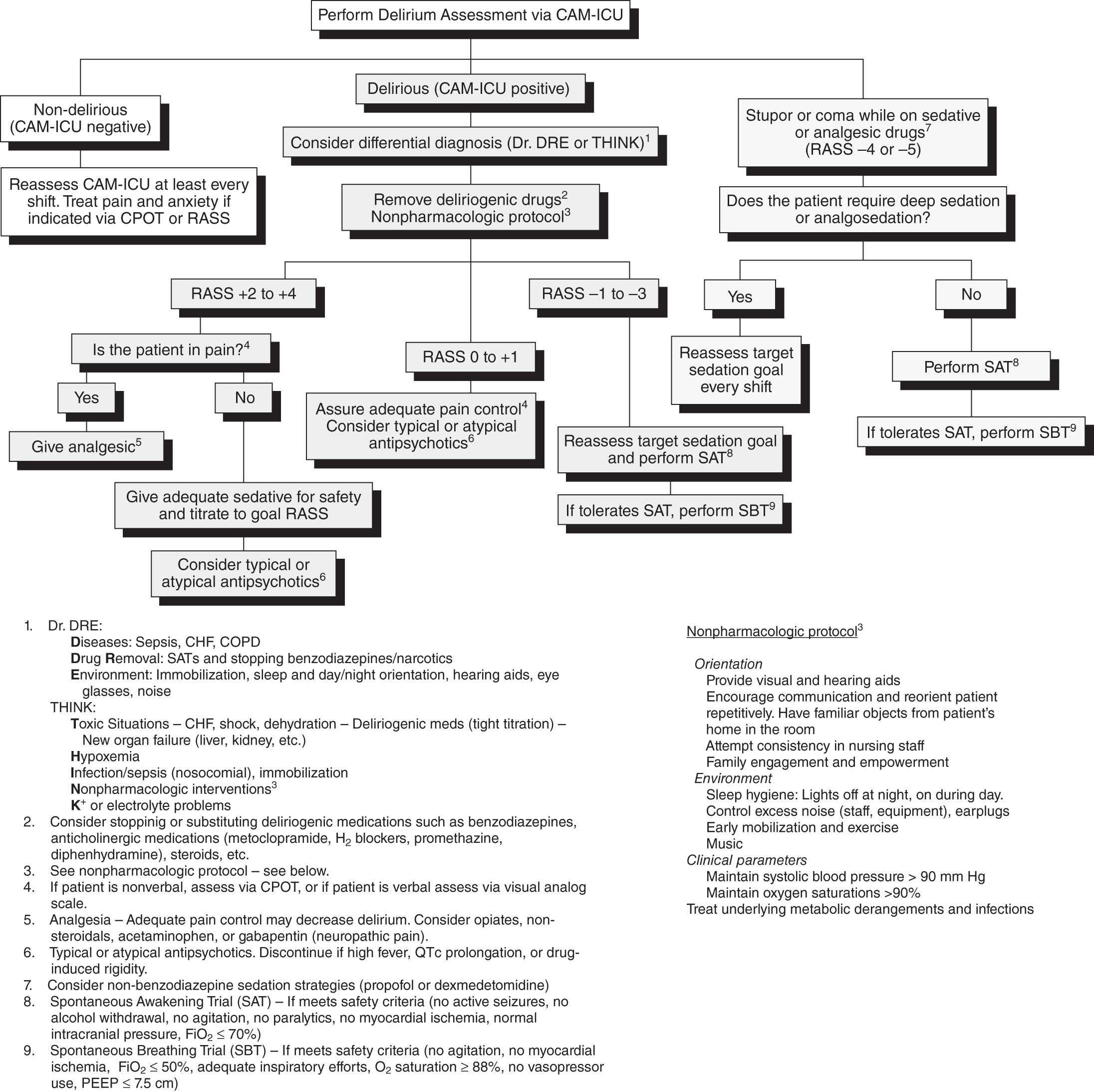
- •Fig. 1 describes a delirium protocol.
- •Table 3 summarizes delirium assessment tools.
Figure 1 Delirium protocol as a part of the ABCDEF Bundle.

(From Vincent JL et al: Textbook of critical care, ed 8, Philadelphia, 2024, Elsevier.)
TABLE 3 Delirium Assessment Tools
| Tool | Structure | Notes |
| Confusion Assessment Method (CAM) | Full scale of 11 items Abbreviated algorithm targeting four cardinal symptoms | Intended for use by nonpsychiatric clinicians |
| Confusion Assessment Method for the Intensive Care Unit (CAM-ICU) | Algorithm targeting four cardinal symptoms | Designed for use by nursing staff in the ICU |
| Intensive Care Delirium Screening Checklist (ICDSC) | 8-item screening checklist | Bedside screening tool for use by nonpsychiatric physicians or nurses in the ICU |
| Delirium Rating Scale (DRS) | Full scale of 10 items Abbreviated 7- or 8-item subscales for repeated administration | Provides data for confirmation of diagnosis and measurement of severity |
| Delirium Rating Scale-Revised-98 (DRS-R-98) | 16-item scale that can be divided into a 3-item diagnostic subscale and a 13-item severity subscale | Revision of DRS is better suited to repeat administration |
| Memorial Delirium Assessment Scale (MDAS) | 10-item severity rating scale | Grades severity of delirium once diagnosis has been made |
| Neecham Confusion Scale | 10-item rating scale | Designed for use by nursing staff and primarily validated for use in elderly populations in acute medical or nursing home setting |
| Delirium Diagnostic Tool-Provisional (DDT-Pro) | 3-item clinical assessment | Interpretation includes sub-syndromal delirium diagnosis |
From Stern TA: Massachusetts General Hospital handbook of general hospital psychiatry, ed 3, Philadelphia, 2025, Elsevier.
Can be multifactorial; often falls into one of the following categories (Table 4):
- •Drugs: Benzodiazepines, narcotics, anticholinergics, beta-blockers, steroids, and certain antibiotics (e.g., cefepime) can cause delirium as can withdrawal states such as those from alcohol and benzodiazepines9
- •Infection or inflammation: states of systemic inflammation, such as associated with burns, long-bone fractures or certain surgeries like abdominal aortic aneurysm repair are highly associated with delirium; there is some evidence that premorbid use of anti-inflammatory medications such as NSAIDs is protective in this regard7
- •States of reduced cerebral perfusion: often seen postoperatively, accompanying cardiac arrythmias, or in sepsis/hypotension; those with history of vascular disease are most vulnerable
- •Metabolic: Hypoxia, kidney or liver failure, thyroid, adrenal, or glucose dysregulation, anemia, vitamin deficiencies such as thiamine, B12, folate, vitamin D, vitamin C
- •Fluids, electrolytes, nutrition (FEN): Dysregulation of calcium, magnesium, potassium, or sodium; dehydration; volume overload; altered pH
- •Brain disorder: Central nervous system (CNS) infection, traumatic brain injury, hypertensive encephalopathy
TABLE 4 Major Causes of Delirium
| Metabolic | Electrolytes: Hypo/hypernatremia, hypo/hypercalcemia, hypo/hypermagnesemia, hypo/hyperphosphatemia | ||
| Endocrine: Hypo/hyperthyroidism, hypo/hypercortisolism, hypo/hyperglycemia | |||
| Cardiac encephalopathy, hepatic encephalopathy, uremic encephalopathy | |||
| Hypoxia and hypercarbia | |||
| Vitamin deficiencies: Vitamin B12, nicotinic acid, folic acid. Most notably Wernicke encephalopathy from thiamine deficiency | |||
| Toxic and industrial exposures: Carbon monoxide, organic solvent, lead, manganese, mercury, carbon disulfide, heavy metals | |||
| Porphyria | |||
| Toxic | Intoxication and overdose | ||
| Serotonin syndrome | |||
| Withdrawal: Alcohol, benzodiazepines, barbiturates, amphetamines, cocaine, coffee, phencyclidine, hallucinogens, inhalants, meperidine, and other narcotics | |||
| Drugs: Anticholinergic, benzodiazepines, opiates, antihistamines, antiepileptics, muscle relaxants, dopamine agonists, monoamine oxidase inhibitors, levodopa, corticosteroids, fluoroquinolone and cephalosporin antibiotics, beta-blockers, digitalis, lithium, clozapine, tricyclic antidepressants, calcineurin inhibitors | |||
| Infectious | Urinary tract infection, pneumonia, sepsis, meningitis, encephalitis, Creutzfeldt-Jakob and other prion diseases | ||
| Neurologic | Vascular: Ischemic stroke, intracerebral or subarachnoid hemorrhage, vasculitis | ||
| Autoimmune and paraneoplastic encephalitides | |||
| Neoplastic: Brain tumors, carcinomatous meningitis | |||
| Seizure related: Postictal state, nonconvulsive status epilepticus | |||
| Trauma: Concussion, subdural hematoma | |||
| Perioperative | Surgery: Thoracic (cardiac and noncardiac), vascular, and hip replacement, anesthetic and drug effects, hypoxia and anemia, hyperventilation, fluid and electrolyte disturbances, hypotension, embolism, infection or sepsis, untreated pain, fragmented sleep, sensory deprivation or overload | ||
| Miscellaneous | Hyperviscosity syndromes |
From Jankovic J et al: Bradley and Daroff’s neurology in clinical practice, ed 8, Philadelphia, 2022, Elsevier.


