section name header

Basic Information

Author: Yiyu Xie, MD and Patan Gultawatvichai, MD

Definition

Disseminated intravascular coagulation (DIC) is an acquired thromboembolic disorder characterized by generalized activation of the clotting pathways, which results in the intravascular formation of fibrin and, ultimately, thrombotic occlusion of small and midsize vessels, reducing blood supply to multiple organs and contributing to end-organ damage.

Synonyms

  • Consumptive coagulopathy
  • DIC
  • Defibrination syndrome
ICD-10CM CODE
D65Disseminated intravascular coagulation [defibrination syndrome]
Epidemiology & Demographics

A 1996 study from Japan showed that about 1% of patients admitted to university hospitals had evidence of DIC. There is no preference for age or gender. More than 50% of cases are associated with gram-negative sepsis or other septicemic infections, and up to 35% of patients with severe sepsis have DIC.

Physical Findings & Clinical Presentation

DIC can occur in acute (decompensated) and chronic (compensated) forms and can present with bleeding, thrombosis, or laboratory evidence of clotting cascade activation and fibrinolysis without evident clinical sequelae. Acute DIC is more common and predominantly manifests as bleeding complications. The risk of bleeding is four- to fivefold higher when the platelet count is below <50 × 109/liter. Sudden procoagulant exposure can prompt coagulation cascade activation and platelet consumption, resulting in thrombosis. In contrast, chronic DIC is more likely to cause thrombotic complications. The diagnosis of chronic DIC can be challenging, as the prothrombin time (PT) and partial thromboplastin time (PTT) are often normal. Multiple pathways are involved in DIC pathophysiology, ultimately leading to consumptive coagulopathy and thrombosis. These include (1) thrombin generation due to the release of tissue factor or other procoagulants, (2) suppression of physiologic anticoagulant (e.g., protein C/S or antithrombin insufficiency), (3) impaired fibrinolysis characterized by an increased level of plasminogen activator inhibitor type 1 (PAI-1) and fibrin degradation products, and (4) activation of inflammatory pathways.

Multiple organs may be affected by DIC, leading to a variety of clinical presentations depending on the organs involved. Symptoms can include:

  • Central nervous system: Altered mental status, transient neurologic deficits
  • Cardiovascular: Hypotension, tachycardia
  • Respiratory: Hypoxia, dyspnea, localized rales, and acute respiratory distress syndrome
  • GI: Intestinal bleeding, bowel infarction
  • Genitourinary: Oliguria, anuria, uremia, acidosis, metrorrhagia
  • Skin: Wound site bleeding, epistaxis, gingival bleeding, hemorrhagic bullae, petechiae, ecchymosis, purpura, skin necrosis
Etiology

  • Traditionally, DIC was thought to be the result of activation of both the extrinsic and intrinsic pathways of coagulation. The classical concept was that the extrinsic pathway was initiated by a tissue-derived component, which activated factor VII, leading to the direct conversion of prothrombin to thrombin. This process would proceed as long as there was tissue damage from systemic infection, trauma, placental abruption, or malignancy. In contrast, the intrinsic or contact pathway of coagulation was initiated by contact activation of factor XII which, together with its cofactors, kallikrein and kininogen, then activated factor XI with subsequent activation of factor IX. The initiators of contact activation were poorly understood until recently but were thought to include collagen and artificial surfaces. In recent years, the molecular mechanisms of coagulation pathways have been defined (Fig. E1). This has provided new insight into the pathogenesis of DIC. In general, current thinking is that thrombin and fibrin generation in patients with DIC is largely driven via the extrinsic pathway; the role of the contact system is uncertain. The development of DIC is counteracted by several mechanisms. First, coagulation inhibitors regulate the coagulation mechanism. These inhibitors include antithrombin (AT), the protein C pathway, and TFPI (Fig. E2). AT, which complexes and inhibits thrombin and factor Xa, is one of the most important inhibitors, and reduced AT levels are a characteristic of DIC. Reductions in AT levels reflect a combination of reduced protein synthesis, increased clearance through the formation of protease-AT complexes, and AT degradation by neutrophil elastase. In addition, cytokines may impair proteoglycan synthesis in the vessel wall, thereby reducing the availability of heparan sulfate for the potentiation of AT activity.1
  • As a result of increased thrombus generation in the small and medium vessels, clotting factors and platelets are consumed more rapidly than the synthetic function of the liver and bone marrow, respectively. Of note, DIC is not a disease in itself. Diseases associated with DIC are summarized in Box E1. Severe infection is the most common inciting etiology; an extensive list of other triggers is known, including:
  • Infections (e.g., gram-negative or -positive sepsis, Rocky Mountain spotted fever, COVID-19, malaria, viral or fungal infection)
  • Obstetric complications (e.g., fetal demise, amniotic fluid embolism, toxemia, abruptio placentae, septic abortion, preeclampsia/eclampsia, placenta previa, uterine atony)
  • Tissue trauma (e.g., polytrauma, burns, hypothermia rewarming)
  • Malignancy (e.g., adenocarcinomas [GI, prostate, lung, breast], especially mucin-producing cancers, lymphoproliferative/myeloproliferative; DIC is a hallmark of acute promyelocytic leukemia and a leading cause of mortality)
  • Quinine, cocaine-induced rhabdomyolysis
  • Liver failure
  • Acute pancreatitis
  • Transfusion reactions
  • Respiratory distress syndrome
  • Toxins (snake bites, amphetamine overdose)
  • Other: Systemic lupus erythematosus (SLE), vasculitis, aneurysms, polyarteritis, hemangiomas with thrombocytopenia, and consumptive coagulopathy (Kasabach-Merritt syndrome)

Figure E1 Pathways Involved in the Activation of Coagulation in Disseminated Intravascular Coagulation
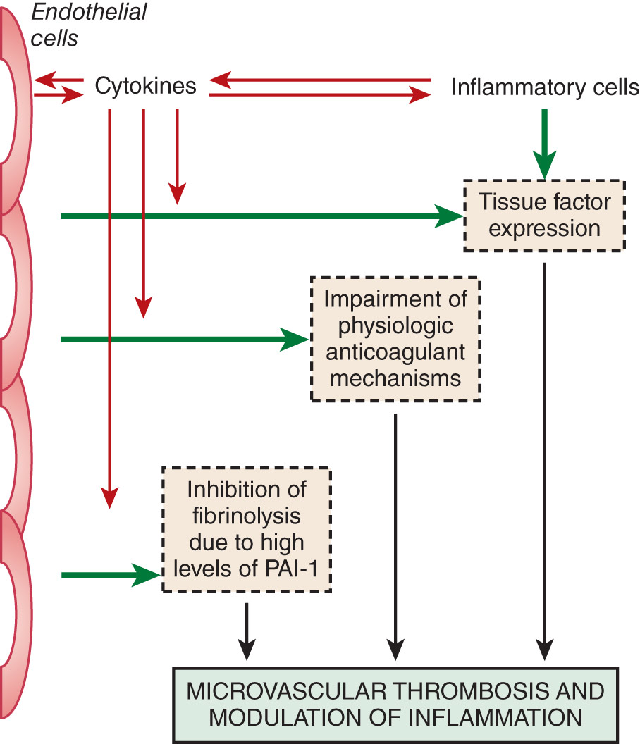
Both perturbed endothelial cells and activated mononuclear cells may produce proinflammatory cytokines that induce tissue factor expression, thereby initiating coagulation. In addition, downregulation of physiologic anticoagulant mechanisms and inhibition of fibrinolysis promote intravascular fibrin deposition. PAI-1, Plasminogen activator inhibitor, type 1.

(From Hoffman R et al: Hematology, basic principles and practice, ed 8, Philadelphia, 2023, Elsevier.)

Figure E2 Physiologic anticoagulant mechanisms in disseminated intravascular coagulation.
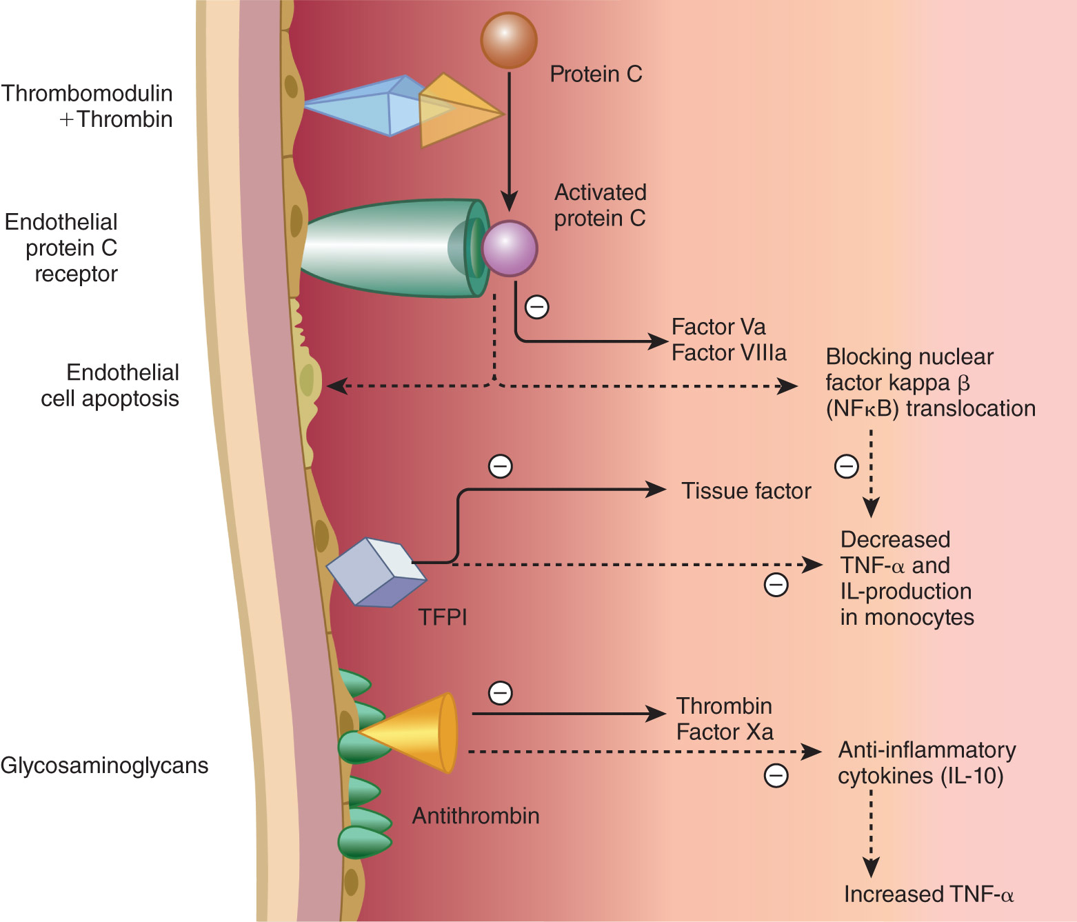
Physiologic anticoagulant mechanisms (activated protein C system, tissue factor pathway inhibitor [TFPI], and antithrombin) are not only involved in blocking thrombin generation and thrombin activity but also affect inflammatory pathways.

(From Hoffman R et al: Hematology, basic principles and practice, ed 8, Philadelphia, 2023, Elsevier.)

BOX E1 Diseases Associated With Disseminated Intravascular Coagulation

Sepsis

  • Typically, gram-negative bacteria contain lipopolysaccharides in their cell membranes, but this can be seen with any microorganism.
Pregnancy and Obstetric Complications

  • Preeclampsia
  • Placental abruption
  • Amniotic fluid embolism
Malignancy

  • Acute promyelocytic leukemia (characteristic feature)
  • Solid tumors
Trauma

  • Burns
  • Fat embolism
  • Severe trauma
Envenomation and Immunologic Reactions

  • ABO-incompatible red cell transfusion
  • Snakebite
Miscellaneous

  • Severe pancreatitis
  • Giant hemangiomas

From Talley NJ et al: Essentials of internal medicine, ed 4, Chatswood, NSW, 2021, Elsevier Australia.

Diagnosis

Differential Diagnosis

  • Severe liver disease: Normal or elevated factor VIII levels
  • Vitamin K deficiency: Normal platelet counts
  • Thrombotic microangiopathy (TMA): Coagulation testing is usually normal in thrombotic thrombocytopenic purpura (low ADAMTS13 activity), drug-induced TMA, and hemolytic uremic syndrome
  • Heparin-induced thrombocytopenia (HIT): History of heparin exposure and positive testing for HIT antibodies
  • Renal failure, SLE, sickle cell crisis, dysfibrinogenemia
  • HELLP syndrome (hemolysis, elevated liver function tests, and low platelets)
  • COVID-19 related coagulopathy: Elevated fibrinogen, SARS-CoV-2 PCR positive
Workup

The diagnostic workup includes laboratory testing to characterize the coagulopathy and its severity and to exclude conditions noted in the differential diagnosis (Table 1, Box 2). Additional workup is guided by the clinical scenario and may involve distinguishing between acute and chronic DIC, identifying the primary manifestations (thrombotic or hemorrhagic), and assessing the extent of involvement (localized or systemic).

BOX 2 Diagnostic Algorithm for the Diagnosis of Overt Disseminated Intravascular Coagulationa

  • Presence of an underlying disorder known to be associated with disseminated intravascular coagulation (DIC) (see Table 1) (no = 0, yes = 2)
  • Score global coagulation test results
    1. 1.Platelet count (>100 = score 0; <100 = score 1; <50 = score 2)
    2. 2.Level of fibrin markers (e.g., D-dimer, fibrin degradation products) (no increase = score 0; moderate increase = score 2; strong increase = score 3)b
    3. 3.Prolonged prothrombin time (<3 sec = score 0; >3 sec but <6 sec = score 1; >6 sec = score 2)
    4. 4.Fibrinogen level (>1.0 g/L = score 0; <1.0 g/L = score 1)
  • Calculate score
  • If 5: Compatible with overt DIC; repeat scoring daily. If <5: Suggestive (not affirmative) for nonovert DIC; repeat next 1-2 days.

From Hoffman R et al: Hematology: basic principles and practice, ed 8, Philadelphia, 2023, Elsevier.

TABLE 1 Differential Diagnosis of Prolonged aPTT and PT in Suspected Disseminated Intravascular Coagulation

Test ResultCause
  • PT prolonged, aPTT normal
  • Factor VII deficiency
  • Mild vitamin K deficiency
  • Mild liver insufficiency
  • Low doses of vitamin K antagonists
  • PT normal, aPTT prolonged
  • Factor VIII, IX, or XI deficiency
  • Unfractionated heparin
  • Inhibitory antibody and/or antiphospholipid antibody
  • Factor XII or prekallikrein deficiency
  • Both PT and aPTT prolonged
  • Factor X, V, II, or fibrinogen deficiency
  • Severe vitamin K deficiency
  • Vitamin K antagonists
  • Global clotting factor deficiency
  • Decreased synthesis: Liver failure
  • Increased loss: Massive bleeding, DIC

aPTT, Activated partial thromboplastin time; DIC, disseminated intravascular coagulation; PT, prothrombin time.

From Hoffman R et al: Hematology: basic principles and practice, ed 8, Philadelphia, 2023, Elsevier.

Laboratory Tests

  • Peripheral blood smear generally shows red blood cell fragments (schistocytes) and low platelet counts.
  • Coagulation testing: Diagnostic characteristics of DIC are decreased fibrinogen level, thrombocytopenia, and increased PT, PTT, thrombin time (TT), fibrin split products, and D-dimer.
  • Coagulopathy secondary to DIC must be differentiated from other coagulopathies that result from the underproduction of clotting factors, such as those seen in liver disease or vitamin K deficiency.
    1. 1.Vitamin K deficiency manifests with prolonged PT but normal PTT, TT, platelet, and fibrinogen levels; PTT may be elevated in severe cases.
    2. 2.Patients with liver disease have abnormal PT and PTT; TT and fibrinogen are usually normal unless severe disease is present; platelets are typically normal unless splenomegaly is present.
    3. 3.Factor VIII is not synthesized exclusively by the liver. It can differentiate between DIC, where it is low, and coagulopathy of liver disease, where it is normal or elevated.
Imaging Studies

Imaging studies are generally not helpful. Imaging may help identify sequelae of DIC, including chest radiographs to exclude infectious processes in patients with pulmonary symptoms such as dyspnea, cough, or hemoptysis.

a According to the International Society of Thrombosis and Haemostasis Scientific Standardization Committee.

b Strong increase, greater than 5× upper limit of normal; moderate increase, greater than upper limit of normal but less than 5× upper limit of normal.

Treatment

Acute General Rx

  • Correcting and eliminating the underlying cause is often sufficient to halt DIC (e.g., antimicrobial therapy for infection, removal of necrotic bowel, evacuation of uterus in obstetric emergencies).
  • Patients with bleeding should be given replacement therapy with fresh frozen plasma (FFP) and platelets:
    1. 1.FFP 10 to 15 ml/kg can normalize the international normalized ratio.
    2. 2.Platelet transfusions are given when the platelet count is <10,000 (or higher if significant bleeding is present).
    3. 3.Cryoprecipitate 1 U/5 kg is given for low fibrinogen. It can also be provided for preventing bleeding in patients with fibrinogen levels <100 mg/dl.
  • Patients with extensive thrombosis (e.g., in acute promyelocytic leukemia, purpura fulminans, acral ischemia) require anticoagulation despite thrombocytopenia and abnormal coagulation parameters. Heparin therapy using unfractionated heparin with a lower PTT goal than is used in venous thrombosis may be helpful to increase the neutralization of thrombin. Low-molecular-weight heparin also may be used in this scenario.
  • The mainstays of supportive treatment of DIC are summarized in Box E3.

BOX E3 Mainstays of Supportive Treatment of Disseminated Intravascular Coagulation

ModalityDetailsExpectations/Rationale
Treating the underlying disorderDependent on the primary diagnosisInhibit or block the complicating pathologic mechanism of disseminated intravascular coagulation (DIC) in parallel with the response (if any) of the disorder
Antithrombotic agentsProphylactic heparin to prevent venous thromboembolic complications (low dose) therapeutic heparin in case of confirmed thromboembolism or if clinical picture is dominated by (micro)vascular thrombosis and associated organ failureRisk of thromboembolism is increased in critically ill patients, trauma patients, or patients with cancer. Prevent fibrin formation; tip the balance within the microcirculation toward anticoagulant mechanisms and physiologic fibrinolysis; allow reperfusion of the skin, kidneys, and brain
TransfusionInfuse platelets, plasma, and fibrinogen (cryoprecipitate) if there is overt bleeding or a high risk of bleedingBleeding should diminish and stop over the course of hours. Platelet count, coagulation tests, and fibrinogen should return toward normal
Anticoagulant factor concentratesRecombinant human activated protein C may be effective in sepsis and DIC (24 μg/kg/h for 4 days); currently withdrawn from the marketRestore anticoagulation in microvascular environment and may have antiinflammatory activity. Latest trials were negative
Fibrinolytic inhibitorsTranexamic acid (e.g., 500-1000 mg q8-12h or Ε-aminocaproic acid 1000-2000 mg q8-12h)May be useful if there is (hyper)fibrinolysis. Bleeding ceases, but there is a risk of microvascular thrombosis and renal failure

From Hoffman R et al: Hematology: basic principles and practice, ed 8, Philadelphia, 2023, Elsevier.

Disposition

The mortality rate in severe DIC exceeds 75%. The high mortality rate is often due to the severity of the underlying trigger and complications, including acute renal failure, intracerebral bleeding, shock, or cardiac tamponade.

Referral

Hematology consultation is recommended in all cases of severe DIC and DIC with hemorrhagic or thrombotic complications.

Pearls & Considerations

Comments

The treatment of chronic DIC is controversial. Low-dose subcutaneous heparin and/or combination antiplatelet agents such as aspirin and dipyridamole may be helpful.

Related Content

  • Disseminated Intravascular Coagulation (Patient Information)

Reference(s)

  1. Levi M : Disseminated intravascular coagulation Hoffman R, editors : Hematology, basic principles and practice. ed 8Elsevier-Philadelphia, 2023.