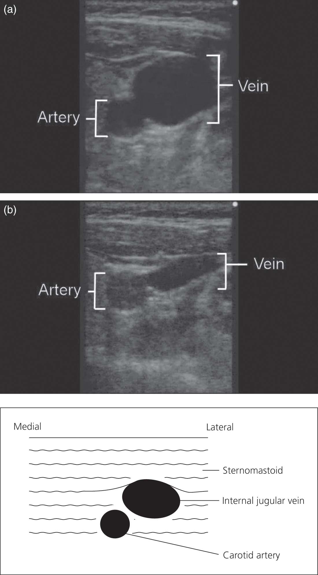
Ultrasonography of the Right Internal Jugular Vein

Ultrasonography of the right internal jugular vein, before (a) and after (b) compression by the ultrasound probe. The vein can be distinguished from the artery by its compressibility. The depth from the skin to the anterior wall of the vein averages 11 mm (range 6–18 mm). The vein is usually 10 mm in diameter, but its calibre is reduced in volume-depletion. Head-down tilt or volume loading increases the diameter of the vein and facilitates venepuncture.