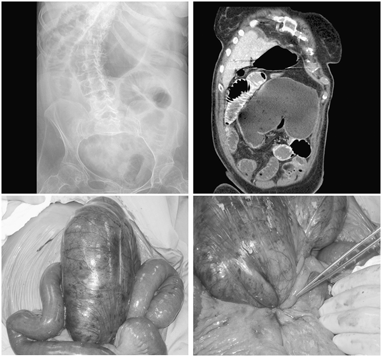
A plain abdominal radiograph (left upper) and a coronal computed tomography (CT) image (right upper) demonstrating cecal volvulus. Intraoperative photographs reveal a dilated and ischemic cecum (left lower), as well as the site of torsion (right lower).