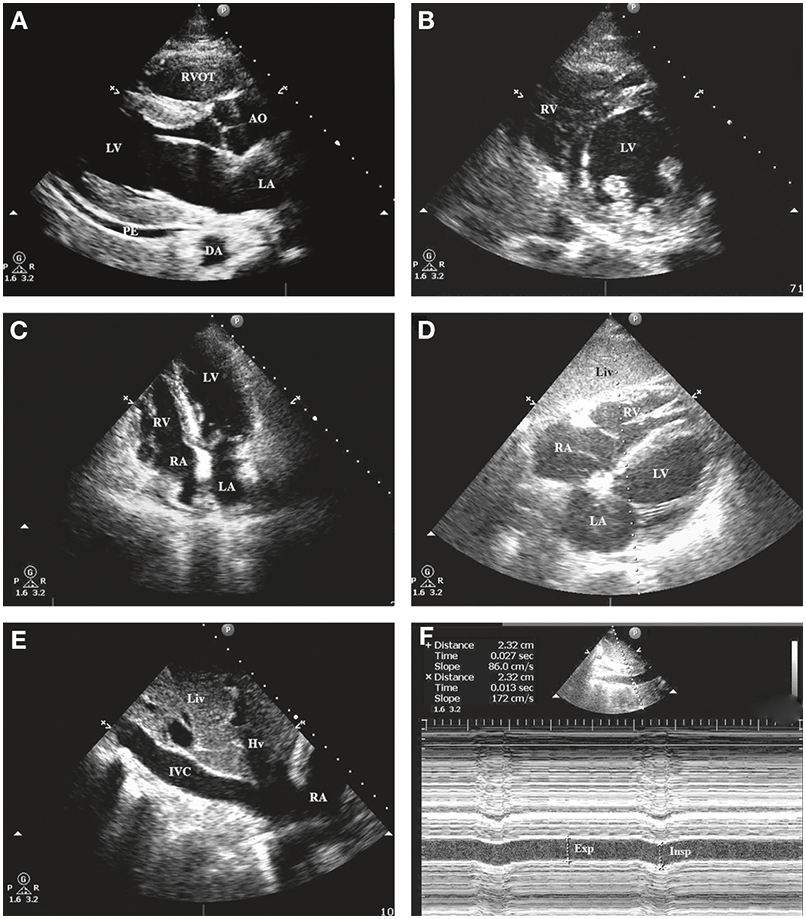
Salient anatomic features are noted on the figures. (A)Parasternal long-axis view in diastole (mitral valve wide open; aortic valve closed). Note the small posterior pericardial effusion (PE). (B)Parasternal short-axis view. (C)Apical four-chamber view. (D)Subcostal four-chamber view. (E)Visualization of the inferior vena cava (IVC). (F)M-mode interrogation of the IVC—no change in diameter between inspiration and expiration. AO, ascending aorta; DA, descending thoracic aorta; Exp, expiration; Hv, hepatic vein; Insp, inspiration; LA, left atrium; Liv, liver; LV, left ventricle; RA, right atrium; RV, right ventricle; RVOT, right ventricular outflow tract.