Ultrasound Examination of the Lung: (A) A-Lines, Which are Consistent with Normal Aeration of the Lung Field Under Examination . (B) Multiple B-Lines Seen in This Sector, Suggesting Excess Interstitial Fluid. Note that A-Lines are Not Visible Because They are Effaced by B-Lines . (C) An Uncomplicated Pleural Effusion. It is Important to Use the Anatomic Landmarks (in This Case, Liver and Diaphragm below and a Portion of Atelectatic Lung above) to Confirm that the Fluid Being Visualized is Pleural Fluid and Not Ascites . (D) M-Mode Interrogation of the Pleural Surface to Show the “seashore Sign” that is Caused by the Movement of the Parietal and Visceral Pleura over Each Other. The Two-Dimensional (2d) Image Being Scanned by M-Mode is Shown to the Right . Rs, Rib Shadow.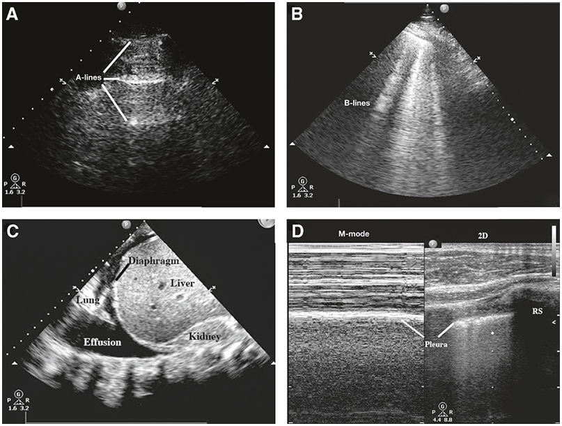
Ultrasound examination of the lung: (A)A-lines, which are consistent with normal aeration of the lung field under examination. (B)Multiple B-lines seen in this sector, suggesting excess interstitial fluid. Note that A-lines are not visible because they are effaced by B-lines. (C)An uncomplicated pleural effusion. It is important to use the anatomic landmarks (in this case, liver and diaphragm below and a portion of atelectatic lung above) to confirm that the fluid being visualized is pleural fluid and not ascites. (D)M-mode interrogation of the pleural surface to show the “seashore sign” that is caused by the movement of the parietal and visceral pleura over each other. The two-dimensional (2D) image being scanned by M-mode is shown to the right. RS, rib shadow.