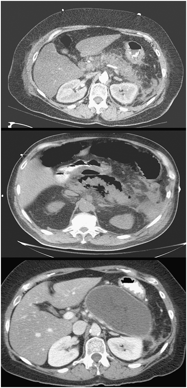
Axial computed tomography (CT) images demonstrating acute pancreatitis with prominent pancreatic inflammation (top), pancreatic necrosis with air around the pancreas (middle), and a giant pancreatic pseudocyst that developed several weeks after the acute episode (bottom).