Introduction ⬇
General Considerations ⬆ ⬇
In the ascending aorta, aneurysms tend to take on three common patterns, as indicated in Figure 37–1. These include the supracoronary aortic aneurysm, annuloaortic ectasia (Marfanoid), and tubular diffuse enlargement.
Figure 37-1. The three common patterns of ascending aortic aneurysm.
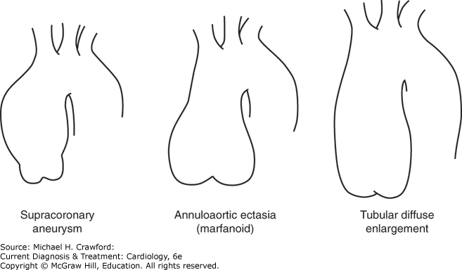
The most common pattern is that of supracoronary dilatation of the ascending aorta. In this pattern of disease, the short segment of aorta between the aortic annulus and the coronary arteries remains normal in size. Sinuses are “preserved,” meaning that the aorta indents normally, forming a “waist,” just above the level of the coronary arteries. For this type of aneurysm, a supracoronary tube graft suffices.
In the second type, annuloaortic ectasia, the aortic annulus itself becomes dilated, giving a shape to the aorta like an Erlenmeyer chemistry flask. In this type of disease, the segment of aorta between the annulus and the coronary arteries is diseased, dilated, and thinned. The sinuses of Valsalva are “effaced,” meaning that the normal indentation, or waist, is lost. When surgery is required, the entire aortic root must be replaced.
In the third type of ascending aortic disease, the configuration is midway between the previous two patterns; that is, there is some dilatation of the annulus and root and some effacement of the sinuses, but these elements are not dramatic. The overall appearance is that of a large tube, rather than a flask. For such aortas, either supracoronary tube grafting or aortic root replacement may be appropriate.
The Crawford classification (Figure 37–2) is used to categorize the appearance of an aneurysm in the descending aorta and thoracoabdominal aorta. This classification, based on the longitudinal location and extent of aortic involvement, has implications for surgical strategy and affects the risk of perioperative complications.
Figure 37-2. The Crawford classification of descending and thoracoabdominal aneurysms. See text for description of each type. (Reproduced with permission from Cohn LH, Edmunds LH Jr, eds. Cardiac Surgery in the Adult, 2nd ed. New York: McGraw-Hill; 2003.)
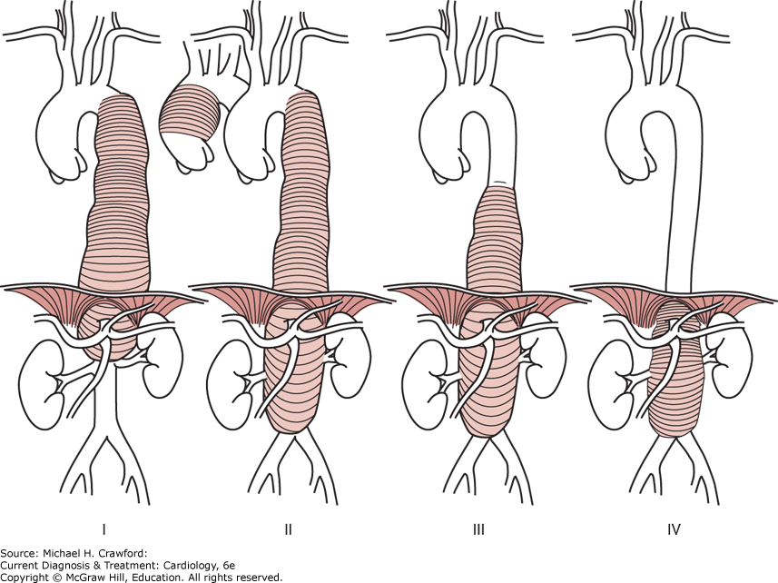
Type I aneurysms involve most of the thoracic aorta and the upper abdominal aorta. Type II aneurysms, the most extensive and most dangerous to repair, involve the entire descending and abdominal aortas. Type III aneurysms involve the lower thoracic and abdominal aortas. Type IV aneurysms are predominantly abdominal but involve thoracoabdominal exposure because of the proximity of the upper border to the diaphragm.
Pathophysiology/Etiology ⬆ ⬇
Etiology
The genetics of Marfan disease, a well-known cause of aneurysms of the thoracic aorta, have been well delineated, with over 1300 mutations identified on the fibrillin gene.
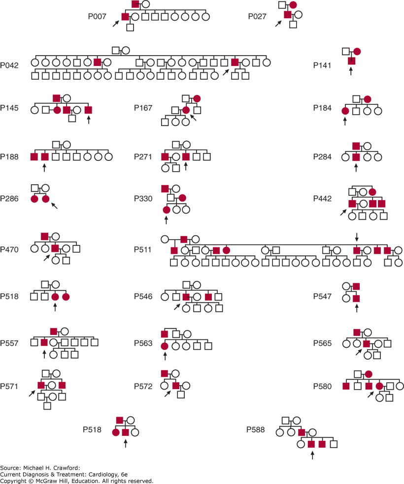
Increasingly, it is being appreciated that patients who do not have Marfan disease also manifest familial clustering of thoracic aortic aneurysms and dissections. Patients with aneurysms often answer one or both of the following questions affirmatively: “Do you have any family members with aneurysms anywhere in their bodies? Did any of your relatives die suddenly or unexpectedly of apparent cardiac causes?” Detailed construction of family trees on over 500 patients with thoracic aneurysm has indicated that 21% of aneurysm probands have a first-degree relative with a known or likely aortic aneurysm. The true number is certainly much higher, as these estimates are based only on family interview and not on head-to-toe imaging of relatives. Figure 37–3 shows the 21 positive family trees of 100 families analyzed. The most likely pattern of inheritance appears to be autosomal dominant with incomplete penetrance. A more recent analysis has shown that the location of the proband’s aneurysm largely influences the location of the aneurysms in the family members. If the proband has an ascending aneurysm, the likelihood is that the family members have ascending aneurysms. If, however, the proband has a descending aneurysm, it is likely that the family members have abdominal aortic aneurysms. These proband–family member observations are in keeping with the general concept that aneurysm disease divides at the ligamentum arteriosum: Ascending and arch aneurysms represent one disease, largely nonatherosclerotic, while descending and abdominal aneurysms represent another disease, largely atherosclerotic.
Figure 37-3. The 21 positive family trees among the first 100 families assessed for genetic patterns of thoracic aortic disease.
This concept of two diseases separated at the ligamentum arteriosum correlates perfectly with recent evidence that the ascending and descending aortic smooth muscle cells arise from different embryologic layers (neural crest and mesoderm, respectively).
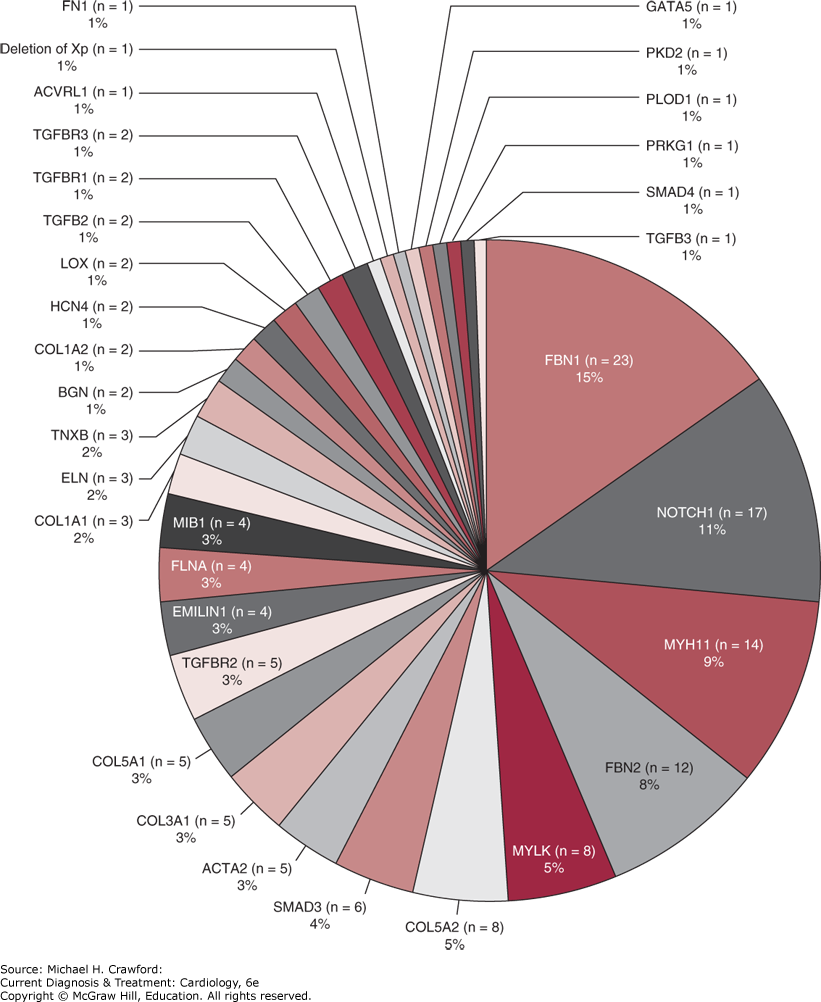
Application of modern molecular genetic techniques is successfully making progress toward determining the specific genetic aberrations responsible for family clustering and for thoracic aneurysms in general. Original progress was made largely by linkage analysis of large families with multiple members affected. Now, as automated genomic analysis has become feasible and affordable, much scientific discovery has been made by direct exome sequencing. Milewicz, Dietz, Loeys, and others have succeeded in identifying specific familial aneurysm patterns and their underlying mutations. Nearly all these familial aortic disorders are transmitted in an autosomal dominant fashion (with decreased penetrance). The ACTA2 disorder presents with aortic dissection at small diameters and the MYLK disorder may dissect without prior aneurysmal enlargement. These mutation-specific categorizations have led to the advent of personalized medicine based on the specific underlying mutation and its accompanying rupture and dissection patterns. Specialized centers have begun routine whole exome sequencing (WES) in all patients with thoracic aortic aneurysm and dissection. Figure 37–4 shows the distribution of variants found at one high-volume center.
Figure 37-4. Frequency distribution of genetic defects in genes implicated with thoracic aortic disease, identified via whole exome sequencing.
It is important to recognize that most genetically triggered thoracic aortic aneurysms are “single gene, single letter” diseases. That is to say, not only are these aneurysmal diseases caused by a single gene, but also by only a single change in a single “letter” (nucleotide or base) among the 2.2 billion letters that comprise the human genome. This stands in contradistinction to many other diseases, which are genetically multifactorial (eg, dozens of genes are known to contribute to coronary artery disease). This single-letter causation is crucially important—providing opportunities for precise diagnosis and, hopefully, future genetically based treatment.
Examination of single nucleotide polymorphisms (SNPs) in the blood of hundreds of patients with thoracic aortic aneurysms via genome-wide surveys using large (> 30,000) SNP libraries has been accomplished. An “RNA signature” in the blood of patients with thoracic aortic aneurysm was found, which can predict with about 85% accuracy from a blood test alone whether the patient harbors a thoracic aortic aneurysm. This “signature” is composed of specific RNAs that are either markedly upregulated or markedly downregulated in aneurysm patients, compared with healthy controls.
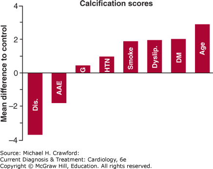
Patients who have a genetic predisposition for aneurysm development, specifically those patients with annuloaortic ectasia or ascending aortic aneurysm, are significantly protected from atherosclerosis (Figure 37–5). Remarkably, they have less arterial medial thickness and less calcification than normal controls—and myocardial infarction is very rare in these patients. It appears likely that the same mutations that promote lysis of the aortic wall also prevent plaque build-up.
Figure 37-5. Difference in overall calcification scores relative to the control group for all risk factors analyzed. Note that patients with ascending aortic dissection or annuloaortic ectasia are significantly protected from arteriosclerosis, manifesting lower calcification scores. AAE, annuloaortic ectasia; Age, age (in 10-year intervals); Dis, ascending aortic dissection; DM, diabetes mellitus; Dyslip, dyslipidemia; G, male gender; HTN, hypertension; Smoke, smoking history.
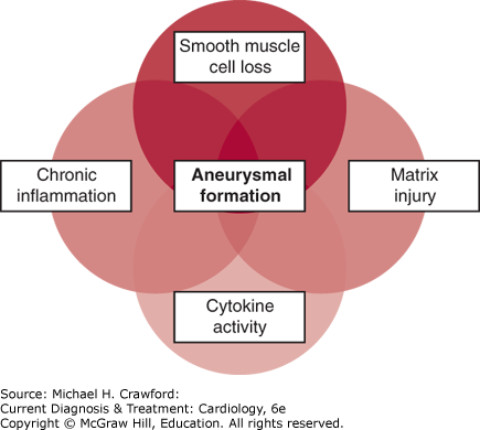
Accepting that most patients with thoracic aortic aneurysms have an underlying genetic predisposition to the condition, how does this genetic programming lead to the development of an aneurysm? Rapid progress is being made in elucidating these mechanisms. Aneurysm formation is currently thought to involve the following processes (Figure 37–6): extracellular matrix proteolysis, chronic inflammation, cytokine activity, and smooth muscle cell loss. A recent recognition is that abnormal mechanosensing and mechanoregulation between smooth muscle cells and the aortic lamellae and matrix also play an important role. The identification of these mechanisms raises the intriguing possibility of interfering pharmacologically with this pathophysiology, so that aneurysm formation or progression can be stopped. The importance of the transforming growth factor-β (TGF-β) pathway in aneurysm formation has been demonstrated; the ability of angiotensin receptor–blocking medications (ARBs) (eg, losartan) to interfere with this pathophysiologic mechanism is being tested, and results of randomized, controlled studies are now becoming available. One recent large study failed to show any clinical advantage of ARBs. At this time, it may be said that no specific pharmacologic strategy exists for delaying aneurysm progression. Results of trials of proteolytic antagonists and β-blockers have been underwhelming. The potential roles of statin medications, anti-inflammatory agents (cyclooxygenase [COX]-2 inhibitors), immunosuppressants (sirolimus), and antibiotics (doxycycline) are being investigated.
Figure 37-6. Diagram illustrating the overlapping cellular and molecular processes that contribute to aortic aneurysm formation.
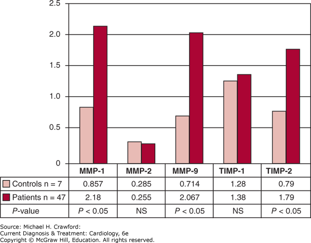
The proteolytic enzymes called matrix metalloproteinases (MMPs) are receiving extensive attention in aneurysm pathophysiology. These powerful enzymes are found in excess in thoracic aortic aneurysms (Figure 37–7) and are thought to play a major role in destroying the substance of the aortic wall, leading to decreased wall strength and, ultimately, dilatation and rupture.
Figure 37-7. Note overabundance of matrix metalloproteinase (MMP)-1 and MMP-9 and TIMP-2 in aortas of patients with thoracic aortic aneurysm, compared with controls. This information suggests an important role for MMP enzymes in the pathophysiology of aneurysm disease. NS, not significant; TIMP, tissue inhibitor of metalloproteinases.
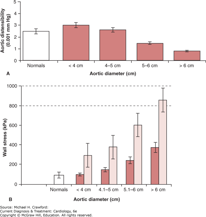
The biologic changes in the aortic wall discussed earlier are vitally important, but hemodynamic forces need to be considered as well. As the ascending aorta reaches a diameter of 6 cm, its distensibility vanishes, so that the aorta becomes essentially a rigid tube (Figure 37–8). Because of this rigidity, the force of systole can no longer be beneficially dissipated by elastic expansion of the aorta, and this translates into increased wall stress. Especially at high blood pressures, this wall stress becomes excessive, setting the stage for disruption of the aortic wall via rupture or dissection. It is instructive to note how closely this mechanical data dove-tails with the clinical behavior of the aorta: The mechanical properties deteriorate at 6 cm diameter, and that is precisely the hinge point for clinically manifest rupture and dissection.
Figure 37-8. A: Distensibility values in normal aortas and aortic aneurysms of different diameters. Distensibility of ascending aortic aneurysms decreases rapidly as diameter increases, to very low values at dimensions 6 cm. At 6 cm, the aorta is essentially a rigid tube, unable to dissipate the force of systole by expanding phasically during the cardiac cycle. B: Exponential relationship between wall stress and aneurysm size in ascending aortic aneurysms. The dark columns represent a blood pressure of 100 mm Hg, and the light columns represent a blood pressure of 200 mm Hg. The lines at 8001000 kPa represent the range of maximum tensile strength of the human aorta. Note that a patient with a 6-cm aneurysm and a blood pressure of 200 mm Hg (as during stress or extreme exertion) flirts with the limits of the ultimate strength of his or her aorta wall.
Clinical Findings ⬆ ⬇
A. Incidence & Prevalence
It is generally acknowledged, based on data from the Centers for Disease Control and Prevention, that aneurysm disease is the 15th most common cause of death for human beings. The incidence of aortic dissection is estimated to be 30 per million population per year. However, new studies are confirming the long-standing suspicion that these figures represent gross underestimates. Very recent data show that, on computerized tomography (CT), aortic dilatation exceeding 4.0 cm is found in 2.8% of individuals older than 50 years. It has been rightly presumed that many individuals dying with chest pain after presenting to an emergency department may have aortic dissection rather than myocardial infarction. Now, evidence is accumulating that substantiates this presumption. Clinical series that have applied routine postmortem computed tomography in patients succumbing to out-of-hospital cardiac arrest have confirmed that 2–7% of such patients actually have died of Type A aortic dissection, with intrapericardial rupture.
B. Natural History
The Yale computerized database now contains information on nearly 4500 patients with thoracic aortic aneurysm, including many thousands of tabulated serial imaging studies and tens of thousands of patient-years of follow-up. This database and these methods of analysis have permitted assessment of multiple fundamental topics and questions regarding the natural behavior of the thoracic aorta and have shed light on appropriate criteria for surgical intervention.
1. How Fast Does the Thoracic Aorta Grow?
Via specifically developed statistical methods designed to account for important potential sources of error, the annual growth rate of an aneurysmal thoracic aorta has been determined to be, on average, 0.12 cm. The descending aorta grows faster than the ascending aorta, at 0.19 cm/year. Also, the larger the aorta becomes, the faster it grows.
2. At What Size Does the Aorta Dissect or Rupture?
Critical to decision-making in aortic surgery is an understanding of when complications occur in the natural history of unrepaired thoracic aortic aneurysms. In the case of the thoracic aorta, the two complications that are vitally important are rupture and dissection. Knowing when these complications are likely to occur permits rational decision-making regarding elective, preemptive surgical intervention to prevent their occurrence.
Size criteria apply only to asymptomatic aneurysms. We are learning increasingly that the aorta can indeed “communicate” with us—via pain. Symptomatic (painful) aneurysms should be resected regardless of size. For ascending aneurysms, this pain is usually felt anteriorly, under the breastbone. For descending thoracic aneurysms, the pain is usually felt in the interscapular region of the upper back. For thoracoabdominal aneurysms, the pain is usually felt lower in the back or in the left flank. Other symptoms may occasionally be produced by thoracic aortic aneurysms, including bronchial obstruction, esophageal obstruction, and phrenic nerve dysfunction; these symptoms also constitute indications for surgical intervention.
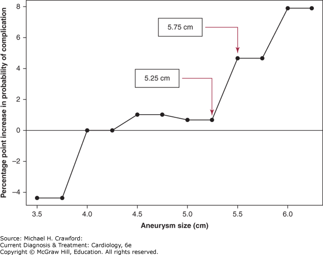
Very recent statistical analysis reveals two sharp “hinge points” (Figure 37–9) in ascending aortic size at which rupture or dissection occurred: at 5.25 and 5.75 cm. An aortic size of 6.0 cm was associated with an 8-percentage point increase in the probability of rupture and dissection relative to the 4.0–4.4 cm reference group.
Figure 37-9. The hinge points (arrows) in the cumulative, lifetime incidence of complications (rupture or dissection) of thoracic aortic aneurysms, based on size. By the time the ascending aorta reaches the dimensions on the x-axis, the percentage of patients shown on the y-axis have already incurred rupture or dissection.
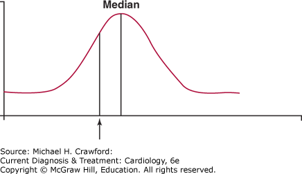
If a surgeon were to wait for the aorta to achieve the median size at the time of complications to intervene, by definition, rupture or dissection would have occurred in half of the patients (Figure 37–10). Accordingly, it is important to intervene before the median value is attained. The following recommendations take this factor into account, permitting preemptive surgical extirpation before rupture or dissection in most patients.
Figure 37-10. A schematic representation of the importance of selecting a criterion for intervention before complications (rupture or dissection) commonly occur. Utilization of the median as the criterion level would allow half the population to realize a devastating complication before preemptive intervention. Accordingly, a criterion below the median is selected (arrow) to allow preemptive intervention before a large proportion of patients have suffered a complication.
Current recommendations, listed in Table 37–1, are based on the hinge points noted in Figure 37–9. Specifically, prophylactic extirpation of the aneurysmal ascending aorta is recommended when the aneurysm measures 5.5 cm; for the descending aorta, which does not rupture until a larger size, surgical intervention is recommended when the aneurysm measures 6.5 cm. At institutions with a large experience in thoracic aortic surgery, where such operations can be accomplished at low risk, it is appropriate to intervene even earlier, at smaller aortic sizes than indicated earlier. Application of these criteria will prevent most ruptures and dissections, without prematurely exposing the patient to the risks and inconveniences of surgery. The efficacy of this management algorithm has recently been documented in a large cohort of prospectively followed patients. As WES progresses, we will develop patient-specific criteria for intervention that are based not only on size, but also on the specific mutation carried by an individual patient.
Table 37–1. Size Criteria for Surgical Intervention for Asymptomatic Thoracic Aortic Aneurysm
| | Marfan | Non-Marfan |
|---|
| Ascending | 5.0 cm | 5.5 cm |
| Descending | 6.0 cm | 6.5 cm |
It is well-known that patients with Marfan disease are prone to unpredictable dissection at an early size. For this reason, earlier intervention is recommended for patients with Marfan disease as indicated in Table 37–1.
For patients with a positive family history, but without Marfan disease, the same criteria are applied as for Marfan disease, because malignant early behavior of the aneurysm in these patients may be seen as well.
If the patient has a positive family history, or if an afflicted family member has suffered rupture, dissection, or death, preemptive surgical extirpation is carried out earlier than otherwise.
Studies of aortic anatomy increasingly recognize that patients with a bicuspid aortic valve also have inherently deficient aortas. About 5% of bicuspid patients will dissect the aorta during their lifetimes. Therefore, although controversial, lower intervention dimensions have often been applied for patients with bicuspid aortic valve, as for Marfan disease. Bicuspid aortopathy was considered, so to speak, as “Marfan’s light.” However, recent data indicate that bicuspid patients are not quite as vulnerable as initially thought, so traditional, non-Marfan criteria can be applied. The inherent propensity of the bicuspid aorta to dissect must also be taken into account in planning surgical management of the ascending aorta in a patient who requires aortic valve replacement for stenosis or regurgitation. In such circumstances, the ascending aorta should usually be resected if it is 4.5 cm or greater in diameter, thus preventing future dissection events. In fact, experienced aortic surgeons, if they are in the chest to perform aortic valve replacement, will often replace the ascending aorta even at 4 cm diameter—to avoid the need for a later operation.
3. What is the Yearly Rate of Rupture or Dissection for Thoracic Aortic Aneurysms?
The preceding data indicate the cumulative lifetime rates of dissection or rupture by the time the aorta reaches a certain size. Determining the yearly risk of complications from the natural history of thoracic aortic aneurysm is more challenging because it requires extremely robust data. Such data must produce enough hard endpoints to permit analysis within a year’s time for different size strata. Calculations of yearly rates of rupture or other complications based on size of the aorta have been produced. These yearly rates are expressed based simply on the size of the aorta (Figure 37–11).
Figure 37-11. Probabilities that rupture, dissection, or death will occur have been calculated for aortic aneurysms in the chest. The likelihood of these events jumps sharply for aneurysms that reach 6 cm or higher.
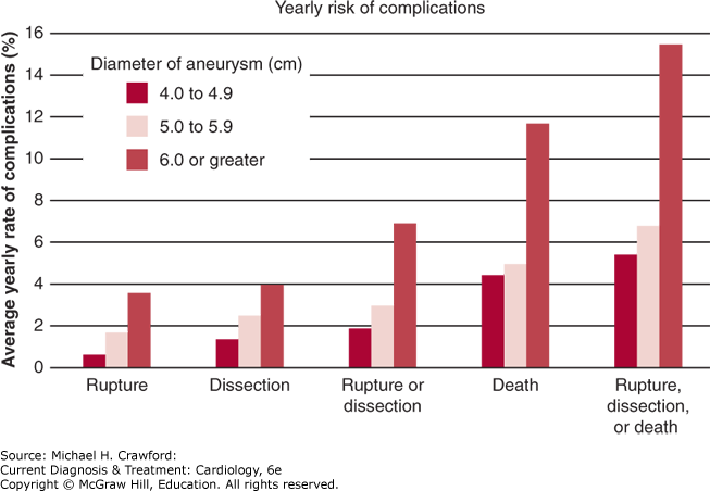
These data all point to a diameter of 6 cm as a very dangerous size threshold. At or above this size, the yearly risk for rupture is about 4%, the yearly risk of dissection is about 4%, and the risk of death is about 11%. (Death is often directly related to catastrophic complications from the aneurysm. In fact, recent studies from Japan, based on routine post-mortem CT scan “autopsies,” show that a staggering 8% of all sudden deaths are due to Type A aortic dissection.) The chance of any one of these phenomena occurring—rupture, dissection, or death—is 14%/year. As a mnemonic point of reference, a 6-cm aneurysm can be equated to about the diameter of a soft drink can. When a thoracic aortic aneurysm reaches the diameter of a soda can, it has certainly reached the point where it poses a major risk to the patient. All current societal guidelines recommend a general criterion for prophylactic surgery of 5.5 cm for the ascending aorta (for individuals of average body size).
These analyses should permit accurate decision-making when seeing a patient during an office visit and considering preemptive surgical extirpation of thoracic aneurysms. These data allow the physician to form a reasonable estimate of the individual patient’s risk of dissection, rupture, or death for each future year of life, if the aorta is not resected. The risk of rupture, dissection, or death based on aortic size is presented graphically in Figure 37–11.
The question arises whether the same surgical intervention criteria should apply for a small woman as for a large man. It is true that a larger individual can be “allowed” a larger aorta, generally speaking. Conversely, even a moderate-sized aneurysm can be quite threatening in an individual of small stature. For this reason, adverse event rates (rupture or dissection) based on aortic size corrected for body surface area (BSA) have been analyzed. By plotting the aneurysm size along the horizontal scale and the BSA along the vertical scale, each particular patient can be classified into low-, medium-, or high-risk categories—thus taking account of the aneurysm size in relation to the patient’s physical size.
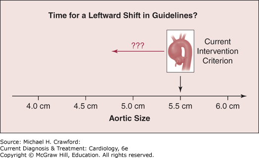
Two important recent developments regarding aortic prediction have occurred. First, it has been found that ascending aortic length (measured from the aortic annulus to the leading margin of the innominate artery) predicts adverse aortic events a bit more accurately than diameter. Elongation, it appears, is another sign of a damaged, vulnerable aorta. Secondly, it has been found, based on CT scans obtained fortuitously just before aortic dissection, that the dissection process itself produces an instantaneous 8-mm enlargement of the aorta. So, the aorta is actually smaller at the moment of dissection than previously appreciated on post-dissection scans. This argues that we should consider shifting our general criterion for intervention to 5.0 cm instead of 5.5 cm (Figure 37–12.) The next iteration of societal guidelines may endorse this “left-shift” to smaller sizes for intervention.
Figure 37-12. Consideration is currently being given as to whether the general criterion for intervention for ascending aortic aneurysm (in individuals of normal body size) should be shifted-left on the aortic size chart—to 5.5 cm.
Very recent studies have shown that enlargement of the aortic root is more “malignant” than similar degree of enlargement of the ascending aorta itself.
C. Symptoms & Signs
Most thoracic aortic aneurysms are asymptomatic and are detected fortuitously during imaging of other thoracic structures. When they are symptomatic, deep visceral pain in the upper anterior chest or interscapular back can occur. The aorta can “talk” to the patient via ascending sympathetic fibers that reach pain centers in the brain. Aortic pain differs from angina pectoris because it is not necessarily precipitated by exertion nor relieved by rest or nitroglycerin. Often, it is rather constant and not influenced by body motion or position. All patients with chest pain should have a screening chest radiograph. Rupture of a thoracic aneurysm usually causes excruciating pain, accompanied by profound dyspnea as the chest fills with blood, and quickly results in shock. A large ascending aortic aneurysm occasionally may result in dysphagia or stridor due to esophageal or large airway obstruction. Rarely, a large aneurysm may cause bone pain due to pressure against thoracic skeletal structures.
D. Physical Examination
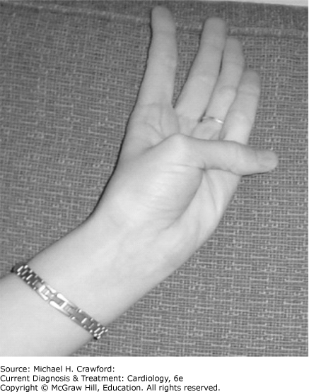
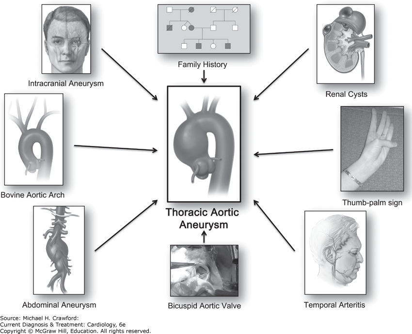
Physical examination is usually unremarkable. The presence of a murmur of aortic regurgitation should raise the suspicion of ascending aortic aneurysm, as should features suggestive of Marfan syndrome or related conditions. The thumb-palm sign, in which the tip of the thumb can be extended beyond the edge of the flat palm, can be a useful indicator of connective tissue laxity, raising the specter of associated aneurysm disease (Figure 37–13). Rarely, an abnormal pulsation will be felt due to a large aneurysm contacting the chest wall.
Figure 37-13. Patient with a positive thumb-palm sign for connective tissue disease. Being able to cross the thumb beyond the edge of the palm indicates that the long bones are excessive and the joints are lax. (Reproduced, with permission, from Elefteriades JA, et al. Guilt by association: paradigm for detecting a silent killer [thoracic aortic aneurysm]. Open Heart. 2015;2:e000169. With permission from BMJ Publishing Group Ltd.)
E. Diagnostic Studies
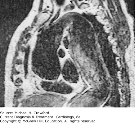
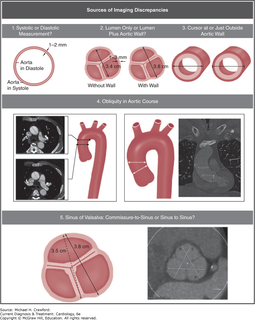
The remarkable strides made in recent decades in three-dimensional body imaging have dramatically advanced the diagnosis and treatment of thoracic aortic aneurysm. Echocardiography (especially transesophageal), computed tomography (CT), and magnetic resonance imaging (MRI) scans all yield images that clarify the presence, location, size, and extent of aneurysmal disease. An example of the precise imaging afforded by MRI is indicated for a specific, very extensive aneurysm in Figure 37–14. Proper interpretation of CT and MRI scans is essential; specifically, axial sections for aortic measurements must be selected carefully so that the aorta is vertical at the chosen site (oblique measurements will be exaggerated by an oblong rather than circular aortic contour). The “centerline method” applies automatic computerized assessment of diameter perpendicular to the long axis of the aorta, thus avoiding obliquity issues.
Figure 37-14. Magnetic resonance scan of a massively dilated aorta, which extends from the aortic valve to the iliac bifurcation. Note that the heart is compressed to a small shadow crushed between the elongated aorta and the diaphragm. This aneurysm was successfully resected in two stages.
It is easy to make errors in measurement of aorta on CT or MRI imaging. Potential sources of errors are indicated in Figure 37–15.
Figure 37-15. Potential sources of error in measurement of the aorta on CT images. These errors plague everyday practice and impede accurate assessment of growth over time. These sources of error include: systole vs. diastole, with or without aortic wall, cursor on or just outside of aortic wall, obliquity of the measuring line relative to the aorta, and sinus-to-commissure or sinus-to-sinus for the aortic root (sinuses of Valsalva). (Reproduced with permission from Elefteriades JA, Mukherjee SK, Mojibian H. Discrepancies in Measurement of the Thoracic Aorta: JACC Review Topic of the Week. J Am Coll Cardiol. 2020;76(2):201217.)
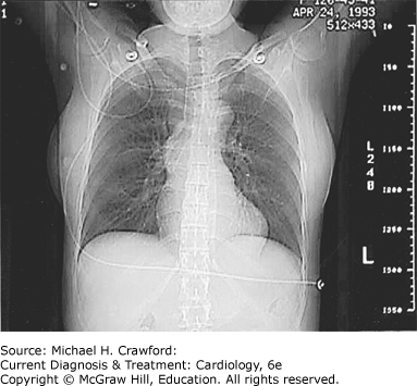
In this era of specialized three-dimensional imaging, it is important not to forget the chest radiograph, which can often yield significant information about the thoracic aorta. An example is provided in Figure 37–16. Ascending aortic aneurysm presents as a bulge beyond the right hilar border. Arch aneurysm produces enlargement of the aortic knob. Descending thoracic aneurysm is often easily seen as a deviation of the stripe of the descending aorta, which normally runs parallel to and just left of the vertebral column.
Figure 37-16. An exemplary chest radiograph indicating that significant information about the aorta can be gleaned from this simple test. Note the bulge of the ascending aorta to the right of the upper mediastinal border. This young patient with Marfan disease suffered dissection at an ascending aortic dimension of 4.8 cm.
Many if not most thoracic aortic aneurysms are found incidentally on three-dimensional imaging studies done for another, unrelated purpose. Most thoracic aneurysms remain undiagnosed in the general population. Recently, a paradigm called “guilt by association” has been proposed for uncovering hidden aneurysms in the general public based on the close association of thoracic aortic aneurysm with a group of related conditions (Figure 37–17). These related conditions include various medical conditions (intracranial aneurysm, abdominal aortic aneurysm, temporal arteritis, autoimmune disorder, renal cysts), certain aortic anatomic variants (bovine aortic arch, direct origin of left vertebral artery from aortic arch, bicuspid aortic valve), and family history of aneurysm disease. The presence of one of these associated conditions warrants imaging to rule out concurrent silent aortic aneurysm.
Figure 37-17. Paradigm of guilt by association for detection of silent thoracic aortic aneurysms. (Reproduced with permission from Elefteriades JA, et al. Guilt by association: paradigm for detecting a silent killer [thoracic aortic aneurysm]. Open Heart. 2015;2:e000169. With permission from BMJ Publishing Group Ltd.)
Treatment ⬆
A. Risks of Aortic Surgery
It is certainly helpful to know numerically and statistically the cumulative and yearly rates of rupture, dissection, and death imposed by an aortic aneurysm of a specific size. On the other hand, the equation is incomplete without the consideration of the risks inherent in elective, prophylactic surgical extirpation of the thoracic aorta. Certainly, these are major operations, and the surgical risks most feared include death, stroke, and paraplegia. However, these operations have become safer, reflecting increased surgical experience, improved perfusion techniques, improved (nonporous) grafts, effective antifibrinolytic agents for perioperative use, improved methods of spinal cord preservation, and the advent of centers specializing in aortic care and surgery. Several contemporary reports emphasize the “safety of thoracic aortic surgery in the present era.” Mortality rates and rates of other complications after aortic surgery are quite low, especially for operations performed electively on stable patients, in whom the safety of ascending aortic and aortic arch surgery is as high as 98%. Table 37–2 shows the pertinent rates of morbidity and mortality.
Table 37–2. Current Risks of Thoracic Aortic Surgery
| | Mortality (%) | Stroke (%) | Paraplegia (%) |
|---|
| Ascending/arch | 2.9 | 3.0 | 0.5 |
| Descending/thoracoabdominal | 2.9 | 4.2 | 5.3 |
B. Indications & Contraindications
By considering the rates of natural rupture, dissection, and death from the thoracic aneurysm itself versus the risks of operation, the physician can make an informed recommendation about elective, preemptive surgery. Once patients and their families are provided the natural history and surgical risk data, they often have strong opinions of their own. Some patients are reluctant to undergo major surgery, with its significant attendant risks, for an asymptomatic problem. Most patients, however, seem to feel they will never be comfortable until the threatening aneurysm is resected.
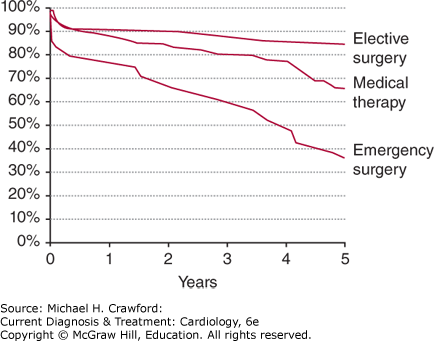
One more very important general point needs to be considered. Once the aorta has dissected, the prognosis is forevermore adversely affected (Figure 37–18). Patients who required emergency surgery not only had a higher rate of early mortality, but their survival curve was dramatically poorer. Patients who elected for planned, nonemergent procedures showed a survival rate very similar to that of a normal population. The poor long-term outlook for patients who required emergency surgery is due largely to the fact that, even after surgical replacement of portions of the aorta, the remainder of this vital organ will always remain dissected. Because the aortic wall was deficient to start with, at half-thickness, after dissection, it is rendered even more vulnerable to subsequent enlargement and rupture.
Figure 37-18. Long-term survival rates based on treatment. Medically treated patients had, of course, smaller and less symptomatic aneurysms. Note particularly that patients having emergent surgery not only manifested a higher likelihood of perioperative mortality, but also had a poorer long-term outlook. On the other hand, patients who received elective surgery showed excellent survival rates, comparable to an age- and sex-matched normal population. These data argue for elective, prophylactic extirpation of the aneurysmal aorta, before rupture or dissection can occur.
C. Surgical Techniques
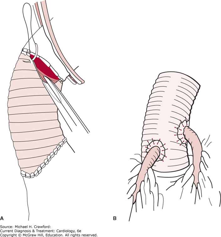
As discussed earlier, the type of operation for the ascending aorta is based on the pattern of aneurysmal pathology. For many patients, a supracoronary tube graft suffices (Figure 37–19A). For others, a composite graft, including both a valve and a graft, with obligate coronary artery reimplantation, is appropriate (Figure 37–19B). Newer valve-sparing aortic replacement procedures (eg, David procedure) have been developed and validated in large clinical series. Although these valve-preserving operations are becoming increasingly popular, the long-term fate of the preserved valve remains somewhat controversial.
Figure 37-19. (A) Supracoronary tube graft replacement and (B) composite graft replacement. (A: Reproduced with permission from Cooley DA, Wukasch DC. Techniques in Vascular Surgery. Philadelphia: WB Saunders; 1979.)
The main debate regarding the conduct of ascending aortic operations and those on the aortic arch concerns the optimal means of protecting brain function during the time that anastomoses in the vicinity of the aortic arch are performed. Deep hypothermic circulatory arrest—a state of suspended animation, which is generally safe for 30–45 minutes or longer—is preferred by many surgeons because of its simplicity and effectiveness. In a study on 500 patients, the effectiveness of this remarkable technique as a sole means of brain preservation was confirmed. Retrograde cerebral perfusion—via the superior vena cava—has its advocates, although the actual amount of effective brain perfusion achieved by this means has been questioned. Direct perfusion of the head vessels—usually via a cannula in the innominate artery or cannulas in both the innominate and the left carotid artery—also has its supporters, despite its added complexity. Direct perfusion is gaining in popularity, and it does provide a margin of protection, especially for very complex arch reconstructions or for surgical teams relatively inexperienced with arch replacement. However, very recent evidence has demonstrated a sobering 100% rate of cerebral embolic events (often subclinical) with antegrade perfusion. No technique has been demonstrated as conclusively superior over the others.
For descending and thoracoabdominal operations, the technique of left atrial to femoral artery bypass has become extremely popular. This method takes strain off the heart by diverting blood away from the left ventricle. This approach mitigates the effect of high aortic cross-clamping on cardiac afterload. It also perfuses the lower body, especially the extremely vulnerable spinal cord. Despite decades of concerted attention, paraplegia from descending and thoracoabdominal aortic replacement continues to be a major clinical problem. The cause is multifactorial, with clamp time, air and particulate embolism, and disconnection of critical intercostal branches all playing a role. Besides the benefits of left atrial to femoral artery perfusion, most authorities feel that routine spinal fluid drainage and deliberate maintenance of a strong postoperative blood pressure (to encourage collateral blood flow) are also effective adjuncts against the complication of postoperative paraplegia.
D. Specific Clinical Scenarios & Issues
1. Patient with Pain, but Aneurysm Smaller Than Criteria
The answer to whether such an aorta should be replaced is a resounding yes. The dimensional criteria are specifically intended for asymptomatic patients. Any and all symptomatic aneurysms need to be resected because symptoms are a precursor to rupture. Aneurysm pain represents stretching or irritation of the aortic adventitia, the adjacent chest wall, the mediastinal pleura, or some other structure impinged on by the expanding aneurysm. Even an aorta smaller than the criterion can rupture or dissect. The size criteria are “fuzzy” lines, not sharp demarcations. A patient with pain but a relatively small aneurysm is of great concern, and preemptive resection is needed. In one case, a patient complained of pain typical of an ascending aortic aneurysm. The aorta was 5.0 cm. Because the medical team thought this was too small for resection, they underestimated the symptoms at presentation. The aorta subsequently ruptured, and the patient died within 48 hours. This point cannot be overemphasized: The size criteria are explicitly intended only for asymptomatic patients; all symptomatic aneurysms need to be resected.
2. Differentiating Aneurysm Pain from Musculoskeletal Pain
This very important point is not always easy to determine, even in the most experienced hands. The patient usually has a good sense of whether the pain is originating from muscles and joints. The clinician usually gets an additional understanding by asking the following questions:
Is the pain influenced by motion or position? (If so, it is probably musculoskeletal.)
Do you have a history of lumbosacral spine disease or chronic low back pain? (If so, the symptoms may not be aortic in origin.)
Do you feel the pain in the interscapular back? (An affirmative answer indicates an almost certain relationship to thoracic aortic aneurysm.) The interscapular region corresponds to the upper descending aorta.
Presume that the pain is aortic in origin if no other cause can be conclusively established. This is the only approach that can prevent rupture.
3. Appropriate Interval for Serial Aortic Imaging
Patients with a thoracic aortic aneurysm should be monitored indefinitely. Stable, asymptomatic patients can undergo imaging about once every 2 years, remembering that the aneurysmal aorta grows at a relatively slow 1 mm/year. In case of new onset of symptoms, imaging should be done promptly, regardless of the interval from the prior scan. For new patients, for whom only one size data point is available, imaging should be done at shorter intervals until the behavior of aorta is understood. Imaging may be done every 3–6 months for new patients with moderately large aortas. Remember to compare the present scan with the patient’s first scan, not with the last prior scan. That is the way to detect growth. Many patients have suffered because scans were only compared with the last prior scan, and major growth went undetected.
4. Choice of Imaging Modality for Serial Follow-Up
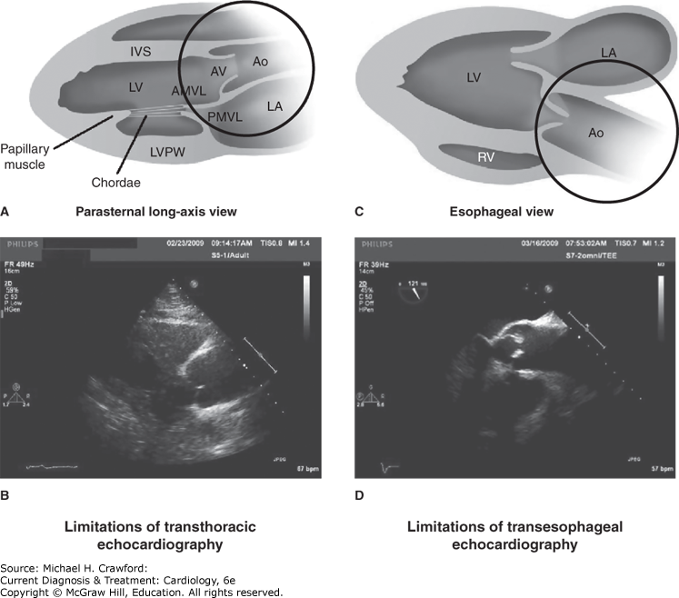
Three quality imaging techniques are currently available: echocardiography, CT scan, and MRI. If echocardiography is chosen, it is important to remember that a standard transthoracic echocardiogram cannot see the distal ascending aorta, the aortic arch, or the descending aorta with conclusive accuracy because of intervening air-containing bronchi and lung tissue. Supplement such studies with a periodic CT scan or MRI, which can visualize the entire aorta. The choice between CT and MRI may depend on ease of availability and radiologic expertise in a particular environment. Both modalities can image the entire aorta extremely well. Elevated creatinine or contrast allergy may contraindicate CT and instead favor MRI. The need to evaluate complex aortic lesions in multiple imaging planes would also favor MRI (although very recently, concerns have been raised about the risk to the kidneys of gadolinium contrast agents used for MRI scanning). Of course, indwelling metallic foreign objects, such as pacemakers or metal artifacts from previous surgery, may make CT the necessary choice instead of MRI. The advantages and limitations of echocardiography are shown in Figure 37–20. Both echocardiography and CT are required for complete evaluation of the entire aorta.
Figure 37-20. A and B: Limited distance above the aortic valve (AV) for which the ascending aorta (Ao) can be seen on transthoracic echocardiography. Schematic (A) and actual echocardiographic (B) image. AMVL, anterior mitral valve leaflet; IVS, interventricular septum; LA, left atrium; LV, left ventricle; LVPW, left ventricular posterior wall. C and D: Limited distance above the aortic valve for which the ascending aorta can be seen on transesophageal echocardiography. The tracheal air column interferes with visualization of the upper ascending aorta. Schematic (C) and actual echocardiographic image (D). RV, right ventricle. (A and C: Reproduced with permission from Mr. Rob Flewell. B and D: Reproduced with permission from Elefteriades JA, et al. Thoracic aortic aneurysm clinically pertinent controversies and uncertainties. J Am Coll Cardiol. 2010;55:841. Copyright © 2010 by the American College of Cardiology Foundation.)
5. Evaluation of Family Members
The data on familial inheritance has become strong enough that the treating physician is obligated to recommend that family members be evaluated. Physicians of family members should be made aware that aneurysm disease has been diagnosed in the family. A CT scan is recommended for adult males and for females beyond childbearing age. For children and for females of childbearing age, echocardiography of the ascending aorta and abdominal aorta is recommended. Investigators hope to identify humoral markers or genetic aberrations that can be used for familial screening of the aneurysm trait in the very near future. Routine genetic testing via Whole Exome Sequencing, while somewhat controversial and inconsistently approved by insurers, is becoming more and more widespread. We now offer and encourage screening of patients and family members by WES for nearly all of our patients. (See Figure 37–4, which presents, in pie diagram form, the most recent specific genetic findings from WES for thoracic aortic aneurysm.)
6. Activity Restrictions
Continuation of any and all aerobic activities, including running, swimming, and bicycling, is recommended. Serious weight lifters, at peaks of exertion, can elevate systolic arterial pressure to 300 mm Hg or higher. This type of instantaneous hypertension is, of course, not prudent for aneurysm patients. Weight lifters should limit themselves to one-half their body weight. The evidence for effort-induced aortic dissection is mounting. Participation in contact sports or those that might produce an abrupt physical impact, such as tackle football, snow skiing, water skiing, and horseback riding, is proscribed.
7. Role of Stent Grafting
A word of caution is appropriate concerning stent grafts. Stent therapy has become routine for many patients (especially those with abdominal or descending aortic aneurysms). It is certainly less invasive and more easily tolerated than open surgical techniques. Specialists, however, must avoid “irrational exuberance.” Owing to the high need for subsequent conventional surgery after abdominal aneurysm stent placement, the large, multicenter Eurostar study questioned the very efficacy and advisability of stent grafting. Endoleak, stent dislodgement, and aneurysm expansion or rupture were disturbingly widespread in medium-term follow-up. It should be remembered that stents were originally designed to keep tissue from encroaching on the vessel lumen, not to keep the vessel from expanding. One noted authority believes that the aneurysmal aorta essentially “ignores” the stent graft, dilating regardless of the stent, at its own pace (personal communication, Dr. L. Svensson). Also remember that the natural history of the thoracic aorta is that aneurysms grow slowly and that hard endpoints (rupture, dissection, and death) take years to be realized. For this reason, short-term stent studies are nearly meaningless. Long-term studies are needed. This newer modality of treatment should be approached with enthusiasm tempered by caution. Its advent should not at this point influence the decision about whether or not to intervene for a specific aneurysm; criteria (see earlier in chapter) should be met before any intervention, including stent therapy, is undertaken. Stent therapy appears especially well suited for patients with traumatic rupture of the descending aorta, in which setting a stent can be acutely life-saving. Those cautions having been stated, it is important to recognize that stent therapy is proliferating rapidly and that incremental improvements in the stent hardware are being made on a regular basis.
BlumelR, PatelK, RizzoJA., Accuracy of the “Thumb-Palm Test”: for detection of ascending aortic aneurysm. Am J Cardiol. 2021 Jul 1;150:114116. doi: 10.1016/j.amjcard.2021.03.041. Epub 2021 May 18. 34020772.
ErbelR, AboyansV, BoileauC,et al. 2014. ESC guidelines on the diagnosis and treatment of aortic diseases: document covering acute and chronic aortic diseases of the thoracic and abdominal aorta of the adult. The Task Force for the Diagnosis and Treatment of Aortic Diseases of the European Society of Cardiology (ESC). Eur Heart J. 2014;35(41):28732926. 25173340
HumphreyJD, SchwartzMA, TellidesG, MilewiczDM. Role of mechanotransduction in vascular biology: focus on thoracic aortic aneurysms and dissections. Circulation Research. 2015;116:14481461. 25858068
KalogerakosP, ZafarMA, LiY,et al. Root dilatation is more malignant than ascending aortic dilatation. J Am Heart Assoc. 2021;10(14):e020645. 34238012
LeshnowerBG, RangarajuS, AllenJW, StringerAY, GleasonTG, ChenEP. Deep hypothermia with retrograde cerebral perfusion versus moderate hypothermia with antegrade cerebral perfusion for arch surgery. Ann Thorac Surg. 2019;107:11041110. 30448484
MansourAM, PeterssS, ZafarMA,et al. Prevention of aortic dissection suggests a diameter shift to a lower aortic size threshold for intervention. Cardiology. 2018;139:139146. 29346780
MoriM, MahmoodSUB, YousefS,et al. Prevalence of incidentally identified thoracic dilatations: insight for screening criteria. Can J Cardiol. 2019; Jul;35(7):892898. 31292088
OstberegNP, ZafarMA, ZiganshinBA, ElefteriadesJA. The genetics of thoracic aortic aneurysms and dissection. Biomolecules. 2020;10:182. [PMC7072177] 31991693
WuJ, ZafarMA, LiY,et al. Ascending aortic length and risk of aortic adverse events: The neglected dimension. J Am Coll Cardiol. 2019;74:18831894. 31526537
ZiganshinBA, BaileyAE, CoonsC,et al. Routine genetic testing for thoracic aortic aneurysm and dissection in a clinical setting. Ann Thorac Surg. 2015;100(5):16041611. 26188975