VT as a cause of morbidity and mortality is grossly underdiagnosed, potentially leading to mismanagement. This may be particularly true when the clinical presentation is unexplained syncope because no concomitant electrocardiographic (ECG) documentation is available. In the case of cardiac arrest or SCD, acute myocardial infarction rather than an arrhythmic problem is often assumed to be responsible. Most persons who have suffered sudden death have no evidence of acute myocardial necrosis, even though the episode often occurs in patients with underlying coronary artery disease. Managing the underlying coronary artery disease with no regard to treating the concomitant VT is inadequate.
If a patient with a wide complex tachycardia is hemodynamically stable, it is often erroneously assumed that the rhythm must be a supraventricular tachycardia (SVT) with aberrant conduction rather than VT. In reality, the clinical presentation of VT can be quite variable, and the rate as well as the hemodynamic tolerance in a patient depends on many factors, including concomitant coronary artery disease, heart failure, the presence of cardioactive drugs, and even the patient’s posture at the time of onset. Therefore, it is prudent not to exclude the diagnosis of VT on the basis of hemodynamic tolerance alone. Approximately 80% of the patients with sustained wide QRS tachycardia have VT. To avoid misdiagnosis, clinicians in emergency settings must become familiar with established ECG criteria (discussed in the next section) that distinguish VT from SVT with aberrant conduction. When there is uncertainty, it is best to simply assume that the rhythm is VT—it is more often the correct assumption, and it is safer to treat SVT as VT than to leave VT untreated, given the risk of degeneration into sudden cardiac arrest.
3. Diagnostic Approach to the Patient with Wide QRS Complex Tachycardia
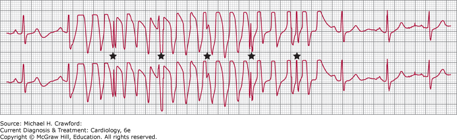
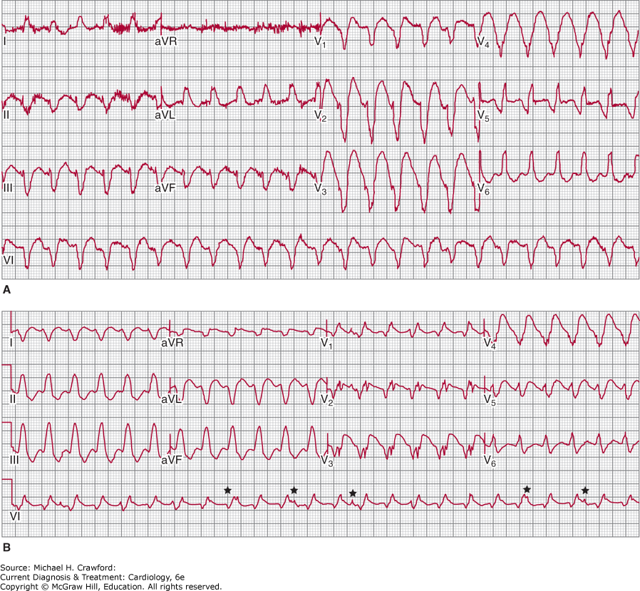
The diagnosis of wide QRS complex tachycardia by ECG analysis has always been a challenge for clinicians. The differential diagnosis includes VT, SVT with aberrant conduction, and preexcited tachycardia in patients with Wolff-Parkinson-White (WPW) syndrome. Figure 13–1 depicts, schematically, the reasons for normal and broad QRS complexes. Preexcited tachycardia results from antegrade activation of the ventricle via an accessory pathway in patients with WPW syndrome, which can present with atrial fibrillation, atrial flutter, atrial tachycardia, atrioventricular nodal reentry tachycardia (AVNRT), or antidromic tachycardia (Figure 13–2). Preexcited tachycardia is a rare cause of wide QRS complex tachycardia (5–8% of cases); however, the QRS pattern of preexcited QRS complex can be difficult to distinguish from VT because in both instances the QRS starts with myocyte-to-myocyte conduction. ECG artifact can also mimic wide QRS complex tachycardia and be misdiagnosed as VT, leading to expensive testing and even placement of implantable cardioverter-defibrillator (ICD). Clues to the diagnosis of artifact include: absence of hemodynamic deterioration, an unstable ECG baseline, association with body movement, and ability to march the normal QRS complexes through the artifact (“notches sign”) at a sinus R-R interval (Figure 13–3). A number of surface ECG criteria, including the atrioventricular (AV) relationship, the QRS complex duration, specific QRS morphology, and the QRS complex axis, have been established to distinguish VT from SVT with aberrant conduction. These criteria are helpful in arriving at an accurate diagnosis if they are used in a systematic fashion.
Figure 13-1. Mechanism of wide QRS. A: Narrow QRS from simultaneous activation of the right and left ventricles. In the three types of wide QRS shown in BD, there is sequential rather than simultaneous activation of the left and right ventricle and a variable amount of muscle-to-muscle conduction. AP, accessory pathway; AVN, atrioventricular node; BBB, bundle branch block; HB, His bundle; LB, left bundle; RB, right bundle. (Reproduced, with permission from Akhtar M, et al. Electrophysiological spectrum of wide QRS complex tachycardia. In: Zipes DP, et al., eds. Cardiac Electrophysiology. From Cell to Bedside. Philadelphia: WB Saunders; 1990.)
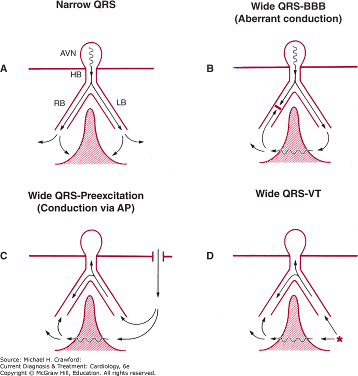
Figure 13-2. A: Preexcited wide complex tachycardia in a patient with Wolff-Parkinson-White (WPW) and atrial fibrillation B: Sinus rhythm electrocardiogram showing short PR interval and delta wave consistent with left posterior WPW. (Reproduced, with permission from Turakhia MP, et al. Wolff-Parkinson-White syndrome: where is the pathway? Indian Pacing Electrophysiol J. 2009;9(2):130133.)
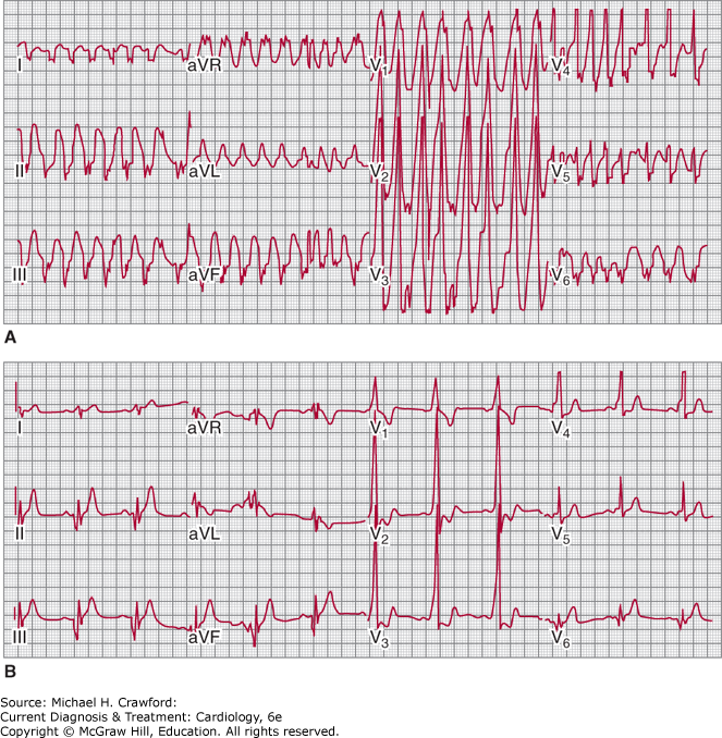
Figure 13-3. Artifact mimicking ventricular tachycardia. The QRS complexes are seen as notches that are marching at the regular sinus interval (*). (Reproduced with permission from Badhwar N, Kusumoto F, Goldschlager N. Arrhythmias in the Coronary Care Unit. Journal of Intensive Care Medicine. 2012;27(5):267289.)
In SVT, the arrhythmia arises in the atria or AV junction and reaches the ventricles through the AV node and His-Purkinje system. Because the atrial arrhythmia is the primary event, either a 1:1 AV response or a varying degree of AV block occurs, but in either case, the atrial rates equal or exceed ventricular rates. During VT, a retrograde block often leads to either AV dissociation or a varying degree of ventriculoatrial conduction ratios, but the ventricular rates equal or exceed the atrial rate. When AV dissociation can be recognized, it is the most reliable criterion for VT (Figures 13–4 and 13–5). This criterion lacks sensitivity, however, because the P waves can be identified on the surface ECG in only 25% of patients with VT. In patients with slower VT and AV dissociation, intermittent ventricular capture can result in fusion with narrow QRS complexes during wide QRS complex tachycardia. This useful but rarely observed finding is also 100% specific for the diagnosis of VT.
Figure 13-4. A: Monomorphic ventricular tachycardia (VT), with a uniform QRS appearance for all complexes. Arrowheads indicate superimposed P waves. B: Polymorphic VT, with a beat-to-beat variation in the QRS morphology; QT-interval prolongation follows the termination of the VT episode. (Reproduced with permission from Akhtar M. Clinical spectrum of ventricular tachycardia. Circulation. 1990;82(5):15611573.)
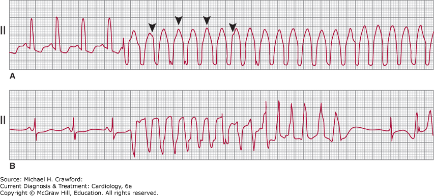
Figure 13-5. A: Scar-related ventricular tachycardia (VT), with a left bundle branch block left axis morphology in a patient with ischemic cardiomyopathy and previous myocardial infarction. B: Right bundle branch block right axis morphology VT in the same patient at the same rate suggesting that both forms of VT have the same circuit (that revolves around the mitral annulus) with different exits causing the difference in morphology. Atrioventricular dissociation is noted in the rhythm strip on V1 (*) that is 100% specific for VT.
For the reasons listed earlier, the QRS complex duration is the widest in VT and narrowest in aberrant conduction. To distinguish VT from SVT with aberrant conduction on the basis of QRS duration alone, however, some specific aspects must be considered. In the absence of cardioactive drugs and extensive myocardial fibrosis, aberrancy rarely results in a QRS duration of more than 140 ms with a right bundle branch block (RBBB) pattern (see Figure 13–5) or more than 160 ms with a left bundle branch block (LBBB) configuration. However, in the presence of intramyocardial conduction delay from drugs (such as class I antiarrhythmic agents) or myocardial fibrosis, the QRS width may exceed these values in SVT with aberrant conduction. Conversely, on a rare occasion, VT can present as a narrow QRS tachycardia (< 120 ms that is narrower than the conducted QRS complex) when there is near-simultaneous activation of the two ventricles, perhaps from the septum.
VT can have both LBBB or RBBB morphology, though because of the myocardial origin of most forms of VT, the QRS contrasts from typical RBBB and LBBB patterns. Many ECG criteria have therefore exploited these differences to distinguish VT from aberrant conduction. The typical RBBB (ie, in the case of aberrancy) is a triphasic complex best seen in V1 as rsR′ or rSR′ pattern and in lead I as qRs, qRS pattern. Similarly, a typical LBBB has no initial q wave in lead I and a small r and a rapid S wave in V1. One general principle that distinguishes the morphology of the QRS complex in SVT versus VT is that the initial portion of the complex in VT is slow, as it originates in myocardial tissue in contrast to the fast initial activation of SVT (which propagates initially down the native conduction system). A study that analyzed the morphology of premature ventricular complexes (PVC) and aberrantly conducted beats of RBBB morphology in V1 found that the triphasic RsR′ pattern with R′> R was predominant in aberrant conduction (70%) compared with PVC (6%). Monophasic pattern or R > R′ was seen in the PVC beats. The limitation of that study is that origin of the anomalous beats (SVT versus VT) was also based on the ECG (presence or absence of preceding P wave). A retrospective ECG analysis of 70 patients with SVT and 70 patients with VT in whom His bundle recordings were used to determine the site of origin of the wide QRS complex tachycardia found that VT was favored by monophasic or biphasic R waves in V1 and an R:S ratio less than 1 in V6 in patients with RBBB morphology and any Q wave in V6 in patients with LBBB morphology. A study of 150 consecutive wide QRS complex tachycardia cases found that 12-lead QRS morphology during wide QRS complex tachycardia was different from that during preexisting bundle branch block in sinus rhythm, favoring a diagnosis of VT. The investigators also noted that a positive QRS concordance (positive complexes V1–V6) is uncommon in aberrancy, but a negative QRS concordance can occur during aberrant conduction in a small percentage of cases. Another study that evaluated wide QRS complex tachycardia with LBBB morphology in V1 found that an R wave of greater than 30 ms, notching in the down stroke of the S wave and an RS (beginning of QRS complex to nadir of S wave) interval greater than 60 ms in V1 or V2, and any Q wave in V6 favored a diagnosis of VT. In a prospective analysis of wide QRS complex tachycardia (with RBBB and LBBB morphology), the following two criteria for a diagnosis of VT were proposed: (1) absence of R-S in all precordial leads and (2) R-S interval greater than 100 ms (measured from the beginning of the QRS complex to the nadir of the S wave) in any precordial lead. Finally, the presence of QR complex in any lead during wide QRS complex tachycardia also favors a diagnosis of VT.
The axis orientation on a 12-lead ECG ranging from normal (−30뀀 to +90뀀), left (−31뀀 to −90뀀), or right (+91뀀 to +180뀀) has significant overlap across the causes of wide QRS complex tachycardia and is of little diagnostic value. The axis range of −91뀀 to 180뀀 (often referred to as a “northwest” axis), however, is usually not seen in aberrant conduction, unless there is a significant structural problem that predisposes the ECG vector to an abnormal axis (eg, severe R ventricular hypertrophy). Similarly, a combination of LBBB pattern with right axis deviation is almost always suggestive of VT over SVT. A previous history of myocardial infarction (MI) and an axis change of more than 40뀀 between sinus rhythm and wide QRS complex tachycardia may independently favor VT over SVT.
History, Physical Examination, & 12-Lead ECG
A detailed history can provide clues to the diagnosis of wide QRS complex tachycardia. A history of prior MI strongly favors VT as the diagnosis in a patient with wide QRS complex tachycardia. The clinician must take note of drug and toxin ingestion to rule out secondary causes of VT. A detailed family history is also important, since it can reveal the presence of inherited arrhythmogenic cardiomyopathies.
There are also several salient features on physical examination that can be helpful. The presence of irregular cannon waves on jugular venous examination and variable intensity of S1 on auscultation suggest AV dissociation and are indicative of VT in the setting of a wide QRS complex tachycardia. Carotid massage (performed carefully after ruling out a bruit) that leads to termination of wide QRS complex tachycardia typically suggests SVT as the mechanism of the arrhythmia (though an exception is idiopathic VT arising from the right ventricular outflow tract (RVOT), which can be responsive to vagal stimulation).
A prior ECG in sinus rhythm showing Q waves, indicative of infarcted tissue and scar, increases the likelihood that a subsequent wide QRS complex tachycardia is due to VT. SVT is the diagnosis if the old ECG shows bundle branch block pattern that matches the 12-lead ECG during wide QRS complex tachycardia. Preexcited tachycardia is inferred from the ECG showing WPW pattern that is similar to wide QRS complex tachycardia ECG pattern.
It is essential to obtain a 12-lead ECG during wide QRS complex tachycardia to compare it to the sinus rhythm ECG and look for subtle findings like AV dissociation and narrow beats (fusion and capture) that might not be evident in some leads. Table 13–1 outlines an approach to diagnosing wide QRS complex tachycardia by analyzing the ECG. The first step is to read the ECG with emphasis on rate, regularity (atrial fibrillation with preexcited tachycardia is irregular), axis (extreme northwest axis suggests VT), and morphology in V1. The next step is to look for AV dissociation (V > A) by marching the sinus P waves at the onset and termination of the wide QRS complex tachycardia if available. Narrow QRS complexes in a wide QRS complex tachycardia (caused by capture and fusion of conducted beats through the AV node-His-Purkinje system) are 100% specific for VT. The next step is to use the Brugada criteria by evaluating the R-S complexes in precordial leads. Absence of R-S complex in all precordial leads or an R-S interval greater than 100 ms in one precordial lead favor a diagnosis of VT.
Table 13–1. Approach to the ECG with Wide QRS Complex Tachycardia
|
AV, atrioventricular; ECG, electrocardiogram; LBBB, left bundle branch block; RBBB, right bundle branch block; SVT, supraventricular tachycardia; VT, ventricular tachycardia.
The last step in the Brugada algorithm relies on QRS morphology criteria (discussed earlier) to differentiate VT from SVT. The onset of QRS complex to peak of the R wave or nadir of S wave in lead II greater than 50 ms favors a diagnosis of VT.
The morphology criteria have certain limitations. Idiopathic VTs (fascicular VT, outflow tract VT) and bundle branch reentrant VT can have a typical bundle branch block pattern on the ECG and can therefore be misdiagnosed as SVT with aberrancy. Furthermore, some antiarrhythmic medications can alter typical conduction and limit generalizability of the morphology criteria developed for the Brugada algorithm, that is, patients with an SVT with aberrancy may have an atypical bundle branch block pattern on the ECG, leading to a misdiagnosis of VT. In addition, patients with preexcited SVT may also have atypical bundle block patterns.
To address some of the limitations in morphology criteria, and to simplify detection, newer criteria have been developed that rely solely on aVR analysis. Criteria in aVR that favor VT include presence of initial R wave, initial r or q wave greater than 40 ms, notch in initial downstroke of QRS complex, and voltage change in initial 40 ms (vi)/voltage change in terminal 40 ms (vt) less than 1. Again, this criterion exploits the fact that the initial portion of the QRS in VT is slower than the terminal portion because the signal originates in slower myocardial tissue, before spreading across the ventricles and involving the faster native conduction tissue.
In clinical practice, traditional criteria and aVR criteria have generally similar sensitivity and specificity for distinguishing VT from SVT. Importantly, if the clinician is unsure of the diagnosis, it is safer to assume the signal is VT, and treat it as such.
CerantolaM, ArklesJ, FrankelDS. Diagnostic approach to wide complex tachycardia. JAMA Intern Med. 2021 Sep 1;181(9):12311233. 34279546. doi: 10.1001/jamainternmed.2021.3189.