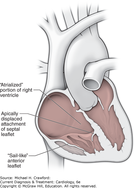
Ebstein anomaly is characterized by deformity of the tricuspid valve with apical displacement of the septal and posterior leaflets (Figure 31–17) and their adhesion to the RV wall. The anterior leaflet is elongated and has been described as sail-like. Tricuspid regurgitation arises from the apically displaced site of leaflet coaptation with considerable variability in the extent of tricuspid leaflet displacement and the degree of tricuspid regurgitation. The portion of the RV proximal to the leaflets is atrialized (thinned), and if the remaining RV is diminutive in size, pump function may be inadequate. Cyanosis may be present as a result of right-to-left shunting across an ASD or patent foramen ovale in the presence of significant tricuspid regurgitation or elevated right atrial pressures. Interatrial septal defects, including patent foramen ovales, are the most common associated anomaly, occurring in 80–90% of patients with Ebstein anomaly.
Figure 31-17. Anatomy of Ebstein anomaly.
Tremendous variability exists in the morphologic abnormalities and clinical presentation of patients with Ebstein anomaly. In severe cases, CHF or cyanosis may be present during infancy. At the opposite end of the spectrum, a mildly affected adult may be asymptomatic or symptomatic only because of supraventricular tachyarrhythmias. The latter are an important feature of Ebstein anomaly, which is associated with preexcitation in 25–30% of patients. The accessory pathway is usually posteroseptal or posterolateral in location.
A. Symptoms & Signs
Cyanosis may be the most important clinical feature in early life, but in older patients, long-standing RV volume overload and right atrial distention result in CHF. Dysrhythmias, including the Wolff-Parkinson-White syndrome, are frequent. Adult patients may have dyspnea, arrhythmias, decreased exercise tolerance, and intermittent or exercise-induced cyanosis (with associated right-to-left shunting across an ASD or patent foramen ovale).
Physical examination reveals right parasternal lift, widely split S1, systolic clicks (from delayed tricuspid valve closure, the “sail” sounds), and the systolic murmur of tricuspid regurgitation. The latter does not usually increase in intensity during inspiration, because the noncompliant RV cannot accept an increase in venous return. On the other hand, the right atrium is compliant, and systemic venous congestion is uncommon; the jugular venous pulse is therefore usually normal. S3 and S4 gallops may be present, as may an early diastolic snap from the opening of the elongated anterior leaflet.
B. Diagnostic Studies
1. Electrocardiography and Chest Radiography
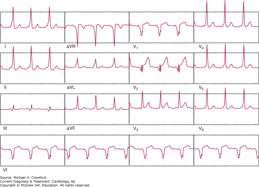
The ECG shows evidence of right atrial enlargement and an RV conduction defect of the RBBB type. The PR interval may be prolonged, except in the presence of an accessory pathway. In 25–30% of patients, ECG findings are consistent with Wolff-Parkinson-White syndrome; the PR interval is short, and delta waves from a posterolateral or posteroseptal bundle of Kent are evident (Figure 31–18). Atrial fibrillation may be present in older patients.
Figure 31-18. Electrocardiogram in Ebstein anomaly with associated Wolff-Parkinson-White syndrome.
The chest radiograph shows normal or reduced pulmonary vascularity without pulmonary artery enlargement; it also shows cardiac enlargement to the right of the sternum caused by right atrial enlargement. The LV and left atrium are normal in size.
2. Echocardiography
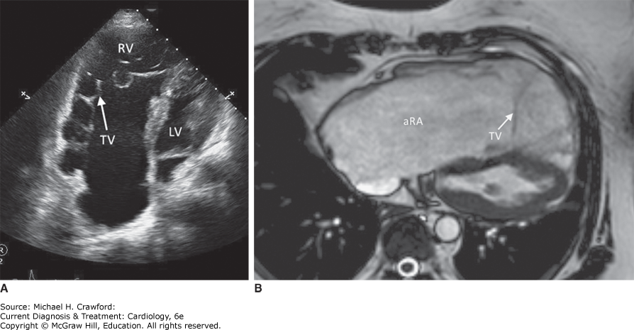
The classic M-mode description of this anomaly included increased excursion of the anterior tricuspid valve leaflet and delayed tricuspid valve closure (> 40 ms) following mitral valve closure. Two-dimensional and Doppler echocardiography are diagnostic in most adults. The four-chamber apical and subcostal views provide most of the necessary information. The right atrium is enlarged and the RV is usually small, consisting of the atrialized portion and the remaining pumping chamber. The septal, and possibly the posterior, leaflet of the tricuspid valve is apically displaced, and color-flow Doppler imaging shows the regurgitant jet arising from the apical point of coaptation (Figure 31–19). The degree of tricuspid regurgitation can be estimated from the extent of right atrial filling by color flow and from the density of the continuous wave Doppler signal. The pulmonary artery systolic pressure estimated from the continuous wave tricuspid regurgitation jet is nearly always normal.
Figure 31-19. A: Transthoracic echocardiogram in a 56-year-old woman with Ebstein anomaly. This four-chamber view shows the apically displaced tricuspid valve (TV) in relation to the normal mitral valve (lower part of left ventricle [LV]). B: Off-axis magnetic resonance imaging of the right ventricle (RV) showing the large atrialized right atrium (aRA).
Although color-flow imaging may reveal a patent foramen ovale or an ASD, it is mandatory to perform a saline contrast examination to reliably exclude these sources of right-to-left shunting. When precordial echocardiography is inadequate, TEE can be used to exclude associated lesions of the atrial septum.
3. Cardiac Catheterization
During right-heart catheterization, simultaneous recordings of an RV electrogram and a right atrial pressure tracing are obtained with a catheter in the atrialized portion of the RV. This finding is considered pathognomonic of Ebstein anomaly, but catheterization is rarely necessary for diagnosis.
Prognosis & Treatment
The chance of surviving up to age 50 is about 50%, with survival dependent on the degree of the anatomic and physiologic abnormalities. As mentioned, 25–30% of patients have supraventricular arrhythmias, many associated with accessory pathways that are now amenable to catheter ablation. Evaluation of these patients with an electrophysiology study prior to consideration for surgery may be necessary. Tricuspid annuloplasty and repair with RV plication have been challenging. The success of these approaches has traditionally been limited, with approximately 50% of patients requiring tricuspid valve replacement. Newer techniques, such as the Carpentier and “cone” techniques, promise to reduce further the need for valve replacement. Improvement in exercise tolerance following tricuspid valve replacement or repair has been observed, especially in patients with associated ASD. In patients with severe morphologic variants, a Fontan-like procedure (see Palliative Surgical Procedures) may be the only suitable choice. In patients who are symptomatic predominantly based on exercise-induced cyanosis, device closure of the interarterial septal defect may be adequate treatment.