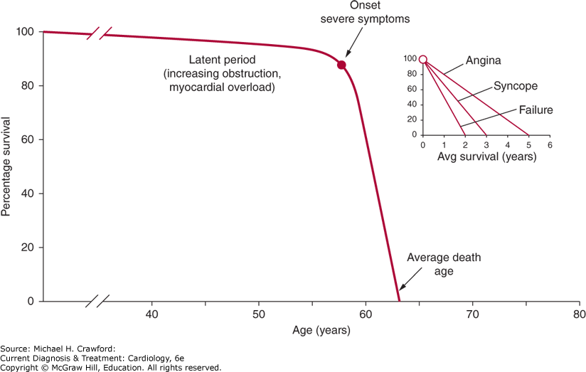
Those with acquired aortic stenosis generally have a long latent period before the onset of the salient clinical manifestations of the disease: effort-related dyspnea (heart failure), angina, and syncope. The most common initial clinical manifestations are a gradual decline in functional capacity and effort-related dyspnea. Regardless of the initial presenting symptom(s), it is imperative to ensure that the pathophysiologic mechanism of the symptoms is attributed to valve disease and not a manifestation of another mechanism such as concomitant coronary artery or lung disease because the onset of even mild symptoms attributed to aortic stenosis heralds a dramatic increase in the mortality rate for these patients if the valve is not replaced. Symptoms are therefore the guidepost for intervention, and understanding them is the key to understanding and managing the disease.
The primary determinants of left ventricular systolic function are contractility and afterload. The load on individual myocardial fibers can best be described as left ventricular wall stress and defined by the Laplace equation:

With acquired aortic stenosis, the obstruction will commonly progress gradually over time. Left ventricular adaptation to increases in systolic pressure is the parallel replication of sarcomeres to increase its thickness (concentric hypertrophy) in an effort to normalize wall stress and maintain systolic performance. As the severity of stenosis progresses, the increase in wall thickness may become insufficient to offset the rise in pressure (“afterload mismatch”), resulting in a rise in wall stress and a decline in ventricular function. The presence of aortic stenosis may also result in true depression of myocardial contractility, the exact mechanism of which is unclear but likely related to a loss of contractile elements secondary to reduced coronary blood flow. Thus, a decline in ejection fraction results from the interplay of varying degrees between excessive afterload and true myocardial depression. Those in whom the decline in function is largely attributed to afterload mismatch are more likely to experience restored ventricular function following aortic valve replacement. The evaluation of left ventricular ejection phase indices should be related to the global wall tension; however, clinically, it is difficult to tease out the extent to which wall stress (afterload) and contractility are contributing to a noted decline in ejection fraction.
1. Dyspnea and Exercise Intolerance
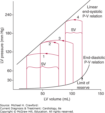
The increase in left ventricular wall thickness, although imposed in an effort to maintain wall stress and systolic performance, does have maladaptive physiologic consequences. The increase in wall thickness makes it harder to fill the ventricle, and therefore, higher filling pressures are required to achieve any given volume and end-diastolic volume will be required to increase to allow the maintenance of a normal ejection fraction (a rightward shift in the ventricular diastolic pressure-volume relationship) (Figure 17–1). In alignment with the concept of hemodynamic continuity, when the mitral valve opens, the left atrium is now exposed to the hemodynamic milieu of the left ventricle. The increased ventricular diastolic pressure is transmitted to atrium and further back into the pulmonary veins and lungs, resulting in pulmonary congestion and an increased work of breathing. In addition, with outflow obstruction, there is a prolongation of the ejection phase; this, in conjunction with an exercise-induced increase in heart rate, will result in a reduction of the diastolic filling time, and the ventricle will reach its limit of “preload reserve.” Once limits of preload reserve are met, stroke volume becomes directly related to ventricular pressure (see Figure 17–1), resulting in an inability to increase cardiac output and further contributing to the presence of dyspnea and exercise intolerance.
Figure 17-1. Left ventricular (LV) pressure-volume (P-V) loops. As LV pressure increases, stroke volume (SV) would decrease if the end-diastolic volume were fixed (loop 2). The ventricle, however, adapts by increasing end-diastolic volume to maintain a normal SV (loop 3). Eventually, the limits of preload reserve are met whereby increases in LV pressure result in a decline in SV (SV is now directly related to LV pressure; loop 4). (Reproduced with permission from Ross J. Afterload mismatch in aortic and mitral valve disease: Implications for surgical therapy. J Am Coll Cardiol. 1985;5(4):811826.)
Angina results from myocardial ischemia, which occurs when there is an imbalance between myocardial oxygen requirements/demand and myocardial oxygen supply. Although epicardial coronary artery disease often coexists with aortic stenosis, symptoms of angina frequently occur in those without epicardial coronary artery disease. Oxygen demand is best estimated clinically by the product of heart rate and wall stress, and as noted earlier, eventually the extent of hypertrophy cannot keep up with pressure demands of the ventricle and wall stress increases. In the absence of epicardial coronary artery disease, myocardial oxygen supply may also be decreased secondary to the rise in left ventricular end-diastolic pressure and a delayed rate of ventricular relaxation, with impaired coronary “diastolic suction” contributing to both reduced coronary perfusion and a decline in coronary flow reserve needed to offset increased oxygen demands during stress or exercise. In addition, with exercise and increased heart rates, there is reduced diastolic coronary perfusion time. Recently, coronary microvascular dysfunction has also been proposed as a mechanism of angina in patients with severe aortic stenosis as determined by adenosine-stress cardiac magnetic resonance.
Syncope is a transient loss of consciousness due to cerebral hypoperfusion. Syncope in those with aortic stenosis usually occurs during exercise. There are two primary mechanisms, not mutually exclusive, that have been theorized to cause syncope. First, the narrowed aortic valve does not permit the appropriate increase in cardiac output necessary to offset the associated reduction in total peripheral resistance associated with exercise, resulting in a drop in blood pressure. Second, the very high ventricular pressure that develops with exercise when sensed by ventricular mechanoreceptors triggers a reflexive vasodepressor response, also leading to a decline in blood pressure. Less commonly, exercise can result in either ventricular or supraventricular arrhythmias, which can lead to a reduction in cardiac output and, consequently, a drop in blood pressure.
The character of a murmur can be described by its timing and shape, intensity and pitch, and location and radiation, along with changes in these characteristics imposed by transient changes in intracardiac hemodynamics.
The murmur of valvular aortic stenosis is a midsystolic murmur, which begins after the first heart sound and ends before the aortic (A2) component of the second heart sound. During systole, as blood flow velocity accelerates across the valve, the intensity of the murmur increases, and as blood flow velocity decelerates, the intensity diminishes. Therefore, there is a classic “crescendo-decrescendo” configuration or shape to the murmur of valvular aortic stenosis. Further insights into the configuration of the murmur may help to discern the severity of disease; the later in systole the peak intensity occurs, generally the more severe is the stenosis.
The intensity of the aortic stenosis murmur relates to the quantity and velocity of transaortic blood flow along with the ability to transmit sound through the chest. The pitch of the murmur relates to the pressure gradient and size of the “aperture” with which the blood flows through. In general, as the stenosis severity increases, the intensity and pitch of the murmur will increase. However, in those with a thick ventricle and low end-diastolic volume or in the presence of a decline in cardiac function, both of which may result in a low stroke volume, the intensity and pitch of the murmur may be lower than expected for the given severity of stenosis. In addition, the ability to transmit the sound through the chest is impaired in those with a pericardial effusion, emphysema, or obesity. Therefore, one should be cautious of excluding significant aortic stenosis solely based on murmur intensity/pitch.
The murmur of aortic stenosis typically is heard loudest in the “aortic area,” the right second interspace at the sternal border, and will often radiate into the carotids and along a line from the aortic area toward the left ventricular apex. At the apex, the intensity and pitch of the murmur may change to more resemble that of the murmur of mitral regurgitation; however, the shape/configuration of the murmur does not change. This is known as the Gallavardin phenomenon.
A transient alteration in intracardiac hemodynamics may influence the characteristics of a murmur helping to further specify its etiology.
Isometric handgrip: Increases systemic vascular resistance, decreasing the intensity of the murmur of aortic stenosis.
Standing/Valsalva maneuver (phase II): Decreases ventricular filling, reducing the intensity of the murmur of aortic stenosis and most other valve murmurs, except that of hypertrophic cardiomyopathy and the duration of the murmur of mitral valve prolapse.
Squatting: Increases ventricular filling but also increases systemic vascular resistance. Its effect on the murmur of aortic stenosis is therefore generally neutral; however, it will reduce the intensity of the murmur of hypertrophic cardiomyopathy.
Cycle length: The intensity of the murmur of aortic stenosis may vary from beat to beat in those with atrial fibrillation or premature contractions, increasing in intensity in the beats following a longer R-R interval where diastolic filling time is increased.
The carotid artery pulse wave contour in those with aortic stenosis is characterized as small and slow rising (parvus et tardus). One may also appreciate a carotid shudder.
The presence of significant aortic stenosis may result in paradoxical splitting of the second heart sound where the A2 component of S2 is delayed, resulting in S2 being more split during expiration than inspiration. Reduced movement of the aortic valve may render A2 inaudible where only a single second heart sound can be appreciated.
As noted earlier, left ventricular hypertrophy imposed by the increase in wall stress reduces ventricular wall compliance. The fourth heart sound is a low-pitch sound heard coincident with late diastolic filling, due to atrial contraction, of a ventricle with reduced wall compliance and increased end-diastolic pressure.
The apical impulse, which in aortic stenosis is generally the point of maximal impulse, remains in its normal position but is sustained due to prolongation of the ejection time. The atrial component of ventricular filling may also be palpable. With simultaneous palpation of the apical and carotid impulses, normally one will appreciate little delay in their peaks; however, they become separated in time proportional to the severity of stenosis.
The clinical severity of aortic stenosis is largely an operational classification based on the presence or absence of symptoms as discussed earlier. However, the indications for consideration of either a surgical or percutaneous aortic valve replacement rely on an estimate of stenosis severity.
Left ventricular hypertrophy is the primary finding noted in those with aortic stenosis. Other common findings include left atrial abnormality along with ST- and T-wave abnormalities. There are, however, no electrocardiogram findings that are either sensitive or specific for aortic stenosis.
With isolated aortic stenosis, the cardiac silhouette is generally normal in size with possible rounding of the left heart border consistent with concentric left ventricular hypertrophy. There may be signs of left atrial enlargement and pulmonary venous hypertension. The aortic shadow may become enlarged, and valve calcification may be appreciated.
Echocardiography is the principal clinical tool used for the evaluation of aortic stenosis, and currently still remains the gold standard imaging modality for the assessment of aortic stenosis despite the advances in other cardiac imaging modalities. Appropriate use criteria deem transthoracic echocardiography appropriate on the initial evaluation of a patient when clinical evaluation provides “unexplained murmur or abnormal heart sounds, reasonable suspicion of valvular or structural heart disease, history of rheumatic heart disease, known systemic or acquired heart disease associated with valvular heart disease, first-degree family history of bicuspid aortic valve, or exposure to medications that could result in the development of valvular heart disease, syncope without other symptoms or signs of cardiovascular disease, hypotension or hemodynamic instability with suspected cardiac etiology, respiratory failure, or initial heart failure evaluation to exclude valvular cause.” Re-evaluation of known valvular heart disease with echocardiography is appropriate “with a change in clinical status or cardiac exam or to guide therapy.” Routine surveillance in the absence of a change in clinical status or cardiac examination is deemed appropriate at intervals of 6 months to 1 year when the aortic velocity is ≥ 4.0 m/s, 1–2 years when the aortic velocity is between 3.0 and 3.9 m/s, and 3–5 years when the aortic velocity is between 2.0 and 2.9 m/s. The echocardiogram should include not only anatomic and hemodynamic measures of stenosis severity, but also an assessment of the left ventricular response to the pressure load, insights into whether there is dilation of the ascending aorta, and assessment for the presence of coexisting valve regurgitation and other cardiac abnormalities. Myocardial imaging and a measure of global longitudinal peak systolic strain may detect adverse ventricular consequences to the hemodynamic load despite a preserved left ventricular ejection fraction.
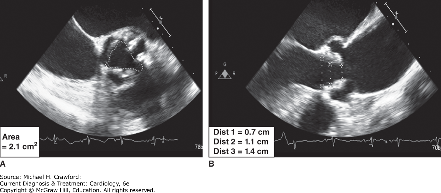
Although most outcome data in those with aortic stenosis are based on the measures of the hemodynamic severity and physiologic orifice area, the anatomic evaluation of the valve is gaining increased importance in the evolving era of percutaneous valve replacement in guiding patient selection and procedural planning. From primarily the transthoracic parasternal views, or transesophageal imaging if the transthoracic images are suboptimal, the number of leaflets, extent of calcification, leaflet thickening, and mobility should be evaluated. An anatomic measure of the geometric valve area can be obtained by planimetry, either from echocardiography or cardiac CT. The fundamental limitations to accurate and reproducible measurements of the geometric orifice area are image attenuation secondary to leaflet calcification and the spatial integration of images required to ensure planimetry of the minimal opening area at the leaflet tips (Figure 17–2). It is for these reasons that the measure of geometric orifice area is reserved clinically primarily for those circumstances where Doppler measurements are unreliable. This holds true for classification of disease severity, however as we will discuss planimetry for procedural planning by cardiac CT has now become routine.
Figure 17-2. Planimetry of the aortic valve. A: Short-axis image of the aortic valve and a calculated anatomic valve area by planimetry. Leaflet calcification and image attenuation limit accurate delineation of the leaflet borders. B: Long-axis image of the aortic valve noting that slight alteration in the tomographic slice obtained through the aortic valve will result in marked variation in the valve area calculated by planimetry. (AV VTI, aortic valve velocity time integral)
The principal measures of the hemodynamic severity include peak transaortic jet velocity, peak instantaneous and mean pressure gradients, and valve area (effective orifice area by the continuity equation).
(1) Transaortic Velocity and Gradient Calculations
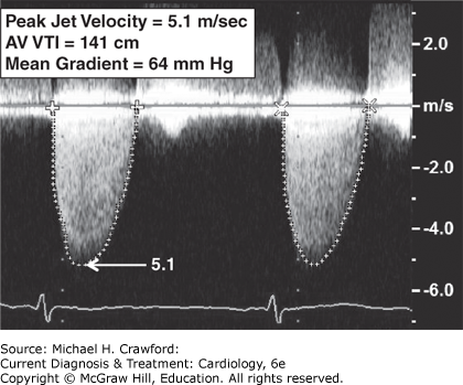
The principle of conservation of energy states that the total amount of energy in a closed system remains constant. Energy can change its location and form, but can be neither created nor destroyed. With respect to flow, as the flow stream approaches a narrowed orifice, its kinetic energy increases and potential energy decreases. Distal to the narrowed orifice, pressure is lost in part due to the dissipation of kinetic energy as heat. This creates a pressure gradient across the valve orifice. Continuous wave Doppler is used to determine the maximum jet velocity through the stenotic valve. Meticulous imaging from multiple acoustic windows is required to ensure that flow velocities are acquired with a parallel intercept angle to the direction of flow, limiting the error of underestimation of the peak velocity. The Bernoulli equation is then applied to the highest jet velocity obtained to calculate the peak instantaneous gradient (Figure 17–3).
Figure 17-3. Continuous wave Doppler of aortic stenosis.
Bernoulli equation:

deltaCap.gif P = pressure gradient, ρ = mass density of blood, V1 and V2 = velocity proximal and distal to obstruction respectively, R = viscous resistance, µ = viscosity
Under most physiologic conditions, the latter two terms (flow acceleration and viscous friction) are negligible and can be ignored and V2>>> V1 and thus V1 can be ignored. Therefore, under most physiologic conditions, a simplified Bernoulli equation can be applied to the peak velocity obtained to derive the peak instantaneous gradient.
Simplified Bernoulli equation: deltaCap.gifP = 4(V2)2
When either V1 is more than 1.5 m/s or V2 is less than 3.0 m/s, the proximal velocity should be included in the simplified Bernoulli equation:  . The final term, R(µ), represents energy losses due to viscous friction. Failing to recall this component may result in an overestimation of gradient in those who are anemic.
The mean gradient is obtained by averaging the instantaneous gradients over the ejection period. Because it is not possible to match each point on the ejection curve between the proximal and distal velocity profiles, it is not possible to “correct” the mean gradient when V1 is significant, and in these circumstances, the measure of mean gradient should not be used to grade stenosis severity.
Valve area calculations are based on the continuity equation, which assumes the principle of conservation of mass where flow across the left ventricular outflow tract (LVOT) is assumed equal to flow across the aortic valve (AV). Stroke volume is calculated as the product of the cross-sectional area (CSA) and velocity time integral (VTI). Therefore aortic valve area (AVA) is calculated as:
AVA = (CSALVOT × VTILVOT)/VTIAV
The calculation of flow across the LVOT assumes the outflow tract is cylindrical in shape with the area of the LVOT therefore equal to π multiplied by its radius squared (πr2); thus, an error in the calculation of LVOT diameter will be exponentially amplified. Note that the calculation of AVA is essentially the estimation of the “flow or effective orifice area” and not the true anatomic/geometric orifice area. As blood flows toward a narrowed orifice, there is flow convergence beyond the anatomic orifice. The narrowest point of the flow stream, the vena contracta, is located just distal to the anatomic orifice, and its area is smaller than the anatomic orifice. The ratio between the anatomic and effective orifice areas is called the correction coefficient.
Based on the principal measures of hemodynamic severity, the American Heart Association/American College of Cardiology guidelines categorize aortic stenosis in those with a normal transaortic flow volume as mild, moderate, or severe (Table 17–1).
Table 17–1. Categories of Aortic Stenosis Based on the Principal Measures of Hemodynamic Severity
| Transaortic Jet Velocity | Mean Gradient (mm Hg) | AVA (cm2) | Indexed AVA (cm2/m2) | |
|---|---|---|---|---|
| Mild | 2.0–2.9 | <20 | 1.51–2.0 | |
| Moderate | 3.0–3.9 | 20–39 | 1.0–1.5 | |
| Severe | ≥4.0 | ≥40 | ≤1.0 | ≤0.6 |
AVA, aortic valve area.
The AVA should be indexed for body surface area in smaller individuals so as not to overestimate the severity of stenosis based on the valve area calculation. The role of indexed AVA in the obese is unclear, and it is not our practice to use this because it is difficult to integrate the relationship of weight in association to body “size” versus “adiposity.”
As noted earlier, an error in the measure of the LVOT diameter will be exponentially amplified notwithstanding the assumption that the LVOT is cylindrical in shape. Therefore, a proposed LVOT independent measure of stenosis severity, the dimensionless index, can be helpful to either confirm or dispute stenosis severity classification based on the effective orifice area calculation. The dimensionless index is defined as the ratio of the LVOT velocity to that of the transaortic jet velocity, and a value of less than 0.25 indicates severe aortic stenosis.
(4) Energy Loss Coefficient (Elco)
The coefficient increases as the extent of pressure recovery (discussed later) increases. The magnitude of pressure recovery is determined by the ratio between the effective orifice area and the cross-sectional area of the ascending aorta (measured at the sinotubular junction) and is most significant when the valve area is less than 1.2 cm2 and the aorta cross-sectional area is less than 3.0 cm.
ELCo = (EOA × Aa)/(Aa − EOA)
where EOA = effective orifice area derived by the continuity equation and Aa = cross-sectional area of the aorta measured 1 cm distal to the sinotubular junction.
The ELCo then provides a value of valve area derived by Doppler echocardiography more equivalent to the valve area derived by the Gorlin equation, which is more representative of the actual energy loss caused by the stenosis and thus the burden it imposes on the ventricle.
The fundamental role of cardiac catheterization in those with aortic stenosis is to evaluate for the presence of coexisting coronary artery disease in those with symptoms of angina pectoris, helping clarify its pathophysiologic mechanism, and in whom AV replacement is being considered. Cardiac catheterization should not be routinely performed for the evaluation of the hemodynamic severity of aortic stenosis except for cases where echocardiographic data are of poor quality or when there remains a discrepancy between the clinical and echocardiographic determinations of stenosis severity.
The principal measures of the hemodynamic severity of aortic stenosis as assessed by cardiac catheterization include peak-to-peak and mean pressure gradients and valve area (derived by the Gorlin equation).
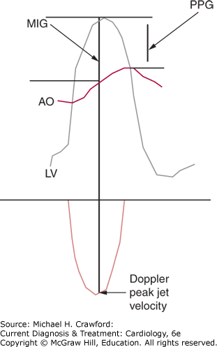
The transvalvular pressure gradient is calculated by placing one catheter into the left ventricle and a second into the proximal aorta. The difference in the pressure from simultaneous recordings from each catheter represents the peak-to-peak pressure gradient. Single-catheter techniques may include a pullback gradient and the use of a dual-lumen catheter. Note that Doppler echocardiography derives a maximum instantaneous gradient at a single point in time and by principle assumes that the pressure drop across the valve is irretrievably lost while the invasive measure derives a peak-to-peak gradient with the upstream pressure partially recovered. Therefore, the invasively derived measure of the peak pressure gradient is always lower than the Doppler-derived measure, and this difference is accentuated as the extent of pressure recovery increases (Figure 17–4). Mean gradients obtained by both Doppler and cardiac catheterization correlate well.
Figure 17-4. Top: Simultaneous left ventricular and aortic pressure tracings. Bottom: Doppler echocardiographyderived transaortic velocity profile. AO, aortic pressure; LV, left ventricle; MIG, maximum instantaneous gradient (the gradient that is derived by applying the Bernoulli equation to the peak jet velocity obtained by Doppler echocardiography); PPG, peak-to-peak gradient (the gradient obtained with invasive catheterization).
During cardiac catheterization cardiac output is measured using primarily either the Fick or thermodilution principal and the pressure gradient measured as discussed earlier with values obtained used to calculate valve area using the Gorlin equation:
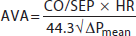
where CO = cardiac output, SEP = systolic ejection period (in seconds), HR = heart rate, and deltaCap.gifP = pressure gradient (mean). The presence of a measure of flow/cardiac output in the numerator of the Gorlin equation highlights the flow dependence on the derived values of AVA. Note that the valve area derived from the Gorlin equation is derived from recovered pressures, and as such, its value is higher than Doppler-derived valve areas by the continuity equation. The current guidelines, however, make no distinction between invasive and Doppler echocardiographic measurements in the characterization of stenosis severity by valve area and pressure gradients (see earlier discussion regarding the energy loss coefficient).
5. Cardiac Computed Tomography (CT)
Cardiac CT can be used to calculate the geometric valve area by planimetry and to quantify the extent and distribution of valve calcification, and although echocardiography remains the gold standard imaging modality, cardiac CT is becoming integral in the care of the patients with moderate to severe aortic stenosis (Figures 17–5 and 17–6). When echocardiography leaves the severity of the diagnosis inconclusive or is discordant between imaging and clinical suspicion, cardiac CT can be an invaluable tool to help classify severity, as it offers evaluation independent of hemodynamics of the patient. Cardiac CT quality continues to improve at an impressive rate. With upgrades in hardware, software, and scanning protocols which optimize gating and slice thickness, reconstruction image resolution continues to improve. These advances all aid in improving the accuracy of cardiac CT and limit the exposure of radiation to the patient. Quantification of AV calcification is strongly, but nonlinearly, associated with the hemodynamic severity of aortic stenosis, and the extent of calcification is predictive of event-free survival (survival without dyspnea, angina, syncope, heart failure, or need for valve replacement). Therefore, the measurement of AV calcium load should be considered for both diagnostic and prognostic purposes. Despite a similar degree of stenosis severity, women have lower valve calcium scores compared to men even after indexing for their smaller body size. For aortic stenosis severity purposes, the interpretation of the calcium load is thus different for men and women, where a score of more than 1300 AU for women and more than 2000 AU for men is considered severe.
Figure 17-5. A: Left; Cardiac CT showing the structures of the left ventricular outflow tract and aorta, along with the heavy calcifications (radio-opaque) on the aortic valve. B: right; Cross-sectional view of aortic valve on cardiac CT. Gating protocols allowing for the valve to be viewed at its opening during systole to allow for planimetry of the valve.
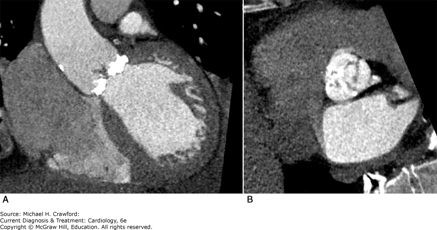
Figure 17-6. Cross-sectional view of a bicuspid aortic valve on cardiac CT. Gating protocols allowing for the valve to be viewed at its opening during systole to allow for planimetry of the valve. Note the diminished AVA along with the heavy (radio-opaque) calcifications.
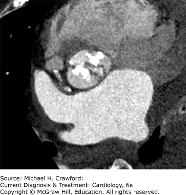
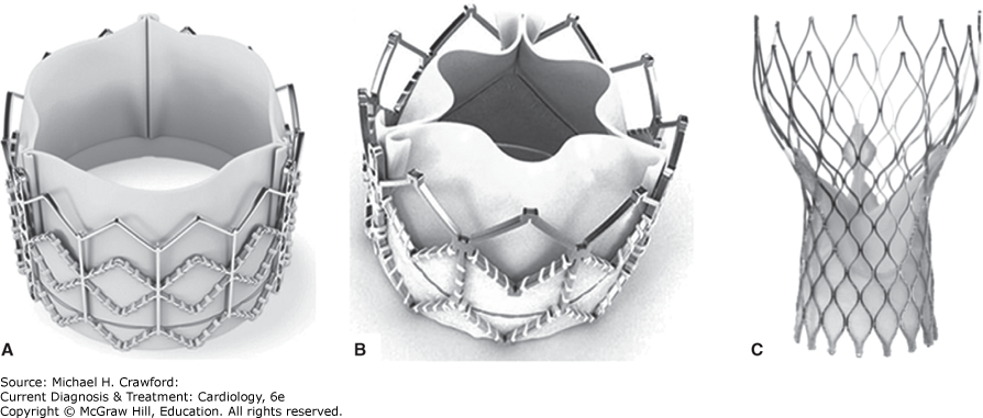
In contemporary clinical practice, cardiac CT, in conjunction with two- and three-dimensional echocardiography, now has an essential role in the evaluation of patients being considered for a percutaneous valve replacement primarily for:
The evaluation of the annular area and perimeter required for the appropriate selection of prosthetic valve size
The assessment of the peripheral vasculature and suitability for arterial access
The assessment of the geometric relationship/distance between the aortic annulus and the coronary ostia to ensure that the implanted valve, when in position, does not blanket the coronary ostia
Characterizing the extent of valve, root, and LVOT calcification, which may impact the technical success, risk of implantation, and paravalvular regurgitation
Allowing the prediction of post-implantation aortic regurgitation
Determining the feasibility of transaortic transcatheter aortic valve replacement when the transfemoral route is not feasible
DohertyJU, KortS, MehranR, SchoenhagenP, SomanP. ACC/AATS/AHA/ASE/ASNC/HRS/SCAI/SCCT/SCMR/STS 2017 Appropriate Use Criteria for Multimodality Imaging in Valvular Heart Disease: A Report of the American College of Cardiology Appropriate Use Criteria Task Force, American Association for Thoracic Surgery, American Heart Association, American Society of Echocardiography, American Society of Nuclear Cardiology, Heart Rhythm Society, Society for Cardiovascular Angiography and Interventions, Society of Cardiovascular Computed Tomography, Society for Cardiovascular Magnetic Resonance, and Society of Thoracic Surgeons. J Am Coll Cardiol. 2017;70(13):16471672. 28870679
PawadeT, ShethT, GuzzettiE, DweckMR, ClavelMA. Why and how to measure aortic valve calcification in patients with aortic stenosis. JACC Cardiovasc Img.2019;12:18351848. 31488252