A. Definition & Prevalence
Hypertrophic cardiomyopathy (HCM) is a disorder of the myocardium caused by mutations of the sarcomere or sarcomere-associated proteins. It was first brought to attention by the British forensic pathologist Donald Teare in 1958 as a disease manifesting with symmetric or asymmetric left ventricular hypertrophy (LVH) more than 1.5 cm (Figure 23–1) in a nondilated ventricle. Additional observations made by him were myocardial clefts and myocyte disarray seen along with hypertrophy in hearts of young and healthy adults who experienced sudden death. Subsequently, pioneering work by Eugene Braunwald has defined the hemodynamics of the disease process as we know it now. The distribution of hypertrophy is variable, can involve the right ventricle, and is not explained by other causes of LVH (see Table 23–1 for differential diagnosis of LVH).
Figure 23-1. Echocardiogram parasternal long-axis view in diastole showing asymmetric septal hypertrophy (white arrow) compared to the inferolateral wall.
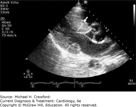
Table 23–1. Differential Diagnosis of Left Ventricular Hypertrophy
Athlete’s heart Systemic hypertension Subaortic membrane/ridge Aortic stenosis Supravalvular aortic stenosis Right ventricular hypertrophy Fabry disease Glycogen storage disease (PRKAG2 cardiomyopathy, Danon disease, Pompe disease) Mucopolysaccharide storage disease Amyloidosis Sarcoidosis |
HCM is relatively common (1:200–1:500) in the general population with about 750,000 people affected in the United States and 15–20 million worldwide. However, only 15–20% of HCM is identified clinically which clearly highlights a troubling reality given that it is the most common cause of sudden cardiac death in individuals less than 35 years of age in North America. On a positive note, most people afflicted with HCM do live a normal life. HCM patients may live well into their sixth to eighth decades, with patients older than age 90 with HCM being reported. Moreover, the first clinical recognition of HCM may occur when patients are in their sixth to eighth decade of life; usually, these patients have milder forms of the disease with the most serious complications being uncommon after age 60. The natural history of HCM can take many paths: sudden cardiac death, symptomatic HCM heart failure, end-stage cardiomyopathy, atrial fibrillation, and stroke. However, if intervened in a timely manner, HCM can potentially have no effect on longevity.
B. Genetics & Histopathology
The genetics of HCM involve an autosomal dominant pattern of inheritance, with 60–70% of patients having an affected family member. HCM is more common in males than females. Offspring of affected individuals have a 50% chance of inheriting mutations and risk of disease. Approximately 70% of genotyped patients have mutations involving β-myosin heavy chain or myosin binding protein C, other mutations include troponin I, and troponin T, but numerous others have been described with over a dozen mutations described in sarcomere-associated proteins. However, in more than 30% of HCM no causal mutations can be identified. The genetic basis of ventricular hypertrophy does not always correlate with prognosis. Patients with tropomyosin mutations have only a mild degree of ventricular hypertrophy, with little or no left ventricular (LV) outflow tract obstruction, but they still carry a disproportionately high risk for heart failure. Studies to date have not been clearly able to associate certain specific mutations to increased risk of sudden death, although multiple mutations may predict earlier disease expression and progression. Although the majority of modifier genes that influence HCM are unknown, certain modifier genes (ACE, RAAS) appear to influence hypertrophy.
C. Phenotype of Hypertrophic Cardiomyopathy
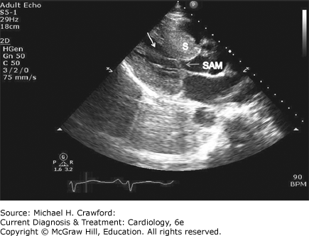
Mutations of the sarcomeric proteins lead to histopathologic evidence of myocardial disarray, which then leads to pathologic hypertrophy and patchy fibrosis of the myocardium. Intramural vessels are also frequently abnormal in HCM with thrombotic obliteration causing ischemia, which may propagate fibrosis regardless of presence of epicardial coronary disease. Asymmetric hypertrophy is most common but multiple variations of hypertrophy may be present (septal 90%, midventricular 1%, posteroseptal and lateral wall 1%, apical 3%, and symmetric 5%). Asymmetric septal hypertrophy is defined as a septal-to-posterior wall ratio more than 1.3 and, in hypertensive patients, more than 1.5. Massive hypertrophy more than 30 mm is a risk factor for sudden death. Hypertrophy in adults is usually stable but can progress in adolescents and can regress in the subset of patients who develop end-stage heart failure making the diagnosis of HCM in the latter group a challenge. An abnormal mitral valve and its apparatus are commonly seen in HCM. Elongated mitral leaflets and abnormal/anteriorly displaced papillary muscles and direct insertion of papillary muscles to the anterior mitral leaflet can be seen and have now been well defined by cardiac magnetic resonance imaging (Figure 23–2). All these factors may contribute to outflow obstruction.
Figure 23-2. Echocardiogram parasternal long-axis view still frame in a patient with obstructive hypertrophic cardiomyopathy. Asymmetric septal hypertrophy (S) is noted, along with systolic anterior motion (SAM; light arrow) and hypertrophied and anteriorly displaced papillary muscle (white arrow) causing further narrowing of left ventricular mid cavity and outflow tract.
PathophysiologyThe following processes contribute to the pathophysiology of HCM.
Abnormal diastolic function
Outflow tract obstruction/systolic anterior mitral valve motion
Mitral regurgitation
Myocardial ischemia
Arrhythmias
Abnormal autonomic function
A. Diastolic Dysfunction
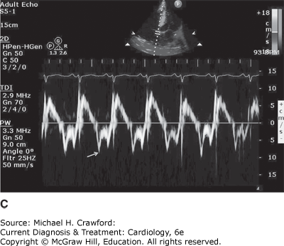
Diastolic filling abnormalities, invariably present in almost all patients with HCM, may precede hypertrophy, and as it worsens, an increased dependence on atrial contribution to ventricular filling occurs. Mitral inflow parameters demonstrate varying degrees of dysfunction, most impaired relaxation, whereas restrictive filling patterns are less common, but manifest in advanced disease states. Myocardial abnormalities documented by tissue Doppler, speckle tracking echocardiography-based strain, and LV twist and torsion abnormalities have been demonstrated in HCM. Figure 23–3 demonstrates impaired relaxation in mitral inflow, delayed propagation slope on color M-mode, and abnormal tissue Doppler, all indicative of diastolic dysfunction in a patient with HCM.
Figure 23-3. A: Doppler echocardiographic diastolic mitral inflow profile in a 32-year-old patient with hypertrophic cardiomyopathy (HCM) indicating impaired relaxation pattern. B: M-mode flow propagation abnormalities in HCM from the same patient. Black slanted line indicates propagation slope of 50 cm/s, indicating impaired relaxation. C: Pulsed tissue Doppler of the septal annulus in a 40-year-old patient with HCM and preserved left ventricular ejection fraction showing low e velocities suggesting underlying myocardial dysfunction.
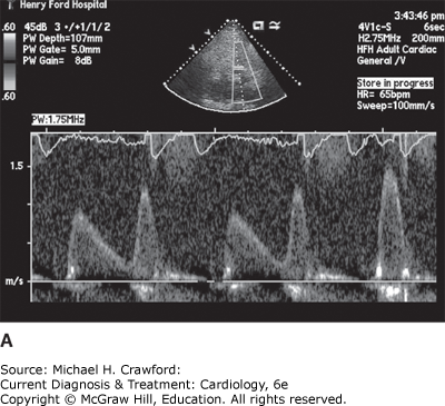
Figure 23-3.
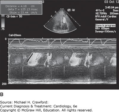
Figure 23-3.
B. Outflow Obstruction/Systolic Anterior Motion
HCM can be broadly categorized into obstructive and nonobstructive types based on presence or absence of an LV gradient (Figure 23–4). The combination of a hypertrophied septum bulging into the LV outflow tract and abnormal systolic anterior motion of the mitral valve in systole (SAM) contributes to outflow obstruction. Two-dimensional Doppler echocardiography remains the cornerstone for evaluation of HCM and quantifying gradients. About two-thirds of patients with HCM have outflow tract obstruction manifesting as resting or provocable gradients of ≥ 30 mm Hg. However, only 25–30% of patients demonstrate obstruction at rest. Simple maneuvers such as the Valsalva maneuver, standing, or administration of amyl nitrite or exercise echocardiography can provoke or exacerbate outflow tract obstruction and gradients and should be performed as part of clinical and echocardiographic evaluation when HCM is suspected and resting gradients are not observed (Table 23–2, Figure 23–5). Outflow obstruction can contribute to exercise intolerance, angina, syncope, and decreased survival. Patients with obstructive HCM are at higher risk of adverse events (heart failure and death) than those without obstruction.
Figure 23-4. Continuous wave Doppler across left ventricular outflow tract at rest demonstrates “dagger-shaped” late-peaking velocity consistent with outflow tract obstruction.
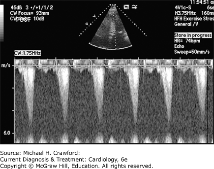
Table 23–2. Factors Influencing Obstruction in Hypertrophic Cardiomyopathy
Obstruction worsens (gradient increases and murmur increases) Tachycardia Hypovolemia Standing Valsalva Inotropes, diuretics, vasodilators
Obstruction lessens (gradient decreases and murmur decreases) Negative inotropes Bradycardia Vasoconstrictors Squatting Isometric handgrip
|
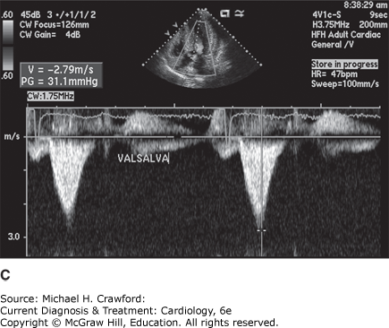
Figure 23-5. Hypertrophic cardiomyopathy outflow tract gradient (A) at rest, (B) standing, and (C) with Valsalva.
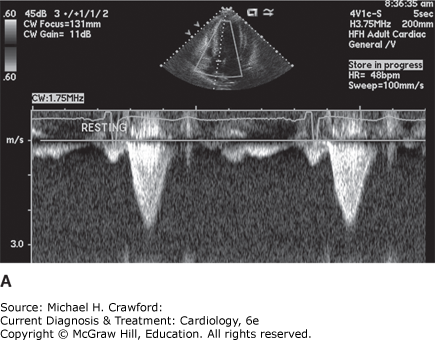
Figure 23-5.
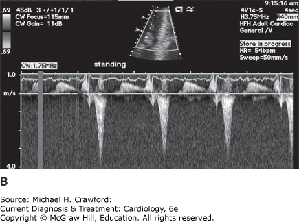
Figure 23-5.
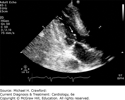
Although the exact cause of SAM is debated, there are two traditional perspectives for the mechanism of SAM. One is a causal mechanism of SAM precipitated by chordal slackening/anterior displaced mitral valve apparatus leading to “drag forces,” and the other explains SAM because of the Venturi effect of accelerated flow across the left ventricular outflow tract (LVOT) (Figure 23–6). However, the “Venturi forces” theory has lately been questioned and increased recognition of anatomic abnormalities of mitral apparatus including elongated mitral leaflets, anterior displacement, anomalous insertion, and bifid papillary muscles all seem to play a role in obstructive physiology and SAM. The severity of obstruction and timing of SAM onset are linearly related. An important caveat is that SAM can be seen in non-HCM states, even when no basal septal hypertrophy is present such as a hyperdynamic small, underfilled ventricle (volume-depleted states), after mitral valve repair, and as a nonspecific finding confined to the chordal apparatus (chordal SAM). More recently increased recognition of dynamic obstructive physiology with SAM has been observed in large anteroseptal infarcts and in Takotsubo cardiomyopathy where the basal septum is hyperdynamic contributing to narrowing of LVOT and drag forces causing SAM and outflow obstruction. Thus, although patients in many different pathophysiologic states may exhibit HCM physiology (gradients, SAM), this does not equate to a diagnosis of HCM.
Figure 23-6. Echocardiographic apical three-chamber still frame showing systolic anterior motion of mitral valve encroaching on the left ventricular outflow tract creating outflow obstruction physiology (white arrow).
C. Mitral Regurgitation
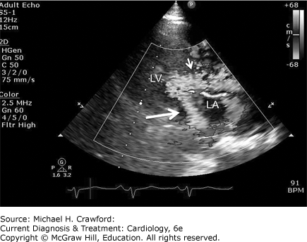
The main etiology of mitral regurgitation (MR) in HCM is malcoaptation of the mitral leaflets secondary to SAM. SAM-related MR is posteriorly directed starting in mid-late systole (Figure 23–7). If other MR jet directions are seen, then intrinsic mitral valve disease should be suspected. Concomitant primary mitral valve disease is reported in about 20% of patients with HCM (mitral valve prolapse, ruptured chordae, mitral annular calcification, and anomalous or anteriorly displaced papillary muscle) (see Figure 23–2). If SAM is relieved by either myectomy or septal ablation, posterior MR tends to resolve or diminish. Nonposterior MR may require surgical repair or replacement of the valve, which may be one reason why surgical myectomy is favored over alcohol septal ablation in patients with concomitant mitral valve apparatus abnormalities as a treatment strategy for symptomatic HCM.
Figure 23-7. Echocardiographic parasternal long-axis view still frame with flow velocity mapping illustrating consequence of systolic anterior motion and left ventricular outflow tract (LVOT) obstruction, which leads to turbulent flow across LVOT (small arrow) during left ventricular (LV) systole and posterior directed jet of mitral regurgitation (large thick arrow) into left atrium (LA).
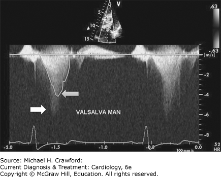
A comprehensive echocardiography assessment of MR is important in HCM. The assessment of peak outflow gradients can be challenging in some patients with HCM in the setting of MR due to contamination of both jets (Figure 23–8). Careful observation of jet profile and coexistent inflow profile and evaluation of peak gradients from different windows will help to differentiate outflow gradients from MR. Aligning the continuous wave doppler parallel to MR jet and away from LVOT flow acceleration may further help avoid contamination of doppler signals.
Figure 23-8. Continuous wave Doppler evaluation of a patient with hypertrophic cardiomyopathy during Valsalva provocation. Two distinct flow patterns are seen: one of outflow obstruction, which has a late peak (light arrow), and a superimposed Doppler profile signal of mitral regurgitation (white arrow), making assessment of peak outflow obstruction velocity challenging.
D. Myocardial Ischemia
The combination of significant LVH with or without outflow obstruction sets the stage for substantial increases in myocardial oxygen demand and can precipitate angina secondary to myocardial ischemia in HCM. Angina has been reported in approximately 30% of HCM patients, and ischemic perfusion defects on myocardial perfusion imaging can be present in a majority of patients with HCM. Although epicardial coronary disease may cause angina/perfusion defects, a supply-demand mismatch in conjunction with abnormal microvasculature is likely the major contributor. Cardiac positron emission tomography assessment of myocardial blood flow and flow reserve has shed further light on abnormalities in flow and flow reserve causing myocardial ischemia and likely related to microvascular dysfunction in the setting of nonobstructive epicardial arteries.
E. Abnormal Autonomic Tone
About 25% of HCM patients demonstrate abnormalities of autonomic function, which if present, portend a poorer prognosis. Abnormalities with exercise, such as systolic drop in blood pressure of ≥ 20 mm Hg, or failure to augment blood pressure can occur from factors such as outflow obstruction and inappropriate vasodilatation despite an appropriate increase in cardiac output.
A. Physical Examination
A general examination is not helpful in diagnosing HCM except when associations with other diseases such as Friedrich ataxia or Noonan syndrome are present. The cardiovascular clinical examination findings of HCM are usually striking unless there is no obstruction at rest. Radial pulse is of brisk character. There is a characteristic brisk carotid upstroke with an abrupt deceleration due to obstruction. The apical impulse is described as bifid and sustained due to the initial powerful contraction followed by obstruction and then by continued contraction of the left ventricle. A systolic thrill may be felt. The first and second heart sounds are normal. An S4 may be heard due to atrial contraction against a noncompliant left ventricle.
The systolic murmur in HCM is a harsh and crescendo decrescendo in character. It is heard all over the precordium but usually is not transmitted to the carotids. The murmur increases in intensity with Valsalva, standing, and inhalation of amyl nitrite, due to a drop in preload and decrease in LV cavity size and vasodilation (amyl nitrite), which can worsen outflow obstruction. The murmur intensity decreases with squatting and passive leg raising. A second murmur of mid-late MR can be heard, induced by SAM of the mitral valve. The combination of powerful ejection followed by obstruction and subsequent mitral insufficiency can be aptly described as the “eject-obstruct-leak” triad of hypertrophic obstructive cardiomyopathy (HOCM). The key differentiation of a HOCM murmur is from that of aortic stenosis (AS) and discrete subaortic stenosis. However, the pulse of AS is low amplitude and slow (parvus et tardus) compared to brisk in HCM. Ejection click (with bicuspid etiology) and aortic regurgitation murmur are more common in AS and not typically seen in HCM. The aortic second sound is abnormal in AS and normal in HCM. Finally, an AS murmur decreases with Valsalva in contrast to the murmur of HCM, which increases.
B. Diagnostic Tests
1. Electrocardiogram
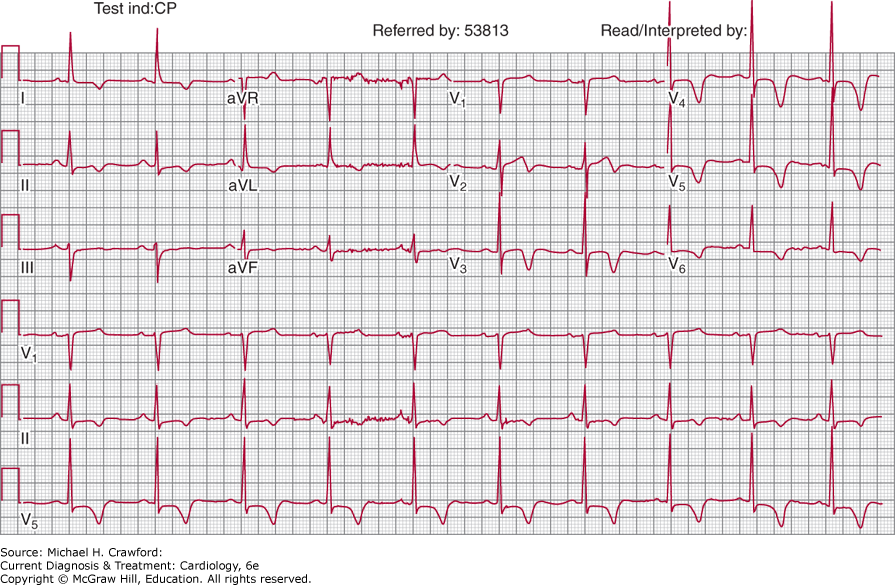
More than 90% of patients with HCM exhibit 12-lead electrocardiography (ECG) abnormalities. Common abnormalities include LVH with strain, prominent Q waves, left atrial enlargement, and left-axis deviation. ST elevation or depression or T-wave inversions can also be seen. Specific patterns such as deep symmetric T-wave inversions in lateral precordial leads (suggestive of apical variant HCM) are also well recognized (Figure 23–9). In general, ECG abnormalities are present in more than 80% of HOCM compared to approximately 50% in nonobstructive types of HCM. Preexcitation patterns are also seen in some patients with HCM, and atrial fibrillation has been documented in up to 25–30% of elderly HCM patients. More recently artificial intelligence (AI) has shown great promise in the recognition of patterns associated with HCM. AI-ECG HCM scores correlated with disease status as measured by decreases over time in left ventricular outflow tract gradients and natriuretic peptide (NT-proBNP) levels in these patients. The longitudinal associations of the AI-ECG HCM score were significant and likely reflected changes in the raw ECG waveform that were detectable by AI-ECGs and correlated with HCM disease pathophysiology and severity. AI-ECG’s potential is enhanced by the advances in that ECGs can now be measured remotely via smartphone-enabled electrodes and may permit remote assessment of disease progression as well as drug treatment response.
Figure 23-9. Twelve-lead electrocardiogram in a patient with palpitations. Note deep symmetric T-wave inversions in the precordial leads. There are also T-wave abnormalities in the limb leads. In the absence of hypertension, these should raise suspicion of hypertrophic cardiomyopathy. Subsequent echocardiography and cardiac magnetic resonance imaging confirmed diagnosis of apical hypertrophic cardiomyopathy.
TisonGH, SiontisKC, AbreauS,et al. Assessment of disease status and treatment response with artificial intelligence-enhanced electrocardiography in obstructive hypertrophic cardiomyopathy. J Am Coll Cardiol.2022;79:10321034. 35272798
2. Echocardiogram
Echocardiography (echo) plays an integral role in diagnosis, follow-up, and management of patients with HCM. Abnormalities can be demonstrated on M-mode echo; two-dimensional (2D) echo; color, pulsed and continuous wave, and tissue Doppler; and strain imaging. Key findings of HCM by echo include the following:
A. M-Mode
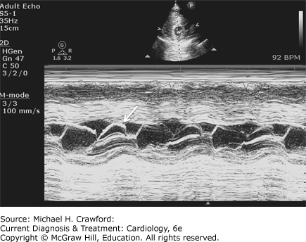
Midsystolic notching of aortic valve is seen in HCM (Figure 23–10). SAM of mitral valve can also be assessed, and extent of SAM and SAM-septal contact distance as a marker of severity can be assessed (Figure 23–11). Right ventricular (RV) and LV septal and posterior wall thickness and chamber sizes can be determined.
Figure 23-10. Midsystolic notching of the aortic valve (arrow) is indicative of dynamic left ventricular outflow obstruction.
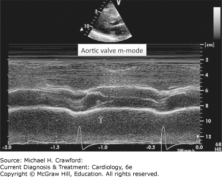
Figure 23-11. Systolic anterior motion of mitral valve on M-mode echocardiography. The duration and extent of mitralseptal contact correlate with extent of outflow obstruction.
B. 2D ECHO
Asymmetric septal hypertrophy is the most common pattern, although other patterns can be seen (see Figure 23–1). RV hypertrophy may also be present. Because echo can underestimate or miss hypertrophy confined to the lateral wall and apex, comprehensive multiple nonforeshortened views of LV are needed. Echo contrast imaging can be very useful to assess apical variant HCM. Mitral valve abnormalities can be seen as described previously. SAM can be assessed, and all chamber sizes can be evaluated. Insights from three-dimensional echo reconstructions of the mitral valve in HCM show a mitral leaflet area twice that of the normal population, and the indexed mitral valve leaflet area has been shown to be predictive of outflow obstruction. Recently, an enlarged left atrium (indexed volume > 34 mL/m2) has been shown to be an independent adverse prognosticator in HCM.
C. Doppler
Pulsed wave and continuous wave Doppler assessment is integral in establishing the level of outflow obstruction and peak gradients, respectively. The modified Bernoulli equation (4peakV2) is used to determine peak gradient. The classic late-peaking “dagger-shaped” velocity profile can be seen when outflow obstruction is present (see Figure 23–4). A peak gradient of more than 30 mm Hg is considered evidence for resting outflow obstruction. Gradients should be assessed at rest and with provocation by Valsalva or standing (see Figure 23–5B). If echo features suggest HCM but no or minimal obstruction is demonstrable at rest or Valsalva, exercise echo with peak/immediate post exercise gradient assessment could help unmask HOCM (latent obstruction) and this is recommended in the 2020 ACC AHA guidelines for HCM in all patients with nonobstructive HCM. In contrast the European Society of Cardiology recommendations suggest exercise echocardiography only in symptomatic patients with nonobstructive phenotype and in selected asymptomatic patients where lifestyle or treatment decisions may be impacted (Color Doppler and continuous wave Doppler are very useful to comprehensively assess severity and mechanism of MR. It is important to reliably distinguish the MR signal from the LVOT obstruction signal to avoid overestimation of severity of outflow obstruction. Diastolic function and tissue Doppler abnormalities of myocardial function should be assessed. In particular, average e′ velocities by tissue Doppler of less than 13.5 cm/s predict genotype-positive HCM with a sensitivity of 75% and specificity of 86%. Reduced longitudinal peak systolic deformation (strain) averaged across all walls less than 10.6% (in absolute value) predicts HCM with a sensitivity of 85% and specificity of 100% and helps differentiate it from hypertensive LVH. A combination of septal/posterior wall ratio more than 1.3 and systolic strain assessment yields a predictive accuracy for HCM of 96%. The estimation of filling pressures using E/e′ at best correlates moderately with invasive wedge pressure measurements, although low e′ in itself has independent adverse prognostic value in HCM. Delayed untwisting of LV has been demonstrated by torsion analysis using speckle tracking techniques, again reflecting diastolic dysfunction-related abnormalities seen in HCM.
3. Cardiac Magnetic Resonance Imaging
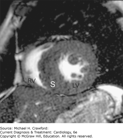
Cardiac magnetic resonance imaging (CMRI) is rapidly becoming a key component in enhancing diagnostic accuracy, morphology, and more importantly, prognostication due to tissue characterization of fibrosis in HCM. With its superior spatial resolution and indefinite choice of imaging planes, it has demonstrated enhanced accuracy in assessing HCM features and identifying patterns of hypertrophy not well seen on echocardiography. Approximately 6% of patients missed by echo (mainly anterolateral hypertrophy) are diagnosed with HCM by CMRI, and 57% of apical aneurysms missed by echo are identified on CMRI. Assessment of LV mass and end-diastolic wall thickness are more accurate with CMRI than echo. Figures 23–12 and 23–13 show CMRI delineation of septal hypertrophy in HCM.
Figure 23-12. Steady-state free precession cine sequence still frame (cardiac magnetic resonance imaging) view of left ventricle (LV) and right ventricle (RV) showing significant asymmetric septal hypertrophy (S) compared to lateral wall (L). Note outstanding delineation of the LV blood to endocardial interface enabling accurate measurements of thickness of myocardial walls.
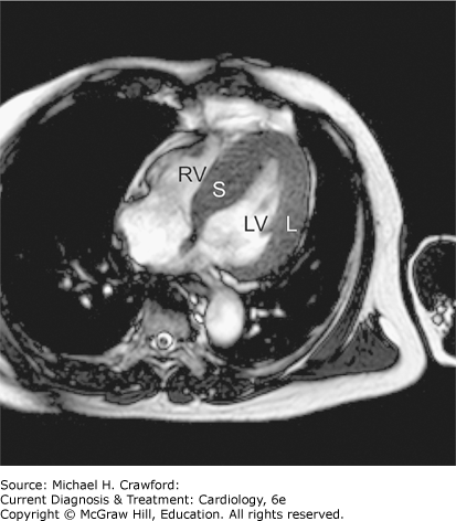
Figure 23-13. A still frame from a steady-state free precession short-axis cardiac magnetic resonance study in a patient with suspected hypertrophic cardiomyopathy. There is severe asymmetric left ventricular (LV) hypertrophy involving the septum (S). Multiple papillary muscle heads are also noted in the LV cavity. RV, right ventricle.
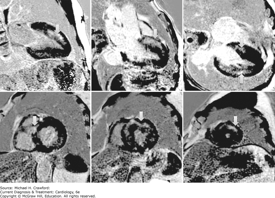
CMRI also provides superb assessment of mitral valve apparatus morphologic abnormalities and gives unparalleled assessment of the right ventricle. The presence of patchy myocardial fibrosis in HCM detected by late gadolinium enhancement techniques in CMRI is increasingly recognized as an adverse prognosticator for arrhythmias and mortality, and ≥ 15% of myocardium with gadolinium enhancement is emerging as a quantitative marker for adverse events. Characteristic patterns include delayed enhancement detected at RV insertion points into the left ventricle, although multiple areas of fibrosis can be seen in the left and/or right ventricle with predominance in the hypertrophied zones of the ventricle (Figure 23–14). CMRI has been used to characterize the presence of “myocardial crypts” or recesses in different areas of the myocardium in phenotype-negative but genotype-positive family members of probands with HCM. Whether these represent early manifestations of an HCM phenotype yet to develop remains to be established, as does any long-term significance.
Figure 23-14. Gadolinium-based delayed-enhancement cardiac magnetic resonance image showing abnormal enhancement (arrows) in multiple areas of the myocardium denoting fibrosis. Note the noncoronary predominant midmyocardial distribution of hyperenhancement seen in nonischemic etiologies.
KampNJ, CheryG, KosinskiAS,et al. Risk stratification using late gadolinium enhancement on cardiac magnetic resonance imaging in hypertrophic cardiomyopathy. A systematic review and meta-analysis. Prog Cardiovasc Dis.2021;66:1016. 33171204
ReichekN. Imaging cardiac morphology in hypertrophic cardiomyopathy: recent advances. Curr Opin Cardiol. 2015;30: 461467. 26154073
4. Cardiac Computed Tomography Angiography, Cardiac Nuclear Perfusion Imaging, and Positron Emission Tomography
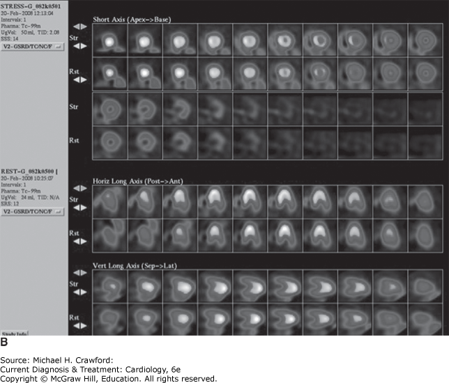
Cardiac computed tomography angiography (CCTA), cardiac nuclear perfusion imaging, and positron emission tomography (PET) are not routinely used in the assessment of HCM but may be useful in select cases. Retrospective gated CCTA can provide LV function assessment apart from LV morphology and coronary anatomy. CCTA may be useful to define coronary anatomy noninvasively in patients with HOCM. It can also accurately delineate septal perforator course noninvasively as a road map for septal ablation. Single-photon emission computed tomography (SPECT) and PET can demonstrate characteristic intense uptake of radioisotope in hypertrophied zones of myocardium and demonstrate perfusion abnormalities mainly related to microvascular flow reserve abnormalities in HCM (Figure 23–15). Quantitative PET-determined coronary flow reserve abnormalities along with ischemic perfusion defects have been shown to predict adverse outcomes in HCM in limited studies. Most recently, sympathetic innervation imaging (iodine-123 [123I] metaiodobenzylguanidine [MIBG]) has shown that there is sympathetic denervation in hearts of HCM patients (decreased heart-to-mediastinal ratio and increased washout of 123I MIBG) and that this is correlated to septal thickness and outflow obstruction. Whether 123I MIBG denervation imaging predicts sudden death above and beyond other risk markers needs further study.
Figure 23-15. A: Regadenoson rubidium-82 rest stress positron emission tomography scan showing moderate reversible ischemic defect (arrow) in the mid-distal septum, apex, and distal lateral wall (stress images, second to last row). Rest images (bottom rows) show intense uptake in mid-distal ventricular walls suggestive of mid-apical hypertrophic cardiomyopathy confirmed by echocardiography and magnetic resonance imaging. Patient had no obstructive coronary artery disease on catheterization. B: Regadenoson single-photon emission computed tomography in a patient with new-onset dyspnea. Images show marked increase in uptake of isotope in both stress (top) and rest (bottom) images in the absence of any ischemia, raising suspicion for mid-apical hypertrophic cardiomyopathy.
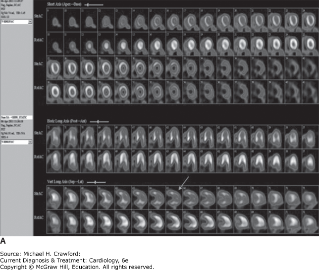
Figure 23-15.
ShariatM, ThavendiranathanP, NguyenE,et al. Utility of coronary CT angiography in outpatients with hypertrophic cardiomyopathy presenting with angina symptoms. J Cardiovasc Comput Tomogr. 2014;8:429437. 25467830
TimmerSA, KnaapenP. Coronary microvascular function, myocardial metabolism, and energetics in hypertrophic cardiomyopathy: insights from positron emission tomography. Eur Heart J Cardiovasc Imaging. 2013;14:95101. 23152441
5. Cardiac Catheterization
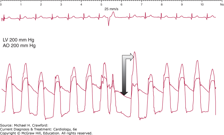
Echocardiography with Doppler is usually sufficient to diagnose HCM, and invasive hemodynamics are usually not necessary. However, if clinical and echo findings are discrepant or the exact severity of obstruction cannot be accurately determined by echo, then catheterization may be needed. A carefully conducted study with continuous documentation of pullback gradients will outline the level and severity of obstruction. A classic “spike and dome” arterial pulse waveform can be recorded representing the rapid ejection followed by sudden obstruction. Pulmonary capillary wedge pressure may be elevated, reflecting high filling pressures from diastolic dysfunction, stiff ventricle, and noncompliant atrium. Depending on the severity of MR, a prominent “v” wave can be recorded. Also, the Brockenbrough-Braunwald-Morrow sign can be demonstrated by inducing premature ventricular beats. Following a ventricular extrasystole, an increase occurs in LV systolic pressure, a decrease in ascending aortic pressure, and increase in gradient. Importantly, a drop in pulse pressure is seen in HCM compared to AS where pulse pressure does not change (Figure 23–16).
Figure 23-16. Brockenbrough-Braunwald-Morrow sign. Following a premature ventricular beat, there is an increase in the gradient but a decrease in aortic and pulse pressure in hypertrophic cardiomyopathy during cardiac catheterization (double arrow). AO, aorta; LV, left ventricle.
6. Exercise Echocardiography
Exercise echocardiography is recommended to provoke obstruction, which is either absent or minimal at rest (< 30 mm Hg), particularly in symptomatic patients with features of nonobstructive HCM at baseline. Furthermore, exercise can unmask abnormal hemodynamic responses such as failure to augment systolic blood pressure or a drop in systolic blood pressure with exercise. Other abnormal signs with exercise include poor exercise capacity, ventricular arrhythmias, ST depression, and/or ischemic wall motion abnormalities (regardless of presence of epicardial disease). Symptomatic patients with HCM with gradients at rest or with provocation ≥ 50 mm Hg usually require therapy (medical and/or invasive), and hence, exercise testing is not indicated in such patients. However, once medical therapy is maximized in patients with HOCM, repeat exercise echocardiography may be helpful to reevaluate symptoms, exercise hemodynamics, and effects of medical therapy on reduction of outflow obstruction/gradients. Since dobutamine can induce outflow obstruction and gradients, it is not recommended for testing in HCM because it can provoke a similar response even in normal individuals.
7. Genetic Testing
Despite initial optimism, the concept of using single-nucleotide sarcomere mutations to identify high-risk patients and prognosticate has not proven useful. The most practical current application for genetic testing in HCM is screening of asymptomatic family members of patients with HCM. If a known mutation exists in HCM patients, focused genetic testing can be done for that mutation in the family. Only about 33% of probands may have a genetic mutation (higher if positive family history). One advantage of genetic testing is identifying the genotype-positive/phenotype-negative family member who may demonstrate subtle ECG or imaging abnormalities and may require closer follow-up to see if he or she progresses to an HCM phenotype. A further use of genetic testing is in differential diagnosis for HCM, including diseases such as PRKGA2, LAMP2 (highly lethal with mortality before 25 years), and Fabry disease, where treatment options such as enzyme replacement are critical. Even if genetic testing is negative in a patient with HCM, continued clinical and surveillance imaging may be needed in at risk relatives. Adolescents and athlete family members should undergo echo annually, and nonathlete family members should undergo echo every 5 years. Pre and post genetic test counselling should be offered to HCM patients undergoing genetic testing. For HCM patients with genetic variants of uncertain significance, serial re-evaluation of test results is recommended to assess for variant reclassification, as this may trigger testing for family members. Preconception and prenatal reproductive and genetic counseling should also be offered.
C. Diagnostic Considerations in Hypertrophic Cardiomyopathy Variants
1. Apical HCM
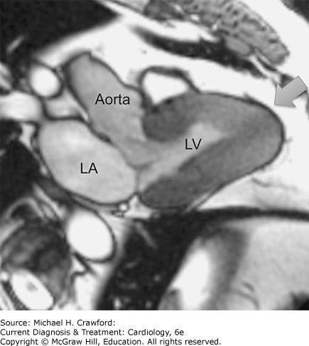
This form of HCM is more frequently reported in Asian populations and is associated with deep symmetric T-wave inversions in the anterior precordial leads (see Figure 23–9). Echo reveals a predominant pattern of hypertrophy distributed toward the apex with relative sparing of the base, although concomitant basal hypertrophy may also be present. In conjunction with the normal contractility, the marked narrowing of the hypertrophied apex in systole produces the “ace of spades” appearance on echo. Apical variant HCM can be missed in echo due to foreshortened views or poor visualization of hypertrophy with low-frequency transducers. Use of higher frequency transducers, color Doppler to delineate full extent of blood flow, and contrast echo can overcome these limitations. CMRI is an excellent alternative to echo and is a superior technique to diagnose apical HCM (Figure 23–17). In general, isolated apical HCM is devoid of outflow obstruction, and these patients have a more benign prognosis than the classical HCM patients.
Figure 23-17. Steady-state free precession left ventricular (LV) outflow tract view cardiac magnetic resonance imaging still frame showing marked LV myocardial hypertrophy with obliteration of apex in systole (arrow). Note characteristic “ace of spades” appearance of LV myocardium in systole. LA, left atrium.
2. Midventricular HCM
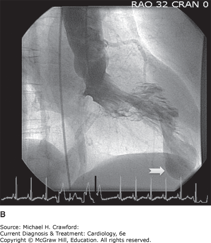
This variant involves predominant hypertrophy of the mid ventricle, resulting in midcavitary obstruction and separation of the ventricle into two compartments in systole. Flow acceleration and obstructive gradients can be demonstrated in the mid-ventricle where most obstruction exists, although concomitant outflow obstruction may be present. Abnormal early diastolic color flow can be seen during isovolumic relaxation due to differential intraventricular pressure gradients across the obstruction. Coexistent hypertension may exacerbate midventricular hypertrophy. In the pure midventricular HCM, SAM may be absent, and no outflow obstruction may be detected. Continuous wave Doppler can delineate maximal obstructive midventricular gradients (Figure 23–18A). Long-standing obstruction in mid ventricle can cause apical necrosis with aneurysm formation due to chronic subendocardial ischemia (Figure 23–18B). This subset of patients is more prone to ventricular arrhythmias and thrombus formation. CMRI is an excellent technique to demonstrate these apical aneurysms, which can be missed by echo.
Figure 23-18. A: An example of a continuous wave Doppler signal across the left ventricle (LV) showing the mitral regurgitation signal and concomitant late-peaking signal of midcavitary obstruction with a peak gradient of 41 mm Hg. B: A still frame left ventriculogram in a patient with severe midventricular hypertrophic cardiomyopathy with apical aneurysm outpouching (arrow).
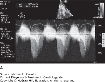
Figure 23-18.
3. Hypertensive HCM in the Elderly
First described in the 1980s, this entity is increasingly recognized more frequently given the growing population of elderly patients with hypertension. Whether this represents a variant of HCM or mimics the physiology of HCM is still debated. Most patients have a long history of hypertension, small, hypertrophied ventricles, a narrow LVOT with mitral annular calcification, and some anterior displacement of the mitral annulus. This constellation can set the stage for acceleration of flow, SAM, and outflow obstruction, although midventricular obstruction may also be present. Treatment is similar to HCM with regard to relief of outflow obstruction with negative inotropic drugs and adequate control of hypertension. Avoidance of excessive diuresis and preload reduction is also a key component.
4. End-Stage HCM
Seen in less than 5% of patients, end-stage HCM features ventricular dilation, advanced diastolic dysfunction, and a course predominated with symptoms of systolic and diastolic heart failure. Medical treatment consists of angiotensin-converting enzyme (ACE) inhibitors or angiotensin receptor blockers, β-blockers, diuretics, and spironolactone, as well as nonpharmacologic strategies such as implantable cardioverter-defibrillator (ICD) and biventricular pacing if indicated. In refractory cases, heart transplantation should be considered. Survival after heart transplantation in HCM is comparable to the general population.