Introduction ⬇
Essentials of DiagnosisA rare but critical diagnosis in the evaluation of cardiac masses. The history, physical examination, and imaging characteristics can yield valuable diagnostic clues. Diagnostic confirmation requires tissue biopsy.
|
General Considerations ⬆ ⬇
Cardiac tumors, although considered a rare diagnostic entity, provide unique insight into the anatomic and pathophysiologic functioning of the heart. Reminiscent of benign, malignant, and metastatic neoplasms elsewhere in the body, theses tumors herald their presence by the secondary effects produced from enlarging within the confines of cardiac chambers and tissues. Because they rarely cause symptoms, cardiac tumors are generally unsuspected and may be discovered incidentally during evaluation of an unrelated condition. This chapter will help guide the astute clinician, focusing on when to consider this diagnosis and how to obtain the appropriate cardiovascular imaging studies. There are a wide variety of common cardiac tumors (Table 32–1).
Table 32–1. Classification of Common Cardiac Tumors
| Primary Cardiac Tumors |
Benign Tumors Myxoma Papillary fibroelastoma Lipoma Fibroma Hemangioma Teratoma Rhabdomyoma Hamartoma (oncocytic cardiomyopathy) |
Malignant Tumors Sarcoma: includes angiosarcoma, rhabdomyosarcoma, fibrosarcoma, osteosarcoma, leiomyosarcoma, liposarcoma, synovial sarcoma Lymphoma Malignant fibrous histiocytoma Epithelioid hemangioendothelioma |
Benign or Malignant Tumors Mesothelioma (includes cystic tumor of the atrioventricular node) Paraganglioma |
| Secondary Cardiac Tumors |
| Metastasis to the heart and pericardium: commonly from malignant melanoma or neoplasms of the lung, hematopoietic system, gastrointestinal tract, kidney, breast, thyroid, and soft tissue |
Primary tumors of the heart are rare, with a prevalence of 0.02% reported in combined data from multiple large autopsy series. Although cardiac myxoma has been traditionally reported as the most frequent tumor type in adults, increasing utilization of imaging studies has revealed a higher frequency of papillary fibroelastomas than previously known. In the pediatric population, rhabdomyomas represent the most common type of primary cardiac tumor (Table 32–2).
Table 32–2. Primary Cardiac Tumors: Location and Features
| Cardiac Tumor | Typical Location | Interesting Features/Associations |
|---|
| Myxoma | Left atrium | Carney complex; constitutional symptoms |
| Papillary fibroelastoma | Cardiac valves | Can present with sudden death from coronary ostial obstruction |
| Fibroma | Left ventricular myocardium | Very large tumors that exert a mass effect |
| Rhabdomyoma | Cardiac chambers | Tuberous sclerosis |
| Angiosarcoma | Right atrium | Highly aggressive tumor |
| Rhabdomyosarcoma | Cardiac chambers | Commonest pediatric malignancy |
| Fibrosarcoma | Left atrium | White fish flesh tumor |
| Leiomyosarcoma | Smooth muscle cells of pulmonary veins and arteries | Epstein-Barr virus in immunosuppressed patients |
| Lymphoma | Right heart chambers | Immunocompromised patients |
| Paraganglioma | Pericardium; left atrial wall | Majority are hormonally inactive |
| Teratoma | Pericardium | Contain elements from all three germ cell layers |
A. Benign Cardiac Tumors
The overwhelming majority of primary cardiac tumors, approximately 75% to more than 90%, are benign, and may arise from the endocardium, myocardium, or pericardium. Timely diagnosis is key because although the tumors themselves are generally curable, their obstructive and embolic effects can be catastrophic.
1. Myxoma: A Stalked Spherical Tumor of the Left Atrium
A. Demographics
Approximately 50% of all benign primary cardiac tumors are myxomas. They are predominantly found in women, in the 30- to 60-year age group. The mean age at diagnosis is 51 years. Most commonly, myxomas are sporadic and isolated in occurrence; however, up to 10% are familial, typically transmitted in an autosomal dominant pattern. Familial myxomas tend to present earlier in life, with a mean age at diagnosis of 25 years. These patients also tend to have multiple (30–50%) and/or recurrent (12–22%) tumors.
B. Typical Locations
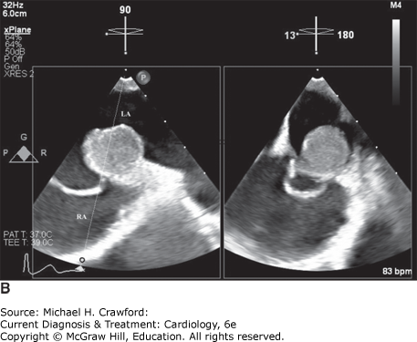
Theoretically, myxomas can occur in any cardiac chamber or on any valve, but the most common site for a myxoma is the left atrium (74%), with 18% occurring in the right atrium and 4% each occurring in the right and left ventricles. The typical site of origin is near the region of the fossa ovalis with a stalk-like attachment to the interatrial septum (Figure 32–1). Ventricular myxomas almost always originate from the ventricular free wall.
Figure 32-1. Atrial myxoma. A: Transthoracic echocardiogram obtained in a 71-year-old woman who presented with palpitations. The apical four-chamber view shows an interatrial septal aneurysm with rightward bowing and an echogenic mass (arrow) near the superior portion of the interatrial septum, later found to be a left atrial myxoma. B: Bicaval view from preoperative transesophageal echocardiogram in the same patient depicts the left atrial myxoma with attachment site near the superior vena cava, while the corresponding biplane view on the right demonstrates rightward bowing of the interatrial septal aneurysm. LA, left atrium; RA, right atrium.
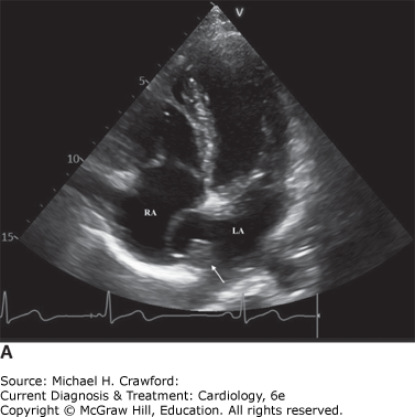
Figure 32-1.
C. Pathology
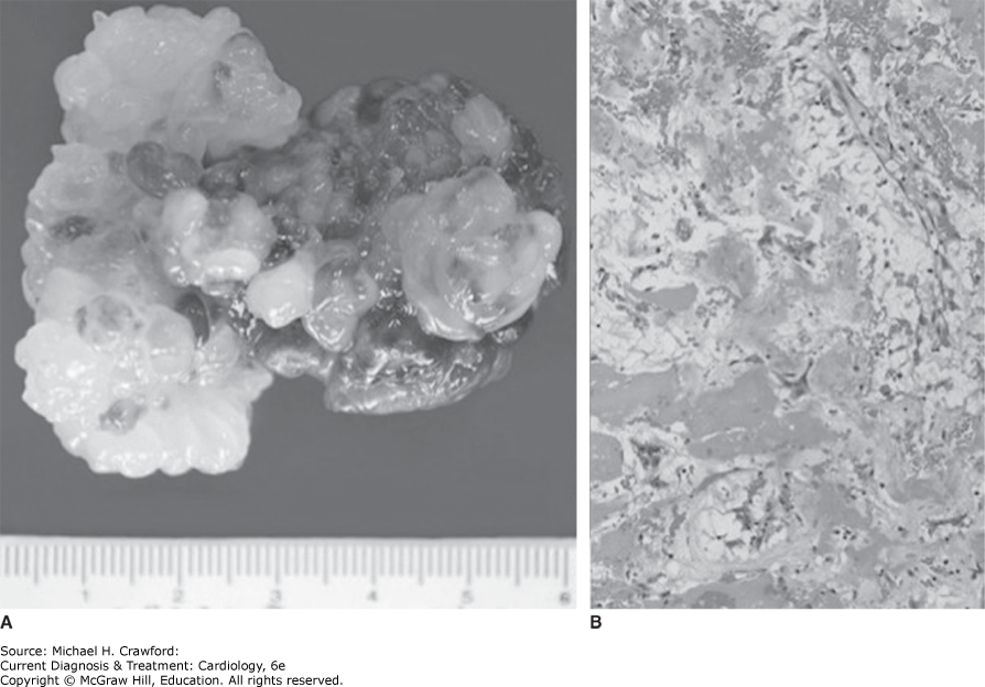
Myxomas vary in size, ranging from 1 to 15 cm in diameter, and are typically pedunculated. They are gelatinous in consistency, with a smooth or villous surface, and may be friable. Histologically, myxomas arise from multipotent mesenchymal cells and produce vascular endothelial growth factor. They are visualized as islands of tumor cells of variable shapes (round, elongated, or polyhedral), scattered within a pale-staining mucopolysaccharide matrix (Figure 32–2).
Figure 32-2. Atrial myxoma. A: Gross pathology: The specimen shows a lobulated, gelatinous tumor with areas of hemorrhage resected from the left atrium of a 58-year-old female patient. The diagnosis was made incidentally on an echocardiogram, which the patient had preoperatively for an unrelated condition. B: Histology: Hematoxylin-eosin stain of the same mass reveals small, perivascular clusters of tumor cells scattered within a myxoid matrix rich in mucopolysaccharides and fibrin, consistent with a cardiac myxoma. (Reproduced with permission from Philip C. Ursell.)
D. Associations
The Carney complex is an inherited autosomal dominant condition characterized by cardiac myxomas, myxoid fibroadenomas of the breast, and testicular and pituitary gland tumors. These occur in association with pigmented lentigines, blue nevi of the skin, and Cushing syndrome, due to primary pigmented nodular adrenocortical dysplasia.
The Carney complex appears to be a genetically heterozygous syndrome with gene localization to loci in 17q22-24, 2p16, and other regions. Inactivating mutations of PRKAR1A, a gene which maps to 17q22-24 and encodes the protein kinase A (PKA) regulatory subunit type 1a, are responsible for most cases of Carney complex. Carney complex should be considered in any patient with a diagnosis of myxoma, particularly if presenting early in life, with multiple tumors, or with a myxoma outside of the left atrium.
BouysL, BertheratJ.MANAGEMENT OF ENDOCRINE DISEASE: Carney complex: clinical and genetic update 20 years after the identification of the CNC1 (PRKAR1A) gene. Eur J Endocrinol. 2021;184(3):R99R109. 33444222. doi: 10.1530/EJE-20-1120
2. Papillary Fibroelastoma: A Sea Anemone on Cardiac Valves
A. Demographics
Papillary fibroelastomas have previously been designated the second-most common primary cardiac neoplasm in adults behind myxomas; however, recent work suggests they may, in fact, be the most common, likely related to the increase in cardiac imaging. While they have been reported in patients age 3–86 years, the typical age at diagnosis is 60 years. Papillary fibroelastomas are often discovered postmortem, with an autopsy incidence of 0.002–0.33%.
B. Typical Locations
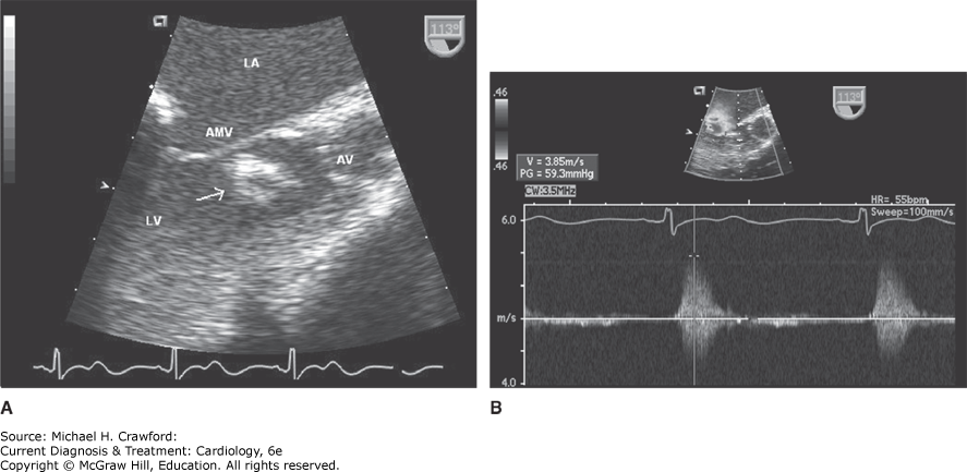
More than 80% of fibroelastomas are found on the valvular endocardium, typically of the aortic and mitral valves, followed by the tricuspid valve. Unlike vegetations, fibroelastomas often occur downstream of the valve (ie, ventricular side of the mitral valve, aortic side of the aortic valve) and normally do not cause valvular dysfunction (Figure 32–3). The remainder of these tumors are located on the subvalvular apparatus as well as within the atria and ventricles.
Figure 32-3. Papillary fibroelastoma. Transesophageal echocardiogram performed in a 75-year-old man presenting with dizziness. A: An irregular mass attached to the ventricular side of the anterior mitral leaflet is shown in this mid-esophageal long-axis view. The location on the downstream side of the valve and the frond-like surface are highly suggestive of a papillary fibroelastoma (arrow). B: Continuous wave Doppler flow demonstrates left ventricular outflow tract obstruction (59 mm Hg gradient) caused by the mass during systole. AMV, anterior mitral valve leaflet; AV, aortic valve; LA, left atrium; LV, left ventricle.
C. Pathology
Known by different names, such as papillary endocardial tumor, cardiac papilloma, or giant Lambl excrescence, the papillary fibroelastoma has been likened to a sea anemone. The description arises from the multiple hair-like fronds of the tumor, which originate from a central core. Histologically, the papillary fronds are comprised of collagen and elastin tissue amid a mucopolysaccharide matrix with an endothelium lining.
D. Associations
Iatrogenic papillary fibroelastomas, which often involve nonvalvular endocardial surfaces, have been reported in the literature after cardiac surgery and chest radiation, leading some to question whether these lesions may be reactive and not truly neoplastic. However, more recently rKRAS mutations were found in nearly 80% of analyzed tumors in one small cohort, arguing that at least a subset of papillary fibroelastomas is neoplastic in origin.
TaminSS, MaleszewskiJJ, ScottCG,et al. Prognostic and bioepidemiologic implications of papillary fibroelastomas. J Am Coll Cardiol. 2015;65(22):24202429. 26046736doi: 10.1016/j.jacc.2015.03.569
3. Lipoma: An Encapsulated Adipose Cell Tumor of Cardiac Chambers
A. Demographics
Lipomas also account for about 8–12% of cardiac tumors, their incidence being slightly lower than that of the papillary fibroelastoma. They most commonly occur in middle-aged and older adults without a particular sex predilection.
B. Typical Locations
Roughly 50% of lipomas originate from the subendocardial layer, protruding into the corresponding cardiac chamber, while the rest arise from the epicardial or myocardial layers and grow into the pericardial space. Rarely, lipomas can occur on cardiac valves.
C. Pathology
Lipomas are usually solitary, well-circumscribed encapsulated tumors with a wide spectrum of size and weight, with some measuring in kilograms! Histopathologically, they are composed of mature adipose cells that may be admixed with other cell types.
D. Associations
Lipomatous hypertrophy of the interatrial septum can mimic a cardiac lipoma given its similar tissue characteristics. However, a primary distinguishing feature is the tendency of lipomatous hypertrophy to spare the interatrial septum in the region of the fossa ovalis, producing a characteristic “dumbbell” shape (Figure 32–4).
Figure 32-4. A: Transthoracic echocardiogram in the subcostal view demonstrating a dumbbell-shaped mass consistent with lipomatous hypertrophy of the interatrial septum (arrows). B: Transesophageal echocardiogram in the mid-esophageal view showing lipomatous hypertrophy of the interatrial septum in a different patient (arrows). LA, left atrium; RA, right atrium.
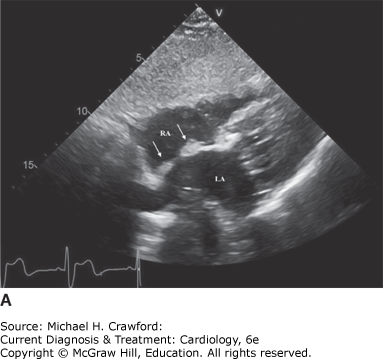
Figure 32-4.
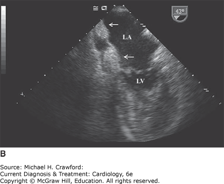
4. Fibroma: A Giant Tumor of the Left Ventricle
A. Demographics
Fibromas are far more common in the pediatric population, being the second or third most common benign cardiac tumor of childhood. Most cardiac fibromas are diagnosed in infants under 1 year of age; however, they have been reported in adults as well older than 50 years.
B. Typical Locations
Fibromas typically arise from the ventricular myocardium, with a predilection for the left ventricle, particularly the left ventricular free wall and the interventricular septum, even at times mimicking hypertrophic cardiomyopathy (Figure 32–5).
Figure 32-5. Cardiac fibroma. A: Coronal T2-weighted spin-echo magnetic resonance image of the heart at the level of the left ventricular anterior wall and the aortic valve. A mass is visualized emanating from the anterior wall of the left ventricle (arrow). The mass is mildly heterogeneous with mild T2 hypointensity relative to the myocardium. B: Transaxial T2-weighted image demonstrates predominantly peripheral enhancement, suggestive of a cardiac fibroma. (Reproduced with permission from C. Higgins.)
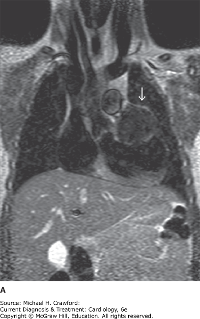
Figure 32-5.
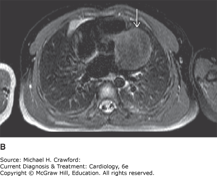
C. Pathology
Typically solitary, fibromas are large tumors with diameters in the 1–10 cm range. As a result, they often exert a mass effect. Fibromas are not distinctly encapsulated though are clearly demarcated from surrounding myocardium and often contain small calcifications.
D. Associations
A small subset of cardiac fibromas (3–5%) are associated with Gorlin syndrome (also known as nevoid basal cell carcinoma syndrome), an autosomal-dominant condition caused by mutations in the PTCH1 gene and characterized by basal cell carcinomas, skeletal abnormalities, and ectopic calcification of the central nervous system. Fibromas have also been associated with familial adenomatous polyposis.
BaidounM, ElgendyM, LokerJ.Cardiac fibroma with cardiac arrest: a rare clinical presentation of Gorlin syndrome in an 8-month-old infant. BMJ Case Rep. 2021;14(6):e241519. Published 2021 Jun 23. 34162607. doi: 10.1136/bcr-2020-241519.
5. Rhabdomyoma: The Spontaneously Regressing Tumor of Childhood
A. Demographics
Rhabdomyomas are the most common benign cardiac tumor in children, accounting for 60% of all pediatric cardiac tumors, and are often detected in utero or in the first year of life.
B. Typical Locations
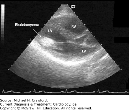
Rhabdomyomas are frequently multiple, occurring predominantly in the right and left ventricular walls, though also found in the atria and occasionally on the atrioventricular valves (Figure 32–6).
Figure 32-6. Cardiac rhabdomyoma. Transthoracic echocardiogram in the parasternal long-axis view showing a rhabdomyoma in the inferolateral left ventricular wall of a patient with tuberous sclerosis. LA, left atrium; LV, left ventricle; RV, right ventricle.
C. Pathology
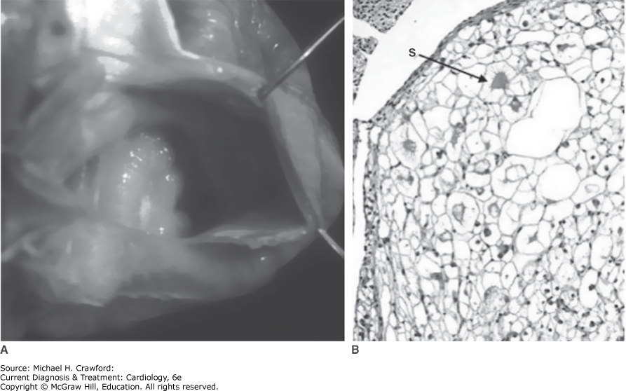
These tumors range in size from a few millimeters to several centimeters and grossly have a yellow or white appearance (Figure 32–7). Most rhabdomyomas spontaneously regress and are typically monitored with serial echocardiograms.
Figure 32-7. Cardiac rhabdomyoma. A: Gross pathology: The specimen shows a small, well-circumscribed grayish-white tumor seen at the right ventricular inlet in a newborn infant. B: Histology: Hematoxylin-eosin stain of the tumor reveals large round and polygonal cells with clear glycogen-rich cytoplasm and attenuated strands of cytoplasm, known as spider cells (arrow). Mitotic activity is absent, and these findings are consistent with the diagnosis of rhabdomyoma. (Reproduced with permission from Philip C. Ursell.)
D. Associations
More than 80–90% of rhabdomyomas are associated with tuberous sclerosis complex, an autosomal dominant genetic disorder caused by a pathogenic variant in one of two genes, TSC1 or TSC2, both of which lead to overactivation of mammalian target of rapamycin (mTOR) and thereby abnormal cell proliferation. TSC is characterized by the development of benign tumors in multiple organs, including the brain, heart, and, kidneys, as well as hypopigmented cutaneous macules.
6. Hemangioma: The Benign Vascular Neoplasm
A. Demographics
Hemangiomas account for about 5–10% of all benign cardiac tumors and can affect any age group, from newborns to older adults.
B. Typical Locations
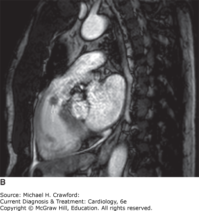
Hemangiomas can be isolated, but about one-third are found in multiple locations. These tumors can occur in any cardiac chamber (Figure 32–8).
Figure 32-8. Cardiac hemangioma in a 59-year-old man presenting with new atrial flutter and heart failure found to have a mass in the right ventricular outflow tract. (A) Parasternal short-axis view demonstrates a smooth, mobile mass in the right ventricular outflow tract and subsequent (B) magnetic resonance images with steady-state free precession imaging show a mass (arrow) of intermediate intensity, slightly higher than that of the myocardium, with smooth contours and no evidence of ventricular invasion. LA, left atrium; RA, right atrium; RVOT, right ventricular outflow tract.
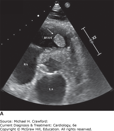
Figure 32-8.
C. Pathology
Hemangiomas are composed of mature blood vessels, and their gross appearance is that of a vascular lesion with reddish-purple coloration. Microscopically, they are composed of dilated vessels, bland endothelial cells, and fibrous connective tissue. There are three histologic subtypes: capillary, cavernous, or arteriovenous.
MaleszewskiJJ, BoisMC, BoisJP, YoungPM, StulakJM, KlarichKW.Neoplasia and the heart: pathological review of effects with clinical and radiological correlation. J Am Coll Cardiol. 2018;72(2):202227. 29976295. doi: 10.1016/j.jacc.2018.05.026.
TyeballyS, ChenD, BhattacharyyaS,et al. Cardiac tumors: JACC CardioOncology State-of-the-Art Review. JACC CardioOncol. 2020;2(2):293311. 34396236. Published 2020 Jun 16. doi: 10.1016/j.jaccao.2020.05.009
7. Cardiac Hamartoma (Histiocytoid Cardiomyopathy): A Tumor of Purkinje Cells
A. Demographics
Also known as oncocytic cardiomyopathy or Purkinje cell hamartoma, these tumors are rare with fewer than 150 reported cases and typically affect infants in the first year of life.
B. Typical Locations
These tumors can arise on the endocardium or myocardium and may involve the sinoatrial and atrioventricular nodes, often producing arrhythmias and various conduction blocks.
C. Pathology
Grossly, cardiac hamartomas appear as multiple, small pale nodules. Histologically, they are composed of vacuolated, rounded myocytes with numerous mitochondria and evidence of disordered myofibrils.
BurkeA, TavoraF.The 2015 WHO Classification of tumors of the heart and pericardium. J Thorac Oncol. 2016;11(4):441452. 26725181. doi: 10.1016/j.jtho.2015.11.009
MaleszewskiJJ, BassoC, BoisMC,et al. The 2021 WHO classification of tumors of the heart. J Thorac Oncol. 2022 Apr;17(4):510518. 34774791. doi: 10.1016/j.jtho.2021.10.021. Epub 2021 Nov 10.
B. Malignant Cardiac Tumors
Up to a quarter of all primary cardiac tumors have malignant features.
1. Sarcoma: The Invasive Connective Tissue Tumor of the Heart
A. Demographics
Cardiac sarcomas are the most common malignant tumor of the heart in adults, comprising 50–75% of all primary cardiac neoplasms. They are typically diagnosed between the third and fifth decades of life, and metastatic spread at the time of diagnosis is common.
B. Specific Tumor Subtypes
Cardiac sarcomas exist as various histologic subtypes, with angiosarcoma being the most common differentiated sarcoma, followed by rhabdomyosarcoma, myxofibrosarcoma, osteosarcoma, and leiomyosarcoma. However, undifferentiated pleomorphic sarcoma (UPS), previously referred to as malignant fibrous histiocytoma, is the most common primary malignant neoplasm of the heart and accounts for roughly 25% of cardiac sarcomas.
Undifferentiated pleomorphic sarcoma affects males and females equally and has a mean age of presentation of 44–47 years. These tumors typically arise from the posterior wall of the left atrium and are invasive with diffuse wall involvement. This group of sarcomas lacks specific histological patterns or immunohistochemical features, and pathology shows undifferentiated spindle cells with frequent mitotic activity.
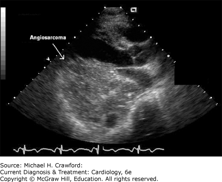
Angiosarcomas characteristically arise from the right atrium or the right atrioventricular groove. They are composed of malignant cells that fashion into vascular channels and aggressively infiltrate surrounding structures (Figure 32–9), often demonstrating gross areas of hemorrhage.
Rhabdomyosarcoma: These malignant tumors of striated muscle are the most common pediatric cardiac malignancy, though remain rare, accounting for less than 5% of all primary cardiac sarcomas. They are large, invasive tumors which arise from the myocardium and often have valvular involvement. Due to their aggressive nature, they typically present with symptoms related to cardiac invasion or obstruction and are frequently metastatic at the time of diagnosis.
Myxofibrosarcomas generally arise in the left atrium and have a yellowish-white or grayish-white appearance on gross pathology. Histologically, myxofibrosarcomas demonstrate myxoid areas, often with curvilinear thin-walled blood vessels and spindle-shaped tumor cells. Unlike benign myxomas which typically arise from the interatrial septum, these tumors are often attached to the posterior wall of the left atrium and present due to symptoms of mitral valve obstruction.
Osteosarcomas may arise from any part of the heart but most commonly originate from the posterior wall of the left atrium, near the entrance of the pulmonary veins. They are highly invasive and often infiltrate into the adjacent myocardium or into the pericardium. These tumors exhibit a similar gross and microscopic appearance as osteosarcoma of the bone, demonstrating malignant spindle or ovoid cells mixed with osteoid or bone. Although cardiac osteosarcomas are predominantly osteoblastic, they often exhibit chondroblastic or fibroblastic differentiation as well, with areas resembling chondrosarcoma or myxofibrosarcoma. Osteosarcomas can metastasize to the thyroid, skin, lymph nodes, and lungs.
Leiomyosarcoma: These high-grade tumors are thought to arise from the smooth muscle cells of the pulmonary veins and arteries with subsequent myocardial infiltration and are most commonly seen in the posterior left atrium. Tumor pathology exhibits spindle or epithelioid cells arranged as compact bundles with regions of necrosis. Leiomyosarcomas are extremely aggressive with a high rate of local recurrence and systemic metastases.
Liposarcoma: Primary cardiac liposarcomas typically originate from the right-sided chambers, particularly the right atrium, though have also been reported on the mitral valve and other left-sided structures. Microscopic examination demonstrates fat cells mixed with fibrous tissue and lipoblasts.
Figure 32-9. Cardiac angiosarcoma. Transthoracic echocardiogram in a 60-year-old woman presenting with a 2-month history of dyspnea and palpitations. The parasternal long-axis view shows an infiltrating mass involving the left ventricular posterior wall and left atrium. Tumor biopsy confirmed the diagnosis of angiosarcoma.
C. Associations
In the acquired immunodeficiency syndrome (AIDS), a specific type of cardiac angiosarcoma called Kaposi sarcoma has been well described. Another interesting association has been made between leiomyosarcomas and the Epstein-Barr virus in immunosuppressed patients, such as those with AIDS or after cardiac transplantation.
SunD, WuY, LiuY, YangJ.Primary cardiac myxofibrosarcoma: case report, literature review and pooled analysis. BMC Cancer. 2018;18(1):512. 29720127. Published 2018 May 2. doi: 10.1186/s12885-018-4434-2.
2. Lymphoma: An Infiltrative Non-Hodgkin Tumor of the Right Atrium
A. Demographic
Primary cardiac lymphoma is exceedingly rare, representing 1–2% of all cardiac tumors and comprising an even smaller percentage of all extranodal lymphomas. However, approximately 25% of patients with lymphoma will develop cardiac involvement.
B. Typical Locations
Cardiac lymphoma can involve any area of the heart though has a predilection for the right-sided chambers, particularly the right atrium.
C. Pathology
Primary cardiac lymphoma belongs to the extranodal subtype of non-Hodgkin lymphoma. The majority of primary cardiac lymphomas are aggressive B-cell neoplasms with the most common being diffuse large B-cell lymphoma. Like extracardiac lymphoma, these tumors are infiltrative in nature, often involving the pericardium and encasing the vasculature, including the coronary arteries.
D. Associations
Primary cardiac lymphomas are predominantly found in immunocompromised individuals.
AsadianS, RezaeianN, HosseiniL, ToloueitabarY, Hemmati KomasiMM.The role of cardiac CT and MRI in the diagnosis and management of primary cardiac lymphoma: A comprehensive review [published online ahead of print, 2021 Aug 25]. Trends Cardiovasc Med. 2021;S10501738(21)00094-3. 34454052. doi: 10.1016/j.tcm.2021.08.010.
ChenH, QianS, ShiP, LiuL, YangF.A presentation, treatment, and survival analysis of primary cardiac lymphoma cases reported from 2009 to 2019. Int J Hematol. 2020;112(1):6573. 32285360. doi: 10.1007/s12185-020-02881-2.
3. Mesothelioma: The Pericardial Tumor
A. Demographics
Primary cardiac mesothelioma is extremely rare though comprises half of all pericardial tumors. It typically affects adults with a slight male predominance.
B. Typical Locations
These tumors are neoplasms of the mesothelial lining of the visceral or parietal pericardium and often invade the superficial myocardium. By definition, primary pericardial mesothelioma necessitates lack of pleural involvement or primarily pericardial disease.
C. Pathology
Cytology may be obtained from pericardiocentesis though is often unrevealing. On histology, mesotheliomas can be epithelial, sarcomatous, and biphasic subtypes.
D. Associations
Unlike pleural mesotheliomas, pericardial mesotheliomas have not been clearly linked to asbestos exposure.
C. Benign or Malignant Cardiac Tumors
1. Paraganglioma: A Catecholamine-Secreting Neuroendocrine Tumor of the Heart
A. Demographics
Cardiac paragangliomas are exceedingly rare, neuroendocrine tumors that originate from the paraganglionic cells surrounding the great vessels, coronary arteries, and atria. They can be either secretory or nonsecretory, depending on whether or not they produce catecholamines. Only 1–2% of all paragangliomas, or extra-adrenal chromaffin cell tumors, occur in the chest, with primary cardiac paragangliomas comprising only a subset of these.
B. Typical Locations
Most paragangliomas are secretory and tend to arise from the visceral autonomic paraganglia of the left atrial wall or adjacent to the pulmonary trunk and ascending aorta.
C. Pathology
Histologically, paragangliomas are composed of discrete small nests of tumor cells in a “Zellballen” pattern. A subset of paragangliomas produce a characteristic melanin pigment. Approximately 10% of these tumors are malignant; however, this is not determined by pathology or immunohistochemistry but rather by occurrence of tumor in tissues not typically affected by paragangliomas. As these are highly vascular tumors often involving critical structures, surgical resection is challenging.
ZubairMM, El NihumLI, HaleySL,et al. Large, Hormonally Active Primary Cardiac Paraganglioma: Diagnosis and Management. Ann Thorac Surg. 2022;113(3):e167e170. 34111385. doi: 10.1016/j.athoracsur.2021.05.042
2. Teratomas: The Germ Cell Neoplasm of Childhood
A. Demographics
Approximately half of cardiac teratomas are diagnosed in utero with the other half typically diagnosed in children under 15 years of age. With a similar incidence as fibromas, cardiac teratoma is the second or third most common primary cardiac tumor in pediatric series. There are rare reported cases of cardiac teratomas manifesting in adults.
B. Typical Locations
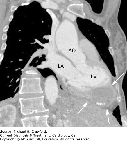
Teratomas typically arise within the pericardium but occasionally have an intramyocardial origin, with a few reports of teratomas occurring in the ventricular or atrial septum (Figure 32–10).
Figure 32-10. Cardiac teratoma. Contrast-enhanced, multidetector computed tomography image of the heart in coronal section, performed in a 40-year-old woman with a history of ovarian cyst, presenting with chest pain. A cystic mass with areas of calcification involving the myocardium and pericardium was noted incidentally (arrows). The mass was resected and found to be a teratoma. AO, aorta; LA, left atrium; LV, left ventricle. (Reproduced with permission from Karen Ordovas.)
C. Pathology
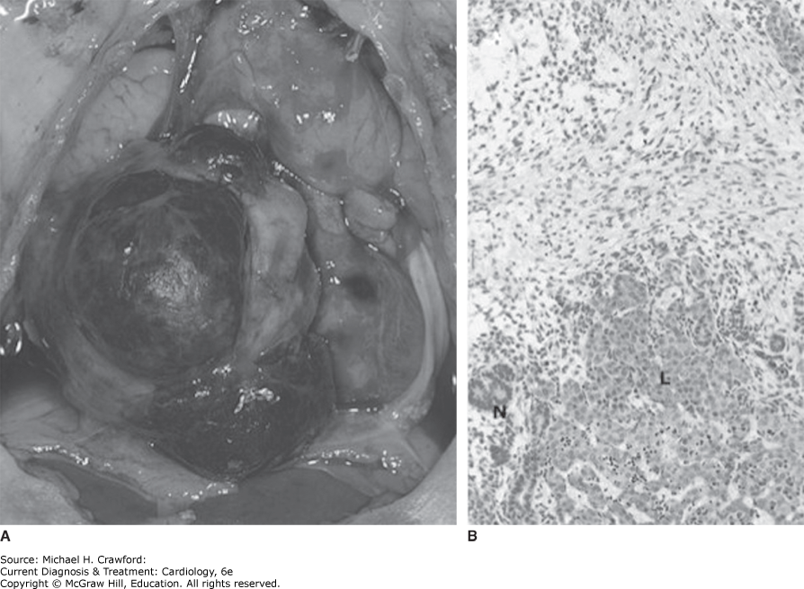
Teratomas are germ cell neoplasms that contain endodermal, mesodermal, and ectodermal elements. The composition of each teratoma varies, but if more than half the tumor is comprised of well-differentiated germinal elements, it is designated a mature teratoma (Figure 32–11). Cardiac teratomas can be either benign or malignant in nature.
Figure 32-11. A: Gross pathology: Postmortem specimen from a newborn infant with hydrops showing a large, encapsulated tumor arising from the pericardium and exerting a mass effect on the anterior cardiac chambers and the great vessels. B: Histology: Hematoxylin-eosin stain of the mass demonstrates liver (L) and neural (N) tissue, as well as glandular epithelium, consistent with a teratoma. (Reproduced with permission from Philip C. Ursell.)
TzaniA, DoulamisIP, MylonasKS, AvgerinosDV, NasioudisD.Cardiac tumors in pediatric patients: a systematic review. World J Pediatr Congenit Heart Surg. 2017;8(5):624632. 28901236. doi: 10.1177/2150135117723904.
YuanSM.Fetal primary cardiac tumors during perinatal period. Pediatr Neonatol. 2017;58(3):205210. 28043830. doi: 10.1016/j.pedneo.2016.07.004
D. Secondary Cardiac Tumors: The Far-Roaming Settlers
1. Demographics
Cardiac metastases are 20–40 times more common than primary cardiac tumors. Roughly 10% of patients with metastatic cancer have cardiac metastases. Secondary cardiac tumors affect a wide range of ages given the number and variety of cancers with a tendency toward secondary cardiac involvement.
2. Typical Locations
Extracardiac malignancies can spread to the heart by one of four routes, including direct local invasion, lymphatic, hematogenous, and transvenous spread, as in the case of renal cell carcinoma which classically features extension of tumor thrombus into the inferior vena cava. Pericardial metastases are the most prevalent and often present with pericardial effusion and tamponade. Myocardial, coronary, and intracavitary involvement occur uncommonly, in order of decreasing frequency.
3. Common Primary Tumors Associated with Cardiac Metastasis
Malignant melanoma has the highest predilection for cardiac spread, which is seen in as many as 50–65% of melanoma cases (Figure 32–12). Other neoplasms that commonly metastasize to the heart include carcinomas of the thorax such as breast, lung, and esophageal cancers, as well as hematologic tumors (eg, lymphoma and leukemia), renal cell carcinoma, and soft tissue sarcomas.
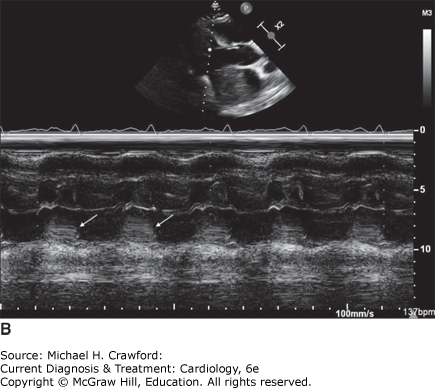
Figure 32-12. Metastatic melanoma A: Transthoracic echocardiogram in a 35-year-old man with widely metastatic melanoma who presented with worsening respiratory distress. The parasternal long-axis view demonstrates a large, fairly homogenous left atrial mass, most likely representing metastatic melanoma, that occupies the majority of the left atrium and protrudes across the mitral valve during diastole. B: M-mode of the mitral valve showing the mass prolapsing into the left ventricle during diastole (arrows). LA, left atrium; LV, left ventricle; RV, right ventricle.
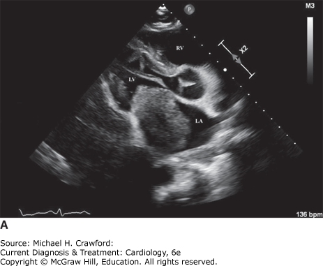
Figure 32-12.
Clinical Findings ⬆ ⬇
Cardiac tumors pose a significant challenge to diagnosis because they present with a wide array of signs and symptoms, many of which are nonspecific. Furthermore, it is often the size and anatomic location of the tumor, rather than its histopathologic features or malignant potential, that determine the clinical findings and the delay to presentation (Table 32–3).
Table 32–3. Clinical Manifestations of Cardiac Tumors
Endocardial Involvement Thromboembolism: cerebral, coronary, pulmonary, systemic Cavity obliteration, outflow tract obstruction Valve obstruction, valve damage |
Myocardial Involvement Arrhythmias: ventricular, atrial Conduction abnormalities: sinus node, atrioventricular node, bundle branches Left ventricular dysfunction: systolic, diastolic Coronary artery involvement: angina, infarction Electrocardiographic changes |
Pericardial Involvement Pericarditis Pericardial effusion, tamponade Pericardial constriction |
Valvular Involvement Valvular damage, obstruction, or regurgitation Congestive heart failure Sudden death or syncope |
Vascular Involvement Vessel dissection Downstream embolism, infarction |
Constitutional Symptoms Fever Night sweats Anorexia Weight loss |
A. Symptoms & Signs
Cardiac tumors present variably depending on their location within or around the heart. Tumors located in the pericardium often present with pericardial thickening, effusion, and/or tamponade and can manifest with pericardial constriction in the long term. Intracavitary tumors may obstruct blood flow or cause valvular obstruction or regurgitation ultimately leading to heart failure. Depending on the size and location of the intracavitary tumor, sudden cardiac death can be precipitated by obstruction, embolization into the coronary artery, or ventricular arrhythmias. Infiltrative cardiac tumors or those arising from the myocardium can cause heart failure symptoms from a restrictive or infiltrative cardiomyopathy. These tumors can also serve as a focus for atrial and ventricular arrhythmias, as well as create conduction system disturbances necessitating cardiac pacing.
Mechanical and obstructive manifestations of large cardiac tumors can produce effects on surrounding organs such as the respiratory and upper gastrointestinal tracts. Friable tumors, as a result of their unique tissue characteristics, are prone to causing both systemic and pulmonary emboli, and this can be further compounded by the presence of an atrial level shunt. Finally, like noncardiac malignancies, cardiac tumors can be associated with constitutional symptoms. Following are key examples of specific neoplasms and their typical presentations.
1. Heart Failure
Malignant tumors or large-sized benign neoplasms like fibromas often present with heart failure from obstructive or restrictive pathology. Because angiosarcomas usually occur in the right atrium or pericardium, right-sided heart failure is often the presenting symptom. Cardiac lymphoma, due to its infiltrative nature, can lead to congestive heart failure symptoms from restriction. Cardiac or pericardial metastases may present with cardiomegaly, a new or changing murmur, or symptoms of heart failure.
2. Arrhythmia and Sudden Cardiac Death
Left ventricular fibromas are associated with ventricular arrhythmias and can be associated with an increased risk of sudden cardiac death. Rhabdomyomas and cardiac hamartomas can also present with rhythm disturbances, such as heart block or ventricular tachycardia. Cardiac metastases may produce various arrhythmias and conduction delays depending on their location. Furthermore, an increased incidence of atrial arrhythmias has been noted in patients with atrial myxomas following surgical resection.
3. Angina/Myocardial Infarction
Papillary fibroelastomas can present with myocardial infarction and even sudden cardiac death if they obstruct a coronary ostium. In addition, any tumor that impinges on an epicardial coronary artery can predispose to anginal symptoms or frank myocardial infarction, as can be seen with cardiac lymphoma encasing the coronary arteries.
4. Obstructive and Mechanical Phenomena
Larger myxomas with smooth surfaces tend to present with obstructive cardiovascular symptoms, particularly at the level of the mitral valve, mimicking mitral stenosis. Fibromas exert a mass effect due to the large tumor burden, typically causing obstruction of left ventricular outflow. Similarly, rhabdomyomas may present with symptoms of obstruction. Most invasive cardiac malignancies and cardiac metastases can cause pericardial disease, often presenting with a pericardial effusion that may produce signs of tamponade and occasionally pericardial constriction. Malignant pericardial mesotheliomas can also present with pericarditis, tamponade, or constriction. Angiosarcomas, due to their predilection for the right atrium, can have vena caval obstruction as the presenting sign.
5. Embolic Phenomena
Friable or villous cardiac myxomas are associated with a higher risk of embolization. Papillary fibroelastomas can form a nidus for platelet and fibrin aggregation and can present with embolic complications. Given the tumor’s predilection for left-sided valves, papillary fibroelastomas more often lead to cerebrovascular and systemic emboli, though can also present with pulmonary embolism in the case of right-sided tumors.
6. Constitutional Symptoms
Approximately 30% of patients with cardiac myxomas report constitutional symptoms such as fever, weight loss, arthralgias, and fatigue, which are thought to be secondary to various cytokines and growth factors secreted by the tumor such as interleukin-6 and tumor necrosis factor. Malignant primary cardiac tumors can produce constitutional symptoms such as cachexia, anorexia, and night sweats, similar to other noncardiac malignancies. However, these tumors usually present with heart failure, rhythm disturbances, or obstructive symptoms far before the onset of constitutional symptoms.
B. Physical Examination
The physical examination findings associated with cardiac tumors are generally nonspecific and often related to a secondary effect of the tumor, such as heart failure or an embolic complication. One of the characteristic physical examination findings ascribed to an intracardiac tumor, however, is the “tumor plop”—an auscultatory phenomenon resulting from the mechanical obstruction of the mitral valve orifice by the mass. The tumor plop should be heard in early diastole prior to the S3 but after the opening snap, making it difficult to distinguish on auscultation. The coexistence of auscultatory features of mitral stenosis in the absence of a history of rheumatic fever should raise the possibility of an obstructive left atrial tumor.
Differential Diagnosis ⬆ ⬇
Diagnostic StudiesOnce limited to chest radiographs and angiography, the diagnostic armamentarium for cardiac neoplasms has grown expansively and continues to evolve with increasing sophistication of standard modalities like echocardiography, computed tomography (CT), cardiovascular magnetic resonance imaging (CMR), and positron emission tomography (PET). Advances in these noninvasive imaging techniques have facilitated the early, often incidental diagnosis of cardiac tumors. These imaging modalities can provide a wealth of information to both the cardiologist and the cardiac surgeon, aiding not only diagnosis but also therapeutic intervention and long-term surveillance. In many instances, particularly with benign cardiac tumors, a diagnosis can be reached without cardiac biopsy by using a structured imaging approach and relying on classic imaging features, location of the mass, and age at presentation to guide diagnosis.
A. Echocardiography
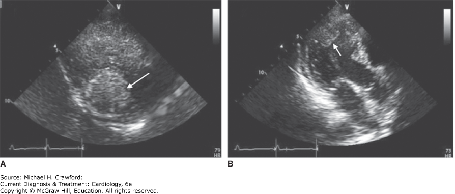
Due to the ease of image acquisition, lack of radiation, widespread availability, and relatively low cost, transthoracic echocardiography (TTE) is the favored initial diagnostic modality for cardiac tumors (Figure 32–13). TTE provides assessment of tumor size, location, and pericardial involvement. It has good spatial resolution making it the ideal imaging modality for assessing small, subcentimeter mobile masses or masses involving valves. Furthermore, TTE is crucial for understanding the hemodynamic consequences of a mass and any secondary valvular disease.
Figure 32-13. Cardiac fibroma. Transthoracic echocardiography parasternal short-axis view at the level of the cardiac apex (A) and apical three-chamber view (B) depicting an intramural mass in the left ventricle in a 57-year-old woman. Magnetic resonance imaging confirmed a T1 and T2 hypointense mass demonstrating delayed enhancement, consistent with the diagnosis of fibroma. (Reproduced with permission from Elyse Foster.)
TTE is often followed by transesophageal echocardiography (TEE) when better characterization of atrial masses or valvular lesions is needed. Intraoperative TEE is crucial in aiding cardiac biopsy, guiding surgical intervention, assessing residual tumor burden, and detecting involvement of adjacent structures. TTE and occasionally TEE are also used as follow-up imaging modalities, both to look for recurrence after tumor resection and to monitor benign tumors that do not necessitate intervention.
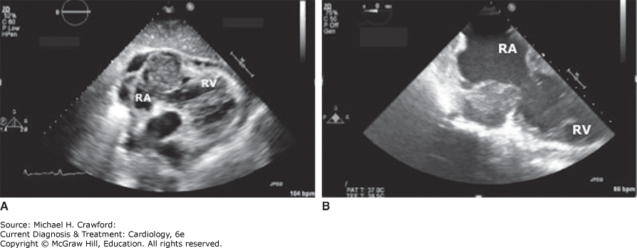
In general, the sensitivity of both TTE and TEE is highest for endocardial lesions, as the mass is easily distinguished from the echolucent cardiac chamber. Their sensitivity is slightly lower for intramyocardial lesions and lowest for pericardial tumors. Despite high sensitivity, there are several important limitations of TTE, including poor acoustic windows, particularly in obese patients or those with chronic lung disease, difficulty in identifying tumor origin and extent given limited visualization of certain anatomy (eg, left atrial appendage, superior vena cava, extracardiac structures), and lack of tissue characterization. Although TEE offers improved spatial resolution and visualization of valves and posterior cardiac structures, it also remains the more invasive imaging modality (Figure 32–14).
Figure 32-14. Right atrial malignancy. A: Subcostal four-chamber view obtained on a transthoracic echocardiogram in a 54-year-old man with a malignant right atrial tumor. B: Transesophageal echocardiogram from the same patient; note the improved resolution of the tumor and the surrounding cardiac structures on this modality. RA, right atrium; RV, right ventricle.
Another valuable facet of echocardiography is the ability to perform real-time three-dimensional (3D) echocardiography, which is a volumetric method and thereby typically allows for visualization of the entire cardiac mass as opposed to a thin slice as seen in two-dimensional imaging. This leads to more accurate assessment of tumor size, shape, and anatomical relationships to adjacent structures. 3D echocardiography has been shown to be comparable to CMR for characterization of cardiac anatomy; however, unlike CMR, 3D echocardiography cannot delineate the vascularization of cardiac masses.
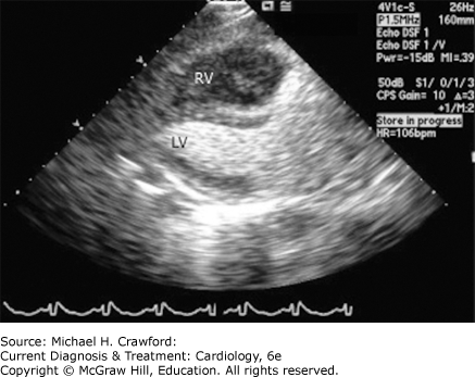
The use of ultrasound-enhancing agents, or echocardiographic contrast, allows for improved detection of smaller tumors and better visualization of intracavity masses, particularly in regard to their points of attachment and any invasion into surrounding cardiovascular structures (Figure 32–15). Perfusion contrast enhancement of cardiac masses can be used to help distinguish between vascular and nonvascular tumors. A mass demonstrating greater enhancement than the adjacent myocardium suggests a malignant, highly vascular tumor, while a poorly enhancing or nonenhancing mass suggests a benign tumor or a thrombus in the case of absent perfusion. However, this is not diagnostic.
Figure 32-15. Enhancement of intracardiac chambers using echocardiographic contrast agents. Transthoracic echocardiogram from a 59-year-old woman with endometrial cancer who presented with palpitations. Parasternal long-axis view using echocardiographic contrast demonstrates normal filling of the left ventricle (LV), with almost no filling of the right ventricle (RV), which is almost completely occupied by metastatic tumor from the endometrial cancer.
PalaskasN, ThompsonK, GladishG,et al. Evaluation and management of cardiac tumors. Curr Treat Options Cardiovasc Med.2018;20(4):29. Published 2018 Mar 20. 29556752. doi: 10.1007/s11936-018-0625-z
Zaragoza-MaciasE, ChenMA, GillEA.Real time three-dimensional echocardiography evaluation of intracardiac masses [published correction appears in Echocardiography. 2012 May;29(5):i. Zaragosa-Macias, Elisa [corrected to Zaragoza-Macias, Elisa]]. Echocardiography. 2012;29(2):207219. 22283202. doi: 10.1111/j.1540-8175.2011.01627.x
B. Cardiac Magnetic Resonance Imaging
CMR has emerged as an increasingly important diagnostic modality for cardiac tumors in recent years. Previously used to provide supplementary information to echocardiography and CT scans, CMR now plays a pivotal role in the primary diagnosis of neoplastic lesions of the heart. The high degree of natural contrast between the blood pool and cardiovascular structures permits clear delineation of cardiac masses. Furthermore, the direct multiplanar imaging capability of CMR is advantageous for demonstrating the complex relationships between cardiac masses and surrounding structures, while the lack of ionizing radiation is favorable. CMR can readily assess the dimensions, morphology, location, and extension of a cardiac mass; however, where it shines is its ability to provide tissue characterization.
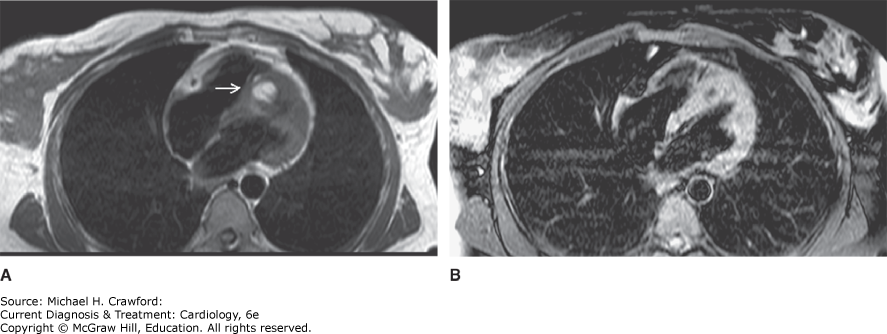
Various signaling properties of a mass aid in histopathological characterization by indicating the presence of fatty infiltration, calcification, necrosis, hemorrhage, etc, which then can be correlated with specific diagnoses. For example, cardiac tumors demonstrate increased signal intensity on T2-weighted images, whereas fibrosis produces a low signal intensity. Delayed gadolinium enhancement with gadolinium in the context of a cardiac tumor indicates necrosis or nonviable tissue, often seen at the core of a tumor where cells have outgrown their blood supply. Gadolinium injection can also enhance highly vascular masses, as can the inversion recovery scouting sequence. The fat suppression technique on CMR can additionally detect adipose-rich tumors such as lipomas (Figure 32–16). Lastly, in patients with a suspected cardiac tumor, CMR has excellent diagnostic accuracy. In a large multicenter study of adult patients undergoing CMR for evaluation of suspected tumor, CMR diagnosis of pseudomass (ie, prominent normal structure or common variant), thrombus, benign tumor, and malignant tumor was accurate in more than 98% of patients. Accuracy of CMR for diagnosis of cardiac masses has also been demonstrated in pediatric populations.
Figure 32-16. Cardiac lipoma. A: T1-weighted transaxial spin-echo magnetic resonance image of the heart through the level of the interventricular septum. A high-signal-intensity mass (arrow) is seen embedded in the distal anteroseptum. B: During application of fat saturation pulse, the mass loses its bright signal, suggesting the diagnosis of a cardiac lipoma. (Reproduced with permission from C. Higgins.)
The main limitation of CMR compared with echocardiography is its lower temporal resolution. As such, it is not typically indicated for evaluation of valvular vegetations. Additional disadvantages include long acquisition times often requiring breath-holds, limited availability compared with echocardiography and CT, potential for claustrophobia, artifact from implantable cardiac devices, and contraindications such as older generation implantable cardiac devices.
BeroukhimRS, GhelaniS, AshwathR,et al. Accuracy of Cardiac Magnetic Resonance Imaging Diagnosis of Pediatric Cardiac Masses: A Multicenter Study. JACC Cardiovasc Imaging. 2022;8:13911405. doi: 10.1016/j.jcmg.2021.07.010
ShenoyC, GrizzardJD, ShahDJ,et al. Cardiovascular magnetic resonance imaging in suspected cardiac tumour: a multicentre outcomes study. Eur Heart J. 2021;43(1):7180. 34545397. doi: 10.1093/eurheartj/ehab635fta
C. Computed Tomography
Cardiac CT is a rapid acquisition imaging technique that provides high-quality images with superior spatial resolution to CMR. Electrocardiographic (ECG) gating used with cardiac CT minimizes motion-related artifacts, resulting in improved image quality, and CT is an excellent option for patients with implanted devices who cannot undergo CMR. The superior spatial resolution of CT allows for accurate delineation of tumor size and location, degree of local invasion, and involvement of coronary arteries and other vasculature. In particular, CT is optimal for the evaluation of calcified masses and provides anatomic detail that is often crucial for preoperative planning.
In recent years, advances in multidetector CT hardware and post-processing software have made 3D reconstructions a useful tool in the evaluation of cardiac masses. These reconstructions can allow for better appreciation of tumor margins and relationships with surrounding structures, making them useful adjuncts in surgical planning. Finally, 3D printing of cardiac models is an emerging technology being used primarily for preoperative planning in patients with congenital heart disease, though its use has also been reported in cases of cardiac masses. While volumetric 3D echocardiography and CMR can also be used to generate 3D models, CT has been the predominant imaging modality for 3D printing largely due to its submillimeter spatial resolution.
Notable disadvantages of CT include radiation exposure, particularly in the pediatric population, a small risk of contrast-induced nephropathy, limited tissue characterization abilities, and lower temporal resolution as compared with CMR.
Al JabbariO, Abu SalehWK, PatelAP, IgoSR, ReardonMJ. Use of three-dimensional models to assist in the resection of malignant cardiac tumors. J Card Surg. 2016;31(9):581583. 27455392. doi: 10.1111/jocs.12812
LiddyS, McQuadeC, WalshKP, LooB, BuckleyO.The assessment of cardiac masses by cardiac CT and CMR including pre-op 3D reconstruction and planning. Curr Cardiol Rep. 2019;21(9):103. 31367849. Published 2019 Jul 31. doi: 10.1007/s11886-019-1196-7
VukicevicM, MosadeghB, MinJK, LittleSH.Cardiac 3D printing and its future directions. JACC Cardiovasc Imaging. 2017;10(2):171184. 28183437. doi: 10.1016/j.jcmg.2016.12.001
WuCM, BergquistPJ, SrichaiMB.Multimodality imaging in the evaluation of intracardiac masses. Curr Treat Options Cardiovasc Med. 2019;21(10):55. 314869926. Published 2019 Sep 5. doi: 10.1007/s11936-019-0756-x
D. Positron Emission Tomography
Whole-body 18F-Fluorodeoxyglucose PET (18F-FDG PET) is routinely used to look for distant metastases in patients with high-risk cancers, such as renal cell carcinoma or lung cancer. It can be useful in detecting cardiac metastases in an asymptomatic patient during staging of the primary malignancy. Moreover, 18F-FDG PET can be helpful in differentiating benign from malignant cardiac masses on the basis of metabolic activity and can identify metastatic lesions which are more amenable to biopsy than cardiac masses.
Hybrid imaging using 18F-FDG PET/CT has been shown to be both sensitive and specific for distinguishing benign from malignant tumors preoperatively in multiple small studies. More recently, integrated imaging with 18F-FDG PET/CMR was demonstrated to be highly sensitive and specific for classification of benign versus malignant lesions in appropriately selected patients. While the role of 18F-FDG PET in assessing response to chemotherapy or radiation and evaluating postoperative residual disease has been established for several malignancies and is commonly done in the case of secondary cardiac tumors (eg, lymphoma with cardiac involvement), there is a lack of data with regard to primary cardiac malignancies.
MartineauP, DilsizianV, Pelletier-GalarneauM.Incremental value of FDG-PET in the evaluation of cardiac masses. Curr Cardiol Rep. 2021;23(7):78. 34081218. Published 2021 Jun 3. doi: 10.1007/s11886-021-01509-z
E. Coronary Angiography
Coronary angiography can be useful in the workup of selected cardiac tumors. Cardiac neoplasms may have their own blood supply as a result of tumor angiogenesis, but they can also invade and involve epicardial coronary arteries. Evaluation of cardiac tumors frequently includes cardiac catheterization to evaluate coronary anatomy and to determine whether contrast enhancement, or “tumor blush,” is present. The appearance of tumor blush has been described in many cardiac tumors such as myxomas, hemangiomas, rhabdomyomas, and angiosarcomas. Tumor blush itself is diagnostic of a cardiac tumor and can indicate which type of neoplasm is likely present. Since certain tumors characteristically appear in specific locations, the associated epicardial coronary blood supply to the lesion and the territory of the blush can aid substantially in tumor localization and identification.
Marok R, Klein LW. Tumor blush in primary cardiac tumors. J Invasive Cardiol. 2012;24(3):139–140. [PMID: 22388311]
Several normal and abnormal intra- and extracardiac structures can confound the diagnosis of cardiac tumors. Imaging features that favor a neoplastic process are a mobile, pedunculated appearance and an associated pericardial effusion. Masses that cross anatomic planes, from myocardium to pericardium or endocardium, are more likely to be tumors as well (Table 32–4).
Table 32–4. Differential Diagnoses of Cardiac Tumors
Intracardiac Structures, Normal Variants Lipomatous hypertrophy of the interatrial septum Eustachian valve Chiari network Septum spurium |
| Thrombus |
Vegetation Infective endocarditis Nonbacterial thrombotic endocarditis Libman-Sacks endocarditis |
| Caseous Calcification of the Mitral Annulus |
| Pericardial Cysts |
| Calcified Amorphous Tumor (CAT) |
Extracardiac Structures Diaphragmatic hernia Loculated pleural effusion Mediastinal mass/tumor |
A. Normal Variant Structures
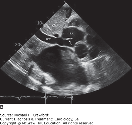
A sizable number of intracardiac structures that occur as anatomic variants can be mistaken for cardiac neoplasms. These include the Eustachian valve (Figure 32–17), seen at the junction of the inferior vena cava and the right atrium, and the Chiari network, a remnant of the sinus venosus, also encountered in the right atrium. The septum spurium is a superior fold at the coronary sinus opening, which can mimic an intracardiac mass, along with the Thebesian valve, which guards the opening to the coronary sinus. Lambl’s excrescences have been described as filiform frond-like structures that are noted at sites of valve closure, particularly on the aortic and mitral valves.
Figure 32-17. Eustachian valve. A: Transthoracic echocardiogram in the subcostal view demonstrating a prominent Eustachian valve (arrow). This appearance has been erroneously called triatriatum dexter. B: Additional subcostal view showing the Eustachian valve (arrow) as a linear structure at the junction of the inferior vena cava and the right atrium. IVC, inferior vena cava; LA, left atrium; LV, left ventricle; RA, right atrium; RV, right ventricle.
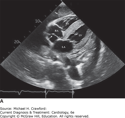
Figure 32-17.
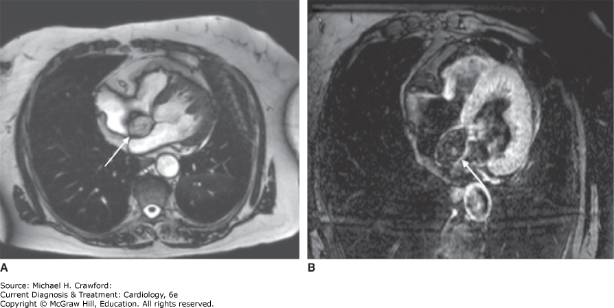
Although lipomatous hypertrophy of the interatrial septum can imitate a tumor (Figure 32–18), it is easily distinguished by its characteristic location along the interatrial septum and its dumbbell-shaped appearance (See Figure 32–4). The shape is due to the thin fossa ovalis that separates the lipomatous atrial septum on either side. No intervention is required unless the hypertrophy causes arrhythmias or obstruction.
Figure 32-18. Lipomatous hypertrophy of the interatrial septum. A: Steady-state free procession cine magnetic resonance image of the heart, demonstrating diffuse fatty infiltration of the right atrial wall, interatrial septum, and right ventricular free wall. There is sparing of the fossa ovalis, characteristic of lipomatous hypertrophy of the interatrial septum (arrow). B: After application of a fat saturation pulse, the infiltrative process in image A is no longer bright, indicative of fat content in the tissues. (Reproduced with permission from Kanae Mukai.)
B. Thrombus
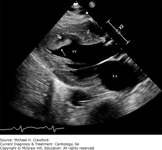
Intracardiac thrombi are fairly easy to differentiate from cardiac tumors if an astute assessment is made of the company they keep. For instance, left ventricular thrombi are associated with regional wall motion abnormalities and cardiomyopathies, particularly in the setting of an akinetic left ventricular apex (Figure 32–19). Left atrial thrombi may be associated with atrial fibrillation, left atrial enlargement, or mitral stenosis, whereas right atrial thrombi can be seen attached to indwelling catheters or pacemaker wires. Right ventricular thrombi, on the other hand, have been described in the setting of impaired right ventricular function and are often associated with pulmonary emboli.
Figure 32-19. Left ventricular thrombus. Transthoracic echocardiogram performed in a 67-year-old man with known coronary artery disease who presented with an embolic stroke shows akinesis of the left ventricular septal and anterior walls with a large, mobile echodense mass (arrow) at the left ventricular apex, consistent with thrombus. LA, left atrium; LV, left ventricle.
C. Vegetation
A vegetation is defined on echocardiography as an independently mobile, oscillatory, echodense mass. Intracardiac vegetations can be infected or sterile and have certain imaging characteristics that can provide clues to their etiology. The vegetations associated with infective endocarditis are typically large and involve the valvular surface or endocardium in the trajectory of the regurgitant jet. These vegetations are associated with other clinical, laboratory, and echocardiographic criteria for infective endocarditis and are more prevalent in certain demographic groups such as intravenous drug users, the immunocompromised, and those with prosthetic cardiac material. Nonbacterial thrombotic endocarditis (NBTE) and Libman-Sacks endocarditis are two distinct entities both comprised of sterile vegetations. The vegetations associated with NBTE, or marantic endocarditis, are small and located along the closure lines of valves; they are often associated with wasting conditions, such as cancer. Libman-Sacks endocarditis is one of the classic cardiovascular lesions associated with systemic lupus erythematosus, in which the sterile vegetations are located on both surfaces of involved cardiac valves.
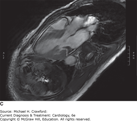
D. Caseous Calcification of the Mitral Annulus
Caseous calcification of the mitral valve is a rare form of mitral annular calcification that typically affects the posterior mitral annulus (Figure 32–20). The condition, also referred to as liquefaction necrosis, usually runs a benign course, although it has been associated with valvular dysfunction and cerebral embolization of necrotic material. Differential diagnosis includes cardiac tumor and valvular abscess, warranting complementary imaging techniques for accurate assessment of this pathology.
Figure 32-20. Caseous mitral annular calcification. A: Contrast-enhanced axial cardiac computed tomography demonstrates heterogeneous calcification along the posterior mitral annulus (arrow). B: Transthoracic echocardiogram in the same patient shows a large echogenic lesion on the posterior mitral valve leaflet (arrow) with an area of central lucency. The lesion extends into the posterior subvalvular apparatus. C: Steady-state free precession cine magnetic resonance image in the three-chamber view demonstrates mass-like, low intensity thickening of the posterior mitral valve leaflet (arrow) and proximal posteromedial papillary muscle, compatible with caseous mitral annular calcification.
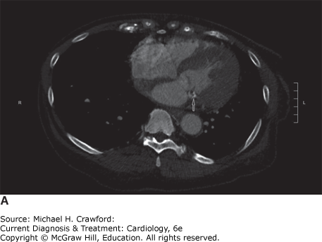
Figure 32-20.
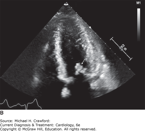
Figure 32-20.
GulatiA, ChanC, DuncanA, RazaS, KilnerPJ, PepperJ.Multimodality cardiac imaging in the evaluation of mitral annular caseous calcification. Circulation. 2011;123(1):e12. 21200011
E. Pericardial Cysts
Pericardial cysts are usually congenital abnormalities but can be acquired after cardiac surgery. They are typically located in the right or left costophrenic angles and can be identified on a chest x-ray or echocardiogram by their unilocular nature. Pericardial cysts are often asymptomatic or produce mild symptoms of chest pain and dyspnea. Occasionally, these cysts can rupture into the pericardial space resulting in cardiac tamponade.
F. Extracardiac Structures
A diaphragmatic hernia can mimic a left atrial mass on TTE. TEE, on the other hand, can help diagnose the hernia by showing an extracardiac structure indenting the left atrium posteriorly and the swirling heterogenous echodensities within this structure caused by the motion of the gastric contents. A loculated pleural effusion can also mimic a mass on echocardiography and, depending on its location, may indent either of the atrial chambers. Similarly, mediastinal masses and tumors can indent cardiac structures and produce a mass effect. This confusion can be readily mitigated, however, by using other imaging modalities, such as CT or CMR.
G. Calcified Amorphous Tumor
Calcified amorphous tumors (CATs) are nonneoplastic cardiac masses that consist of calcified nodules or flecks of calcification within a background of eosinophilic, amorphous, or fibrillary material. The true incidence of CATs is unknown, but they appear to compose only a very small portion of the nonneoplastic intracavitary cardiac masses, with approximately 30 cases reported in the literature.
HussainN, RahmanN, RehmanA. Calcified amorphous tumors (CATs) of the heart. Cardiovasc Pathol. 2014;23(6):369371. 25123614
Treatment ⬆ ⬇
Cardiac neoplasms remain a rare group of entities with limited therapeutic options that mostly focus on tumor removal and debulking with a very limited role for chemotherapy and radiation. For certain cardiac tumors that are either very large or highly invasive, cardiac transplantation is an emerging option, with some data suggesting improved survival rates with orthotopic heart transplantation. However, given the concern for recurrence of the primary malignancy on the posttransplantation immunosuppression regimen, further research is warranted at this time.
A. Surgery
Surgical excision is typically the primary treatment option for all cardiac tumors. Surgery can often prove curative for benign tumors, and even for malignant tumors, it can significantly improve symptoms and quality of life. In patients who present with embolic complications or symptoms of obstruction, timely surgical intervention is required regardless of whether a tumor is benign or malignant. While most tumors will require open heart surgery, there have recently been case reports of minimally invasive resection of aortic valve papillary fibroelastomas. In addition, in patients who are exceedingly high risk for surgical excision, there have been reports of successful use of the AngioVac vacuum-assisted thrombectomy device for debulking or extraction of right atrial masses. However, further study is required to establish any clinical benefit.
1. Benign Tumors
Although benign by definition, a significant portion of primary cardiac tumors may require expeditious surgical removal given the heart’s limited ability to tolerate space-occupying lesions. Furthermore, tumors that appear large or friable on imaging studies should be considered for expedited surgery to mitigate the high pulmonary and systemic embolic risk, as well as the risk of sudden cardiac death from intracavitary lesions.
Myxomas and large (≥ 1 cm), left-sided, mobile papillary fibroelastomas are considered to have high embolic potential and should be removed expeditiously (Figure 32–21). Postoperatively, the mortality rates after myxoma resection are very good, although patients are at risk for atrial arrhythmias and atrioventricular nodal conduction abnormalities. Although there is a small but significant risk of myxoma recurrence after removal, recurrence of cardiac papillary fibroelastoma following surgical resection has not been reported in the literature.
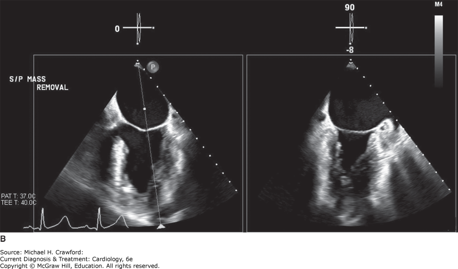
Figure 32-21. Left atrial myxoma. A: Preoperative transesophageal echocardiogram in a 58-year-old woman referred for further work up after a left atrial mass was discovered incidentally on chest computed tomography scan shows a large, heterogeneous mass (arrow) attached to the lateral atrial wall, adjacent to the left atrial appendage. B: Postoperative transesophageal echocardiogram shows complete resection of the atrial myxoma following minimally invasive resection. LA, left atrium; LAA, left atrial appendage; LV, left ventricle.
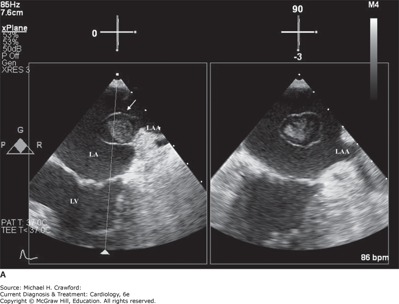
Figure 32-21.
Smaller papillary fibroelastomas and cardiac lipomas (< 1 cm), on the other hand, are often asymptomatic and do not warrant surgical therapy. Cardiac fibromas typically involve large areas of ventricular myocardium and can be a focus for life-threatening ventricular arrhythmias. For this reason, complete and prompt surgical resection is recommended, despite the fact that a large amount of ventricular tissue may be jeopardized in the process, necessitating reconstructive surgery with a synthetic patch.
Rhabdomyomas are best managed conservatively in the asymptomatic patient. Most rhabdomyomas regress spontaneously in childhood or early adolescence. However, symptoms often result from large rhabdomyomas, which can cause inflow and outflow obstruction, warranting immediate intervention. These lesions tend to be multiple, nonencapsulated, and embedded in myocardial tissue, often necessitating extensive surgical dissection. As resection usually involves large territories of myocardium, patients can require inotropic support postoperatively.
Cardiac teratomas, even when benign, can have serious mechanical complications very early in life. Therefore, appropriate treatment includes tumor resection in the fetus or cesarean section delivery and immediate surgical removal of the tumor in the infant.
Paragangliomas can be either benign or malignant, and a subgroup of these tumors are hormonally active, producing catecholamines that give rise to symptoms associated with heightened sympathetic activity. Although surgical excision is the treatment of choice for this tumor, perioperative α- and β-adrenergic blockade is warranted at the time of intervention to prevent catastrophic clinical outcomes from hormonal surges associated with tumor handling.
EnezateT, AlkhatibD, RajaJ, ChintaV, PatelM, OmranJ.AngioVac for minimally invasive removal of intravascular and intracardiac masses: a systematic review. Curr Cardiol Rep. 2022;24(4):377382. doi: 10.1007/s11886-022-01658-9.
NisivacoSM, PatelB, BalkhyHH.Robotic totally endoscopic excision of aortic valve papillary fibroelastoma: The least invasive approach. J Card Surg. 2019;34(12):14921497. 35129741. doi: 10.1111/jocs.14291.
2. Malignant Tumors
Given that the prognosis of malignant cardiac tumors is extremely poor overall, surgery is often performed to relieve symptoms rather than to prolong life. Aggressive and invasive tumors such as cardiac sarcomas often present with mechanical symptoms from obstruction or compression but are not amenable to complete surgical resection. Debulking of the primary tumor can relieve symptoms though is typically only palliative unless adjuvant therapies are effective. Furthermore, given the propensity for primary malignant cardiac tumors to invade surrounding structures, including blood vessels, in-hospital mortality is high.
For metastatic cardiac lesions, surgery may occasionally be performed if there is a high likelihood of complete remission from the primary tumor. However, most commonly, surgical intervention is undertaken for palliative reasons in patients with cardiac metastases.
SvobodovAA, GlushkoLA, ErgashovAY.Surgical treatment of primary cardiac tumors in children systematic review and meta-analysis. Pediatr Cardiol. 2022;43(2):251266. doi: 10.1007/s00246-022-02814-2.
TorabiS, Arjomandi RadA, VardanyanR,et al. Surgical and multimodality treatment of cardiac sarcomas: A systematic review and meta-analysis. J Card Surg. 2021;36(7):24762485. 33797789. doi: 10.1111/jocs.15538
B. Pharmacologic Therapy
Chemotherapy does not have an established role in the management of benign tumors, where cardiac surgery remains the gold standard of treatment. However, in the case of rhabdomyomas associated with TSC, current evidence suggests that sirolimus, an mTOR inhibitor, can increase the regression rate of cardiac rhabdomyomas. Adjuvant chemotherapy regimens containing doxorubicin and paclitaxel have been used to treat malignant cardiac tumors such as sarcomas; however, the data to support a survival benefit with this approach are limited. Chemotherapy has also been used in this context for tumor cytoreduction in preparation for surgical resection and for palliation of unresectable disease.
Primary cardiac lymphoma, on the other hand, is considered more responsive to chemotherapy. A regimen using CHOP (cyclophosphamide, doxorubicin, vincristine, and prednisone), when combined with surgery, radiation, or both, has been shown to induce complete remission in one-third of patients, with a reported median survival of 1 year. This chemotherapy regimen has also been shown to effectively treat distant metastases from primary cardiac lymphomas. Therapy with the monoclonal CD20 antibody rituximab and autologous stem cell transplantation can also yield beneficial results. In patients with primary cardiac lymphoma, it is imperative to perform the first cycle of chemotherapy with close monitoring, as the infiltrative nature of the tumor and the potential brisk response to chemotherapy may result in tumor lysis syndrome.
Rhabdomyosarcomas have been shown to have a better outcome with chemotherapy when compared to other cardiac malignancies, and adjuvant chemotherapy regimens have been tried with varying degrees of success.
ChenXQ, WangYY, ZhangMN,et al. Sirolimus Can Increase the Disappearance Rate of Cardiac Rhabdomyomas Associated with Tuberous Sclerosis: A Prospective Cohort and Self-Controlled Case Series Study. J Pediatr. 2021;233:150155.e4. 33631166. doi: 10.1016/j.jpeds.2021.02.040
GyotenT, DoiT, NaguraS,et al. Primary cardiac malignant lymphoma: survival for 13 years after surgical resection and adjuvant chemotherapy. Ann Thorac Surg. 2015;99(3):10601062. 25742830
C. Radiation
The role of radiotherapy alone or as adjuvant is also limited. The high doses of radiation used to treat sarcomas in other locations are poorly tolerated by the heart, leading to short-term and long-term cardiac toxicity. Radiotherapy has not been shown to systematically prolong survival; however, there are anecdotal reports of its success in cases of cardiac sarcoma, in particular rhabdomyosarcoma. While primary cardiac sarcomas are relatively radio-insensitive, metastatic lesions associated with this tumor can be managed with radiation. With regards to primary cardiac lymphoma, the role of adjuvant radiotherapy remains controversial, with studies showing little to no improvement in overall survival when combined with systemic chemotherapy. However, there are reports of its successful use as salvage therapy in patients not achieving complete remission following chemotherapy.
Cichowska-CwalińskaN, DutkaM, KlapkowskiA, PęksaR, Maciej ZauchaJ, ZauchaR.The role of radiotherapy in the management of primary cardiac lymphoma a case report and the literature review. Leuk Lymphoma. 2019;60(3):812816. doi: 10.1080/10428194.2018.1509321.
D. Cardiac Transplantation
Cardiac transplantation is emerging as a potential therapeutic option for tumors that are not amenable to resection, either because the tumor is too extensive or too infiltrative to allow for complete removal. Fibromas that involve large territories of the left ventricle have been successfully treated with cardiac transplantation, as have vastly invasive cardiac sarcomas in the absence of extracardiac disease. Cardiac transplantation has also been used for certain cases of recurrent atrial myxomas. This approach is limited by the potential for posttransplant recurrence, which may be accelerated by immunosuppressive therapy.
Rodriguez-GonzalezM, Pérez-ReviriegoAA,et al. Primary cardiac fibroma in infants: A case report and review of cases of cardiac fibroma managed through orthotopic heart transplant. Ann Pediatr Cardiol. 2021;14(2):224227. 25742830. doi: 10.4103/apc.APC_78_20.
E. Palliation
With newer and more scientific approaches to palliative care in the current medical milieu, terminal life-limiting cardiac neoplasms can be treated with palliation in cases where surgical outcomes are known to be poor. Specifically, in patients with cardiac sarcomas that are not amenable to complete resection, palliative surgery may be performed to relieve mechanical symptoms. In patients with malignant pericardial mesothelioma, a tumor with a poor response to chemotherapy and radiation, surgical pericardiectomy can serve as a palliative measure. In addition, in carefully selected patients with cardiac metastases, resection of metastatic lesions may provide symptomatic improvement and prolong life.
Prognosis ⬆
The prognosis for cardiac tumors varies significantly with the nature of the neoplasm. For primary cardiac tumors, surgical resection usually results in complete cure if the tumor is benign. However, 1.5% of myxomas can recur within 10–15 years, usually in patients with familial or syndromic myxomas. For this reason, patients should be monitored with serial echocardiograms approximately every 5 years after resection.
Malignant primary cardiac tumors, in contrast, have a dismal long-term prognosis, most commonly because of early local invasion, metastatic spread, or recurrence after removal. Survival time, as expected, correlates inversely with tumor size and the degree of regional tumor extension at the time of surgery. In a large cohort of patients within the past decade, 1-, 3-, and 5-year survival rates were found to be 50%, 24%, and 19%, respectively.
The overall prognosis with cardiac sarcomas is very poor, with mean survival ranging from 9.6 to 16.5 months. Irrespective of histologic type, common features that predict a less aggressive disease course are location in the left atrium, low mitotic activity with scarce cellular pleomorphism on pathology, and absence of metastasis or necrosis. The survival of patients with primary cardiac lymphoma can be as long as 5 years with appropriate therapy but whittles down significantly if left untreated, to as low as 1 month. The median survival with cardiac lymphoma is just 7 months after initial diagnosis and is likely a result of distant metastasis, which can occur even after seemingly successful eradication of the primary cardiac tumor. When paragangliomas are completely resected, a 10-year survival rate as high as 84% has been reported in some series. However, postoperative recurrence rates for these tumors are in the 50% range, making routine surveillance imperative.
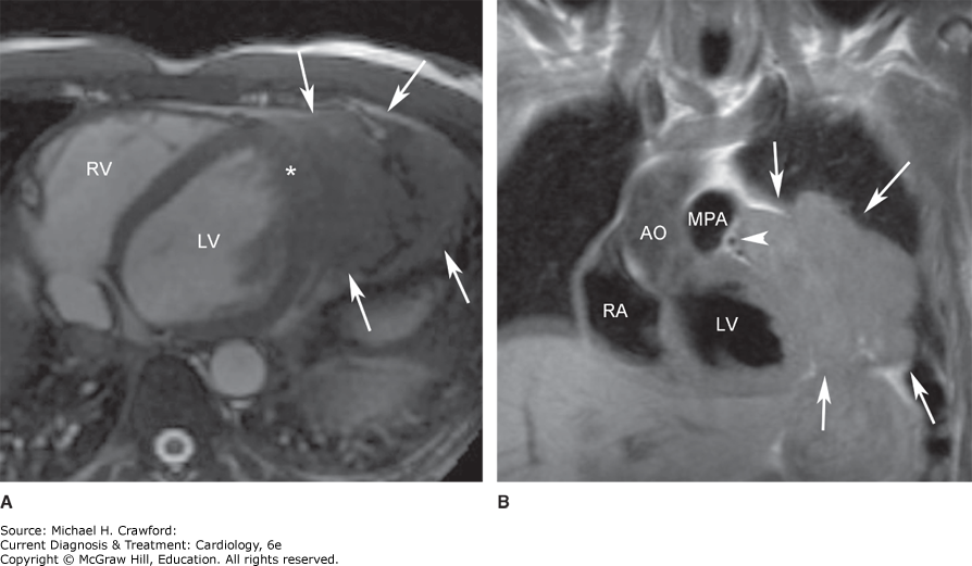
The prognosis for secondary cardiac tumors from metastases is uniformly poor, and long-term survival rates are similar to those quoted for the metastatic spread of each individual neoplasm (Figure 32–22).
Figure 32-22. Metastatic cardiac tumors. A: Magnetic resonance imaging of the heart in a 50-year-old man who presented with dyspnea on exertion 3 months after being diagnosed with a melanoma on his back. A large pericardial mass was demonstrated on this bright blood-cine steady-state free precession magnetic resonance image in the axial plane. The arrows show the extension of the mass into the pleural space and possibly lung parenchyma. The asterisk indicates different texture of the myocardial signal, suggesting myocardial invasion of the mass. B: Coronal black-blood T1-weighted image showing extension of the mass anteriorly and superiorly to impinge on the proximal left anterior descending artery (arrowhead). Biopsy of the mass confirmed metastatic melanoma. AO, aorta; LV, left ventricle; MPA, main pulmonary artery; RA, right atrium; RV, right ventricle. (Reproduced with permission from Karen Ordovas.)
OliveiraGH,et al. Characteristics and survival of malignant cardiac tumors: a 40-year analysis of over 500 patients. Circulation. 2015;132:23952402. 26467256
PadalinoMA, VidaVL, BoccuzzoG,et al. Surgery for primary cardiac tumors in children: early and late results in a multicenter European Congenital Heart Surgeons Association study. Circulation. 2012;126(1):2230. 22626745. doi: 10.1161/CIRCULATIONAHA.111.037226