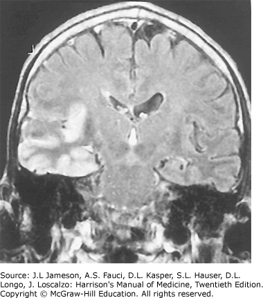
Coronal Flair Magnetic Resonance Image from a Pt with Herpes Simplex Encephalitis

Coronal FLAIR magnetic resonance image from a pt with herpes simplex encephalitis. Note the area of increased signal in the right temporal lobe (left side of image) confined predominantly to the gray matter. This pt had predominantly unilateral disease; bilateral lesions are more common, but may be quite asymmetric in their intensity.