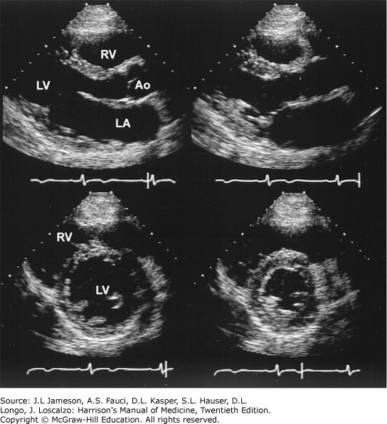
Two-Dimensional Echocardiographic Still-Frame Images of a Normal Heart

Two-dimensional echocardiographic still-frame images of a normal heart. Upper: Parasternal long axis view during systole and diastole (left) and systole (right). During systole, there is thickening of the myocardium and reduction in the size of the left ventricle (LV). The valve leaflets are thin and open widely. Lower: Parasternal short axis view during diastole (left) and systole (right) demonstrating a decrease in the left ventricular cavity size during systole as well as an increase in wall thickness. Ao, aorta. (Reproduced from Myerburg RJ: Harrison's Principles of Internal Medicine, 12th ed, 1991.)