MRI Findings in MS

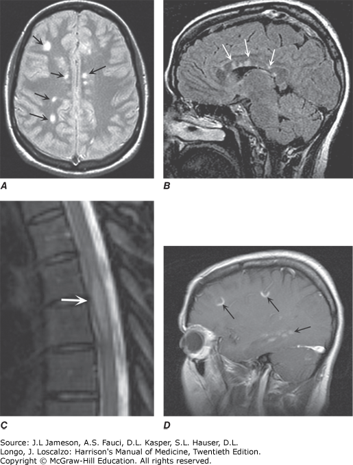
MRI findings in MS. A. Axial first-echo image from T2-weighted sequence demonstrates multiple bright signal abnormalities in white matter, typical for MS. B. Sagittal T2-weighted fluid-attenuated inversion recovery image (FLAIR) in which the high signal of CSF has been suppressed. CSF appears dark, while areas of brain edema or demyelination appear high in signal as shown here in the corpus callosum (arrows). Lesions in the anterior corpus callosum are frequent in MS and rare in vascular disease. C. Sagittal T2-weighted fast spin-echo image of the thoracic spine demonstrates a fusiform high-signal-intensity lesion in the midthoracic spinal cord. D. Sagittal T1-weighted image obtained after the IV administration of gadolinium reveals focal areas of blood-brain barrier disruption, identified as high-signal-intensity regions (arrows).