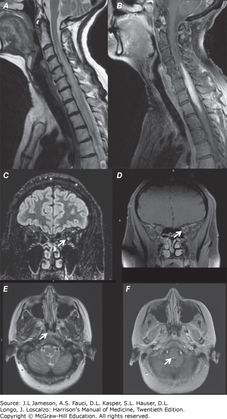
Imaging Findings in Neuromyelitis Optica: Longitudinally Extensive Transverse Myelitis, Optic Neuritis and Brainstem Involvement

Imaging findings in neuromyelitis optica: longitudinally extensive transverse myelitis, optic neuritis and brainstem involvement. (A) Sagittal fluid attenuation inversion recovery (FLAIR) cervical-spine magnetic resonance image (MRI) showing an area of increased signal change on T2-weighted imaging spanning more than 3 vertebral segments in length. (B) Sagittal T1-weighted cervical-spine MRI following gadolinium-DPTA infusion showing enhancement. (C) Coronal brain MRI shows hyperintense signal on FLAIR imaging within the left optic nerve. (D) Coronal T1-weighted brain MRI following gadolinium-DPTA infusion shows enhancement of the left optic nerve. (E) Axial brain MRI shows an area of hyperintense signal on T2-weighted imaging within the area postrema (arrows). (F) Axial T1-weighted brain MRI following gadolinium-diethylene triamine pentaacetic acid (DPTA) infusion shows punctate enhancement of the area postrema (arrows).