West Point View for the Identification of a Glenoid Rim Lesion.
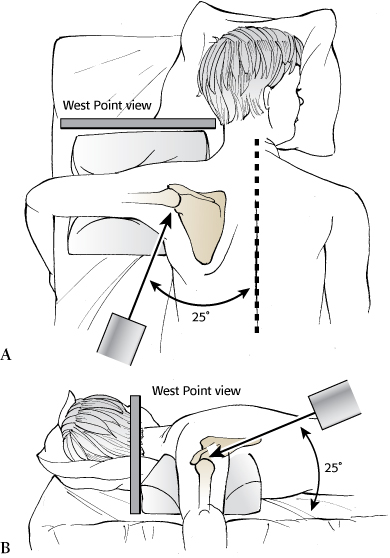
This x-ray is taken with the patient in the prone position. The beam is angled approximately 25 degrees (A) to provide a tangential view of the glenoid. In addition, the beam is angled 25 degrees downward (B) to highlight the anterior and posterior aspects of the glenoid. In this fashion, the entire glenoid rim can be clearly visualized.
(From JawaA, RicchettiET. Glenulohumeral instability. In: Court-BrownCM, KeckmanJD, McQueenMM, et al., eds. Rockwood and Green’s Fractures in Adults. Vol 1. 8th ed. Philadelphia: Wolters Kluwer Health; 2015:1503–1571.)