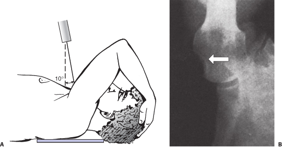
(A) The Position of the Patient for the Stryker Notch View. The Patient is Supine with the Cassette Posterior to the Shoulder. The Humerus is Flexed Approximately 120 Degrees So the Hand Can Be Placed on Top of the Patient’s Head. Note that the Angle of the x-Ray Tube is 10 Degrees Superior. (B) The Radiograph Can Clearly Reveal the Presence of Any Osseous Defects (Arrow).
(From JawaA, RicchettiET. Glenulohumeral instability. In: Court-BrownCM, KeckmanJD, McQueenMM, et al., eds. Rockwood and Green’s Fractures in Adults. Vol 1. 8th ed. Philadelphia: Wolters Kluwer Health; 2015:1503–1571.)