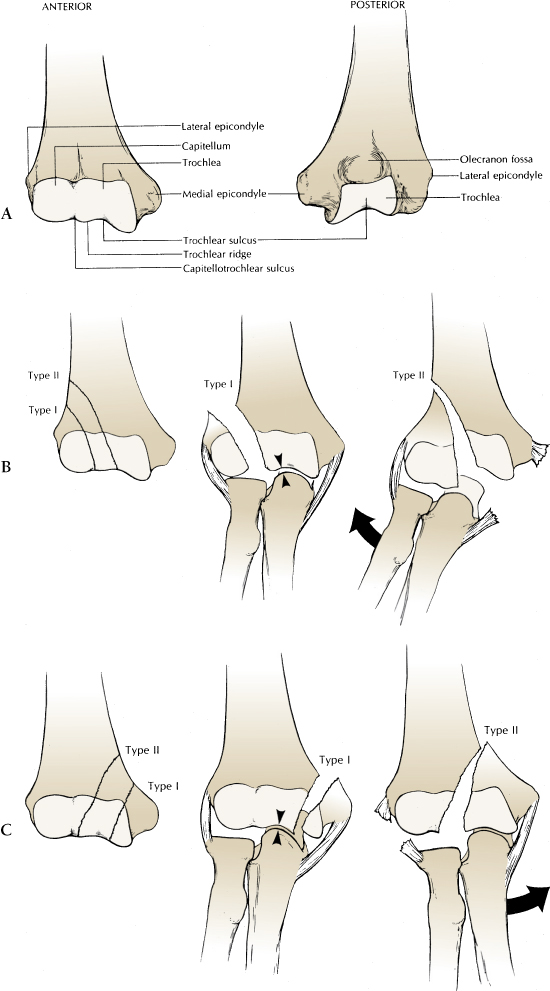
Classification of Condylar Fractures According to Milch and the Location of the Common Fracture Lines Seen in Types I and II Fractures of the Lateral (B) and Medial (C) Condyles. (A) Anterior View of the Anatomy of the Distal Articular Surface of the Humerus. The Capitello Trochlear Sulcus Divides the Capitellar and Trochlear Articular Surfaces. The Lateral Trochlear Ridge is the Key to Analyzing Humeral Condyle Fractures. In Type I Fractures, the Lateral Trochlear Ridge Remains with the Intact Condyle, Providing Medial-to-Lateral Elbow Stability. In Type II Fractures, the Lateral Trochlear Ridge is a Part of the Fractured Condyle, Which May Allow the Radius and Ulna to Translocate in a Medial-to-Lateral Direction with Respect to the Long Axis of the Humerus. (B) Fractures of the Lateral Condyle. In Type I Fractures, the Lateral Trochlear Ridge Remains Intact, Therefore Preventing Dislocation of the Radius and Ulna. In Type II Fractures, the Lateral Trochlear Ridge is a Part of the Fractured Lateral Condyle. With Capsuloligamentous Disruption Medially, the Radius and Ulna May Dislocate. (C) Fractures of the Medial Condyle. In Type I Fractures, the Lateral Trochlear Ridge Remains Intact to Provide Medial-to-Lateral Stability of the Radius and Ulna. In Type II Fractures, the Lateral Trochlear Ridge is a Part of the Fractures Medial Condyle. With Lateral Capsuloligamentous Disruption, the Radius and Ulna May Dislocate Medially on the Humerus.