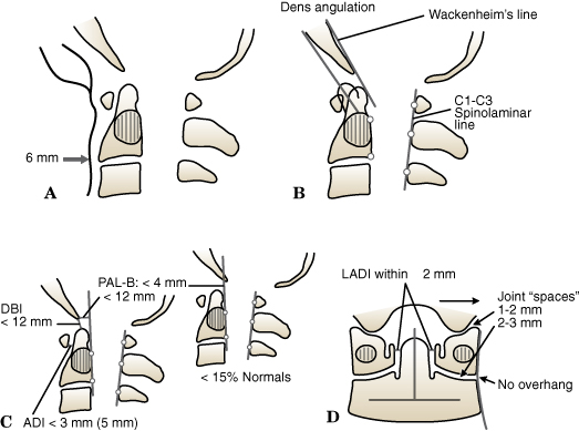
(A) Prevertebral Soft Tissue Shadow. In a Healthy Recumbent Adult Without an Endotracheal Tube, the Prevertebral Soft Tissue Shadow Should Not Exceed 6 Mm. (B) Bony Screening Lines and Dens Angulation. The Anterior Cortex of the Odontoid Should Parallel the Posterior Cortex of the Anterior Ring of the Atlas. Any Kyphotic or Lordotic Deviation Should Be Viewed with Suspicion for an Odontoid Fracture or Transverse Ligament of the Atlas Disruption. Wackenheim Line is Drawn As a Continuation from the Clivus Caudally. The Tip of the Odontoid Should Be Within 1 to 2 Mm of This Line. The C1–c3 Spinolaminar Line’s Reference Points are Drawn from the Anterior Cortex of the Laminae of the Atlas, Axis, and C3 Segments, Which Should Fall Within 2 Mm of One Another. Greater Deviation Should Raise Suspicion of Atlantoaxial Translation or Disruption of the Neural Arches of Either Segment. (C) Ligamentous Injury Reference Lines (Lateral x-Rays). The Atlantodens Interval (Adi) Should Be 3 Mm in an Adult (5 Mm in a Child). The Space Available for the Cord is Measured As the Distance from the Posterior Cortex of the Odontoid Tip to the Anterior Cortex of the Posterior Arch of the Atlas and Should Amount to More Than 13 Mm. The Dens–basion Interval (Dbi) is the Distance Between the Odontoid Tip and the Distal End of the Basion. It Should Be 12 Mm in Adults. The Posterior Axis Line (Pal-B) Should Not Be More Than 4 Mm Anterior and Should Be 12 Mm Posterior to the Basion. (D) Bony Screening Lines (AP Imaging). The Left and Right Lateral Atlantodens Intervals (Ladis) Should Be Symmetric to One Another (with 2-Mm Deviation). The Bony Components of the Atlantooccipital Joints Should Be Symmetric and Should Not Be Spaced More Than 2 Mm Apart on AP Images.
(Courtesy of Fred Mann, MD, Professor of Radiology, University of Washington, Seattle.)