section name header

Symptoms

Red eye, burning, foreign body sensation, pain, tearing, itching mild photophobia, and frequent blinking. The course can be chronic with exacerbations and remissions.

Signs

Critical

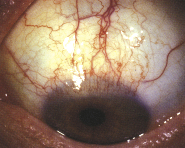
Sectoral thickening, inflammation, and radial injection of the superior bulbar conjunctiva, especially at the limbus. Superior bulbar conjunctivochalasis often present (see Figure 5.4.1).

5-4.1 Superior limbic keratoconjunctivitis.

Gervasio-ch005-image011

Other

Fine papillae on the superior tarsal conjunctiva; fine punctate fluorescein staining on the superior cornea, limbus, and conjunctiva; superior corneal micropannus and filaments. Usually bilateral, frequently asymmetric.
Work Up

Workup
  1. History: Recurrent episodes? Thyroid disease?
  2. Slit lamp examination with fluorescein, lissamine green, or rose bengal staining, particularly of the superior cornea and adjacent conjunctiva. Lift the upper eyelid to see the superior limbal area and then evert to visualize the tarsus. Sometimes the localized hyperemia is best appreciated by direct inspection with room light rather than at the slit lamp, by raising the eyelids of the patient on downgaze.
  3. Thyroid function tests (there is a 50% prevalence of current or remote thyroid disease in patients with superior limbic keratoconjunctivitis).

Treatment

Mild

  1. Aggressive lubrication with preservative-free artificial tears four to eight times per day and artificial tear ointment q.h.s.
  2. Consider punctal occlusion with plugs or cautery because of association with dry eyes.
  3. Treat any concurrent blepharitis.
  4. Consider treatment with cyclosporine 0.05%, cyclosporine 0.09%, or lifitegrast 5% b.i.d. if not responding to lubrication.
  5. In the absence of dry eyes, a therapeutic bandage disposable soft contact lens can be placed to help relieve symptoms and facilitate healing.

Moderate to Severe (in Addition to Preceding)

  1. Autologous serum drops may be tried with intermittent dosing throughout the day.
  2. Consider treatment with topical tacrolimus 0.03% ointment b.i.d. if there is no improvement with aggressive lubrication (off-label in the eye).
  3. If a significant amount of mucus or filaments are present, add acetylcysteine 10% drops four to six times per day. Low potency topical steroids such as loteprednol, rimexolone, or fluorometholone can be used for short courses to treat exacerbations.
  4. Application of silver nitrate 0.5% solution on a cotton-tipped applicator for 10 to 20 seconds to the superior tarsal and superior bulbar conjunctiva after topical anesthesia (e.g., proparacaine). This is followed by irrigation with saline and use of antibiotic ointment (e.g., erythromycin) q.h.s. for 1 week.
  5. A low dose of doxycycline can be a helpful adjuvant to counteract matrix metalloproteinase upregulation caused by superior limbic keratoconjunctivitis.
  6. Botulinum toxin can be injected into the muscle of Riolan for temporary relief of symptoms.
  7. Surgical considerations include conjunctival cautery, cryotherapy, conjunctival resection (with or without amniotic membrane graft), recession of the superior bulbar conjunctiva, or high-frequency radiowave electrosurgery.
NOTE:

Do not use silver nitrate (75% to 95%) cautery sticks, which cause severe ocular burns.

Follow Up

Every 2 to 4 weeks during an exacerbation. If signs and symptoms persist despite multiple medical treatment strategies, surgical options should be considered.