Description
CT uses ionizing radiation and computer-assisted formatting to produce multiple cross-sectional planar images. With the use of multidetector technology, CT now provides direct axial, coronal, and parasagittal images without the need for patient repositioning or data reformatting. Bone and soft tissue windows should always be reviewed in axial, coronal, and parasagittal orientations (see Figures 14.2.1 to 14.2.3). Orbital studies should be obtained using the lowest radiation dose necessary for diagnosis (as low as reasonably acceptable [ALARA]—see below). This is especially important in children; for accreditation by the American College of Radiology, health care facilities must maintain specific pediatric protocols for CT. Radiopaque iodinated contrast allows for more extensive evaluation of vascular structures and areas where there is a breakdown of the normal capillary endothelial barrier (as in inflammation).
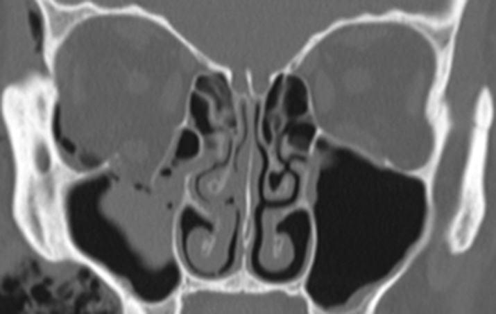
14-2.3 Coronal bone window shows the fracture again.

In bone windows, the soft tissue detail fades, but bone detail is enhanced, allowing for better examination of bony anatomy.
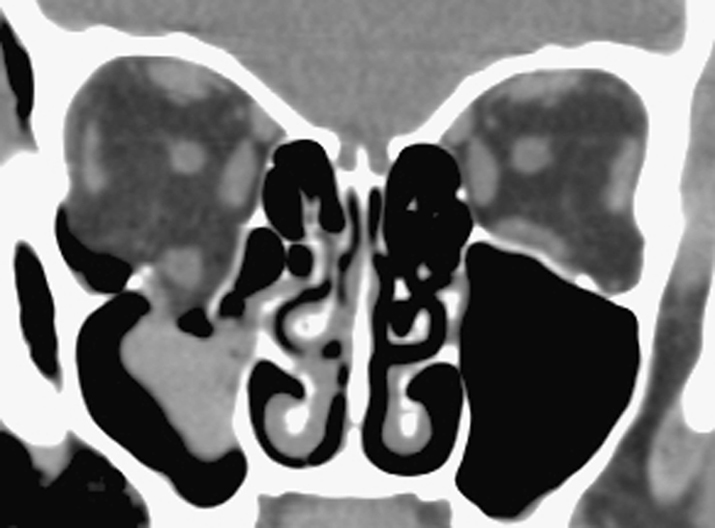
14-2.2 Coronal soft tissue window shows a large blowout fracture of the orbital floor.

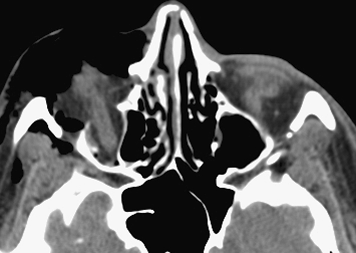
This finding was missed on the axial study in Figure 14.2.1, demonstrating the importance of reviewing both axial and coronal images.
14-2.1 Axial soft tissue window of inferior orbit shows abnormality, which is difficult to assess using this window.

Uses in Ophthalmology
- Excellent for defining bone abnormalities such as fractures (orbital wall or optic canal), calcification, or bony involvement of a soft tissue mass.
- Locating suspected intraorbital or intraocular metallic foreign bodies. Glass, wood, and plastic are less radiopaque and therefore more difficult to isolate on CT.
- Soft tissue windows are good for determining some pathologic features, including orbital cellulitis/abscess, inflammation, and tumors. May be useful in determining posterior scleral rupture when clinical examination is inconclusive, but B-scan ultrasonography is likely more sensitive. CT should never be used to definitively rule out globe rupture; clinical examination and/or surgical exploration are more sensitive modalities.
- Excellent for imaging paranasal sinus anatomy and disease.
- Head CT is very sensitive for locating parenchymal, subarachnoid, subdural, and epidural hemorrhage in either acute or subacute settings. Although CT may show orbital hemorrhage as a nonspecific opacification within radiolucent orbital fat, it is significantly less sensitive for orbital hemorrhage than central nervous system hemorrhage.
- Imaging modality of choice for most cases of thyroid eye disease. See 7.2.1, THYROID EYE DISEASE.
- Loss of consciousness or unwitnessed head trauma with poor history requires CT of the brain unless otherwise recommended by a neurosurgical consultant.
- Note that CT of the brain does not provide adequate detail of the orbital anatomy, and orbital CT does not image the entire brain. Each site has its own specific, separate CT protocol.
Guidelines for Ordering an Orbital Study
- Always order a dedicated orbital study if ocular or orbital pathology is suspected. Always include views of paranasal sinuses and cavernous sinuses.
- Order both direct axial and direct coronal views if older scanners are used. Newer, multidetector CT scanners provide excellent views in coronal and parasagittal planes with very short scan times and no patient repositioning. Multidetector CT has supplanted older technology in the vast majority of hospitals and imaging centers.
- When evaluating traumatic optic neuropathy, request 1-mm cuts of the orbital apex and optic canal to rule out bony impingement of the optic nerve. See 3.11, TRAUMATIC OPTIC NEUROPATHY.
- When attempting to localize ocular or orbital foreign bodies, order 1-mm cuts.
- Intravenous contrast may be necessary for suspected infections or inflammatory conditions. Contrast is helpful in distinguishing orbital cellulitis from abscess. However, it is not mandatory to rule out orbital inflammation or postseptal involvement. Relative contraindications for contrast include renal failure, diabetes, congestive heart failure, multiple myeloma or other proteinemias, sickle cell disease, multiple severe allergies, and asthma. Check renal function and discuss the options with the radiologist for patients in whom renal insufficiency is suspected. Shellfish allergy is not a contraindication for CT contrast.
- Obtain a pregnancy test before obtaining CT scans in females of childbearing age.
- CT angiography (CTA) is helpful in diagnosing intracranial vascular pathology, including aneurysms. It is available on all multidetector CT scanners and may be more sensitive than magnetic resonance angiography (MRA) in specific clinical situations. However, it requires the use of intravenous iodinated contrast. Also note that CTA requires significantly more radiation than CT. CTA should be avoided if at all possible in children; MRA is the preferred modality in the pediatric age group.
- CT scans may be obtained in children with careful consideration of the risk of radiation exposure versus benefits of performing the scan. According to the ALARA paradigm, it is best to avoid CT if possible in children unless there is no reasonable substitute. Check with your radiologist about using a pediatric protocol to limit radiation exposure. Each CT scan exposes children to a cumulative radiation dose that may increase the risk of malignancy over their lifetime. Radiation exposure from CT scans in children is of particular concern when serial imaging is required. In these cases, MRI/MRA is often a better choice for children, although intravenous sedation or general anesthesia may be needed because of the significantly longer scanning time.
The radiologist may recommend premedication with corticosteroids and antihistamines if contrast is required and a prior allergy is suspected. Follow the protocol recommended by your radiology department. |
CT is an extremely valuable tool and is an acceptable imaging modality in children when necessary. Because of its easy availability, rapid scan time, excellent bone resolution, and ability to diagnose acute intracranial hemorrhage, CT is the modality of choice in head and orbital trauma in all age groups, including children. |