section name header

General Information

Definition

Ophthalmoplegia secondary to lesion in the medial longitudinal fasciculus (MLF).

Symptoms

Double vision, blurry vision, or vague visual complaints.

Signs

(See Figures 10.13.1 and 10.13.2.).

Critical

Weakness or paralysis of adduction, with horizontal jerk nystagmus of the abducting eye.

NOTE:

INO is localized to the side with the weak adduction.

Other

May be unilateral or bilateral (WEBINO: “wall-eyed,” bilateral INO). Upbeat nystagmus on upgaze may occur when INO is unilateral or bilateral. The involved eye can sometimes turn in when attempting to read (intact convergence). A skew deviation (relatively comitant vertical deviation not caused by neuromuscular junction disease or intraorbital pathology) may be present; brainstem and posterior fossa pathology should be ruled out. With skew deviation, the three-step test cannot isolate a specific muscle. See 10.7, ISOLATED FOURTH CRANIAL NERVE PALSY. In addition, presence of other neurologic signs, including gaze-evoked nystagmus, gaze palsy, dysarthria, ataxia, and hemiplegia, favors skew deviation rather than fourth cranial nerve palsy.
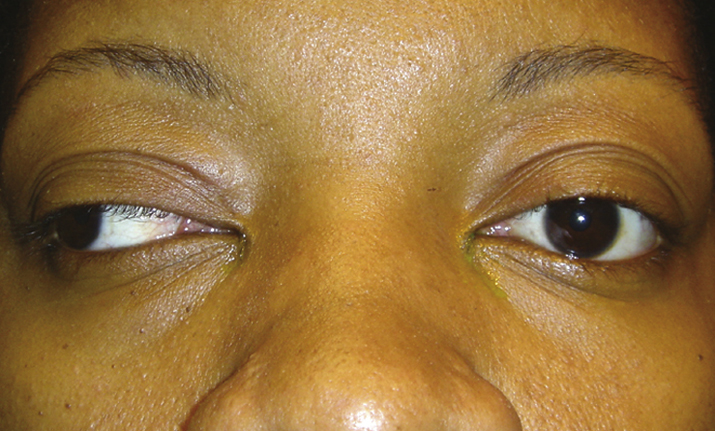
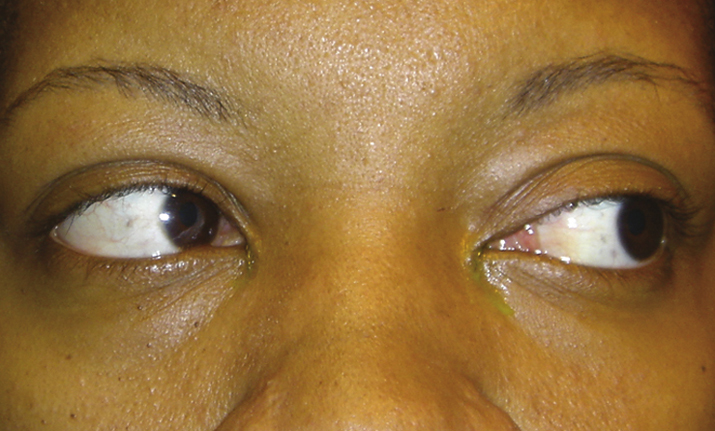
10-13.2 Left internuclear ophthalmoplegia: Right gaze with severe adduction deficit.

Gervasio-ch010-image016

10-13.1 Left internuclear ophthalmoplegia: Left gaze showing full abduction.

Gervasio-ch010-image015

Differential Diagnosis

Differential Diagnosis of Attenuated Adduction

Etiology

  • MS: More common in young patients, usually bilateral.
  • Brainstem stroke: More common in elderly patients, usually unilateral.
  • Brainstem mass lesion.
  • Rare causes: CNS cryptococcosis, tuberculosis granuloma, pyoderma gangrenosum (all shown to cause WEBINO).

Work Up

Workup
  1. History: Age? Are symptoms constant or only toward the end of the day with fatigue? Prior optic neuritis, urinary incontinence, numbness or paralysis of an extremity, or another unexplained neurologic event (concerning for MS)?
  2. Complete evaluation of eye movement to rule out other eye movement disorders (e.g., sixth cranial nerve palsy, skew deviation).
  3. Ice test, rest test, or edrophonium chloride test when the diagnosis of myasthenia gravis cannot be ruled out.
  4. MRI of the brainstem and midbrain.
NOTE:

Ocular motility can appear to be full, but a muscular weakness can be detected by observing slower saccadic eye movement in the involved eye compared with the contralateral eye. The adducting saccade is assessed by having the patient fix on the examiner’s finger held laterally and then asking the patient to make a rapid eye movement from lateral to primary gaze. If an INO is present, the involved eye will show a slower adducting saccade than the uninvolved eye. The contralateral eye may be tested in a similar fashion.

Treatment/Follow Up

  1. If an acute stroke is diagnosed, admit to the hospital for neurologic evaluation and observation.
  2. If concern for demyelinating disease, consider treatment recommendations described in 10.14, OPTIC NEURITIS.
  3. Patients are managed by physicians familiar with the underlying disease.