section name header

Symptoms

Ocular complaints are rare. Patients experience symptoms of cirrhosis, neurologic disorders, psychiatric problems, or renal disease. Onset typically between 5 and 40 years of age.

Signs

Critical

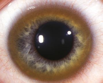
Kayser–Fleischer ring: 1- to 3-mm, brown, yellow, green, or reddish band that represents copper deposition in the peripheral Descemet membrane (See Figure 13.9.1). Present in 50% to 60% of patients with isolated hepatic involvement and more than 90% of patients with neurologic manifestations. First appears superiorly (may only be visible on gonioscopy) and eventually forms a ring involving the entire corneal periphery extending to the limbus. Anterior-segment optical coherence tomography (OCT) may be useful in detecting early Kayser–Fleischer rings that are not readily detected on slit-lamp examination; appears as linear hyperreflective material on Descemet membrane.

Figure 13.9.1: Kayser–Fleischer ring in Wilson disease.

Rapuano9781975243722-ch013_f004.jpg

Other

“Sunflower” cataract: Ring or stellate yellow, brown, or reddish anterior capsule opacity due to copper deposition under the lens capsule.

Differential Diagnosis

Etiology

Autosomal recessive inborn error of copper metabolism that results in impaired copper excretion and toxic accumulation of copper in multiple organ systems (e.g., liver, brain, cornea, kidney). ATP7B gene on chromosome 13. 

Workup

  1. Slit-lamp examination: Deposition at Descemet membrane is apparent with a narrow slit beam.

  2. Gonioscopy if the Kayser–Fleischer ring is not evident on routine slit-lamp examination.

  3. Check serum copper and ceruloplasmin levels, urine copper level (low serum copper and ceruloplasmin and elevated urine copper levels with ocular findings are diagnostic).

  4. Referral to appropriate systemic specialists.

Treatment

  1. Lifelong systemic therapy (e.g., zinc salts, d-penicillamine, trientine) is instituted by the appropriate systemic specialist. Liver transplantation may be required for fulminant hepatic failure or disease progression after medical therapy.

  2. While a patient is on a chelating agent (e.g., d-penicillamine), routine lab monitoring of serum copper, ceruloplasmin, liver enzymes, international normalized ratio (INR), complete blood count with differential, and urinalysis is recommended.

  3. Patients are recommended to start a low-copper diet.

  4. Ocular manifestations usually require no specific treatment.

Follow-Up

  1. Systemic therapy and monitoring.

  2. Successful treatment should lead to resorption of the corneal copper deposition and clearing of the Kayser–Fleischer ring, although residual peripheral corneal changes may remain. This change can be used to monitor treatment response. Reappearance of ring may suggest noncompliance with treatment.

  3. Consider referral of family members for genetic testing for early detection and prevention.