Proper evaluation of the configuration of the anterior chamber requires the use of at least three descriptors: the point at which the peripheral iris is adherent to the cornea or uvea, the depth of the anterior chamber, and the curvature of the peripheral iris. The Spaeth grading system of the anterior chamber angle takes into account all the three attributes.
(See Figure A.14.1.)

A = Anterior to the Schwalbe line (SL)
B = Between SL and scleral spur
C = Scleral spur visible (common in blacks and Asians)
D = Deep: ciliary body visible (common in whites)
E = Extremely deep: >1 mm of the ciliary body is visible
Indentation gonioscopy may be necessary to differentiate false opposition of the iris against the structures in the iridocorneal angle from the true iris insertion. First, make note of the most posterior portion of the inner wall of the eye that can be seen without indentation. The iris is then displaced posteriorly by compressing the cornea. This allows for the determination of the true iris insertion. When the true iris insertion is different from the preindentation appearance, the preindentation appearance is placed in parentheses. For example, a (B)D grade means that without indentation, it is not possible to see any of the scleral spur or ciliary body, but with indentation, the ciliary body can be seen.
The angular width that is measured is the angle between a line parallel to the corneal endothelium at the SL and a line parallel to the anterior surface of the iris.
Pigmentation of the Posterior Trabecular Meshwork (PTM)
Viewing at 12 o’clock in the angle with the mirror at 6 o’clock position, pigmentation graded on a scale of 0 (no PTM pigment seen) to 4+ (intense PTM pigment).
Pigmentation >2+ is usually pathologic though can occur naturally in patients with heavy skin pigmentation.
Examples of the Spaeth Grading System
(B)D30p 0 ptm = open, atypical narrow angle, occludable with dilation.
D40c 4+ ptm = open-angle characteristic of patients with myopia or iris pigment dispersion syndrome.
(See Figure A.14.2.)
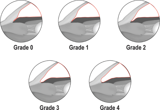
Grade 1: Extremely narrow angle (10 degrees). Only the SL, and perhaps also the top of the trabecula, can be visualized. Angle closure is probable.
Grade 2: Moderately narrow angle (20 degrees). Only the trabecular meshwork can be seen. Angle closure is possible.
Grade 3: Moderately open angle (20 to 35 degrees). The scleral spur can be seen. Angle closure is not possible.
Grade 4: Angle wide open (35 to 45 degrees). The ciliary body can be visualized with ease. Angle closure is not possible.
Van Herick Angle Depth Estimation