(See Figure 10.2.1.)
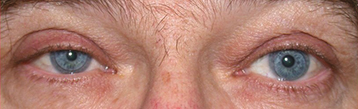
Anisocoria that is greater in dim illumination (especially during the first few seconds after the light is dimmed). The abnormal small pupil dilates less than the normal, larger pupil. Mild ptosis (2 mm) and lower eyelid elevation (reverse ptosis) occur on the side of the small pupil.
Lower intraocular pressure, lighter iris color in congenital cases (iris heterochromia), loss of sweating (anhidrosis, distribution depends on the site of lesion), and transient increase in accommodation (older patients hold their reading card closer in the Horner eye). Involved eye may have conjunctival hyperemia due to decreased episcleral vascular tone. Light and near reactions are intact.
First-order neuron disorder: Stroke (e.g., vertebrobasilar artery insufficiency or infarct), tumor, multiple sclerosis (MS). Rarely, severe osteoarthritis of the neck with bony spurs.
Second-order neuron disorder: Tumor (e.g., lung carcinoma, metastasis, thyroid adenoma, neurofibroma), aortic aneurysm (e.g., tertiary syphilis). Patients with pain in the arm or scapular region should be suspected of having a Pancoast tumor. In children, consider neuroblastoma, lymphoma, or metastasis.
Third-order neuron disorder: Headache syndrome (e.g., cluster, migraine, Raeder paratrigeminal syndrome), internal carotid dissection, varicella zoster virus, otitis media, TolosaHunt syndrome, neck trauma/tumor/inflammation, cavernous sinus pathology.
Congenital Horner syndrome: May also be caused by birth trauma.
Other rare causes: Cervical paraganglioma, ectopic cervical thymus.
Diagnosis confirmed by a relative reversal in anisocoria with apraclonidine (0.5% or 1%). The miotic pupil with Horner syndrome will appear larger than the normal pupil 40 to 60 minutes after apraclonidine instillation. Alternatively, 10% cocaine may be used. Place one drop of 10% cocaine in both eyes. Check in 15 minutes. If no change in pupillary size is noted, repeat drops and recheck the pupils in 15 minutes (repeat until normal pupil dilates). A Horner pupil dilates less than the normal pupil. The test is considered positive if there is >1 mm of anisocoria 40 to 60 minutes after cocaine eye drop instillation.
A third-order neuron disorder may be distinguished from a first-order and second-order neuron disorder with hydroxyamphetamine. Place one drop of 1% hydroxyamphetamine in both eyes. Check in 15 minutes and repeat if no change in pupillary size is noted. Failure of the Horner pupil to dilate to an equivalent degree as the fellow eye indicates a third-order neuron lesion, which may help guide the workup. However, most experts feel the entire sympathetic pathway should be imaged in Horner syndrome regardless of the results of pharmacologic testing. Additionally, hydroxyamphetamine is often unavailable even from compounding pharmacies.
Determine the duration of the Horner syndrome from the patients history and an examination of old photographs. New-onset Horner syndrome requires a more urgent diagnostic workup to exclude life-threatening etiologies (e.g., internal carotid artery dissection, which can present with transient visual loss, head/neck/face pain, pulsatile tinnitus, or dysgeusia [foul taste in the mouth]). An old Horner syndrome is more likely to be benign.
History: Headaches? Arm pain? Previous stroke? Previous surgery that may have damaged the sympathetic chain, including cardiac, thoracic, thyroid, or neck surgery? History of head or neck trauma? Ipsilateral neck pain?
Physical examination (especially check for supraclavicular nodes, thyroid enlargement, or a neck mass).
Computed tomography (CT) or magnetic resonance imaging (MRI) of the chest to evaluate lung apex for possible mass (e.g., Pancoast tumor).
Magnetic resonance angiography (MRA) or CT angiography (CTA) of the head/neck to evaluate for carotid artery dissection (especially with neck pain). Obtain carotid angiogram if MRA or CTA yield equivocal results.
Treat the underlying disorder if possible.
Workup acute Horner syndromes as soon as possible to rule out life-threatening causes. Neuroimaging (as above) should be performed immediately for dissection. Remaining workup may be performed within 1 to 2 days.
Chronic Horner syndrome can be evaluated with less urgency. There are no ocular complications that necessitate close follow-up.